|
| ||||||
Flash Cards Video - Got Skills?
NOTE: You should familiarize yourself with the anatomy of the heart and have a good understanding of the flow of blood through the heart before reading this page. You may want to go to the "Anatomy of the Heart" page to review (link is provided above).
The circulatory system functions to circulate blood through the body. The blood carries vital nutrients, oxygen and hormones to the cells, and also picks up unwanted toxins, waste and carbon dioxide from the cells.
The circulatory system circulates blood around two loops.
1) THE PULMONARY CIRCUIT - Functions to bring blood from the heart to the lungs and back to the heart
2) THE SYSTEMIC CIRCUIT - Functions to bring blood from the heart to all the parts of the body (except the lungs) and then back to the heart
The Components of the Circulatory System are...
1) The Heart
- The function of the heart is to pump the blood around the body. It does this by creating a pressure gradient that forces the movement of the blood.
2) The Blood
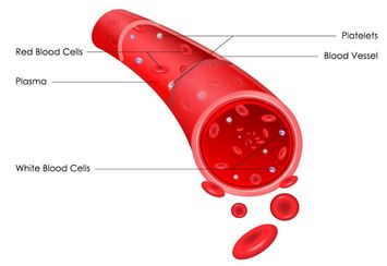
- The function of the blood is to deliver oxygen and nutrients to the cells of the body and to take away the unwanted waste products and carbon dioxide from the cells.
3) The Blood Vessels
- The function of the blood vessels is to provide the network of passage ways that the blood can travel in. In short, the blood vessels allow the blood to travel through the body.
Overview of the Vessels of the Thoracic Cavity
The Aorta
|
We begin our discussion on blood vessels with the AORTA. The aorta is the largest and strongest artery in the body and it marks the beginning of the systemic circuit (HEART --> BODY --> HEART). Oxygenated blood gets pumped out of the left ventricle through the aortic semilunar valve, into the aorta.
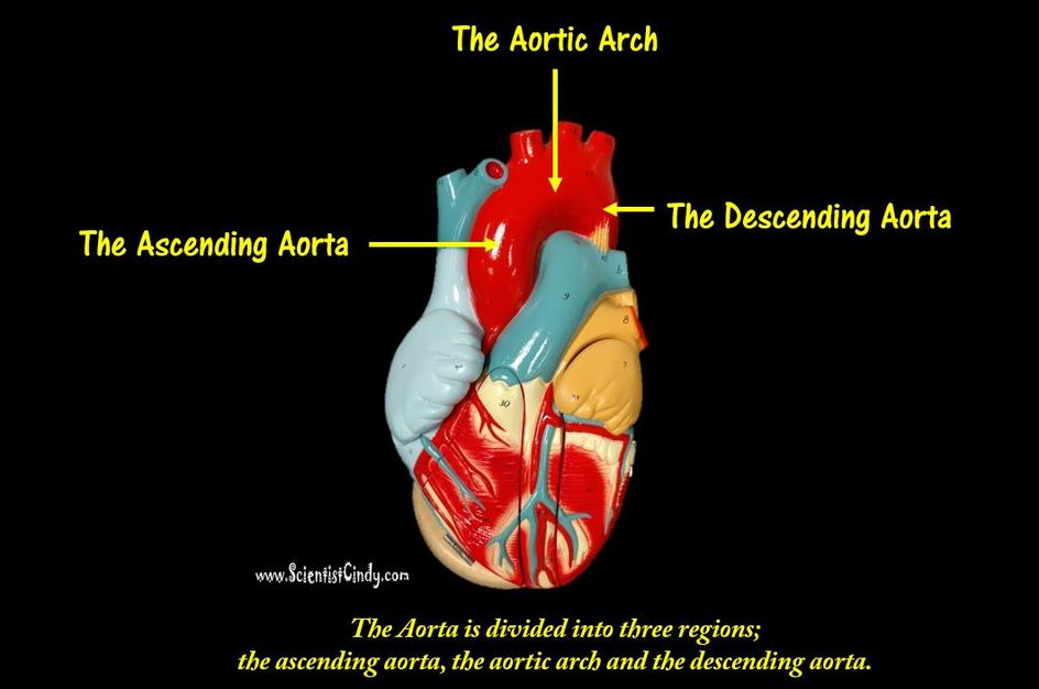
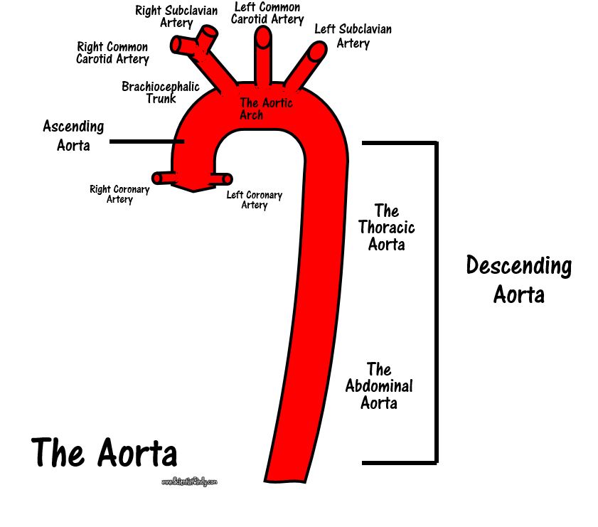
The aorta is separated into 3 segments
|
|
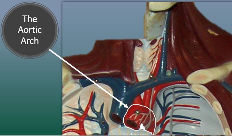
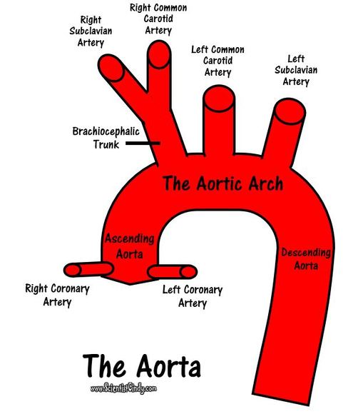
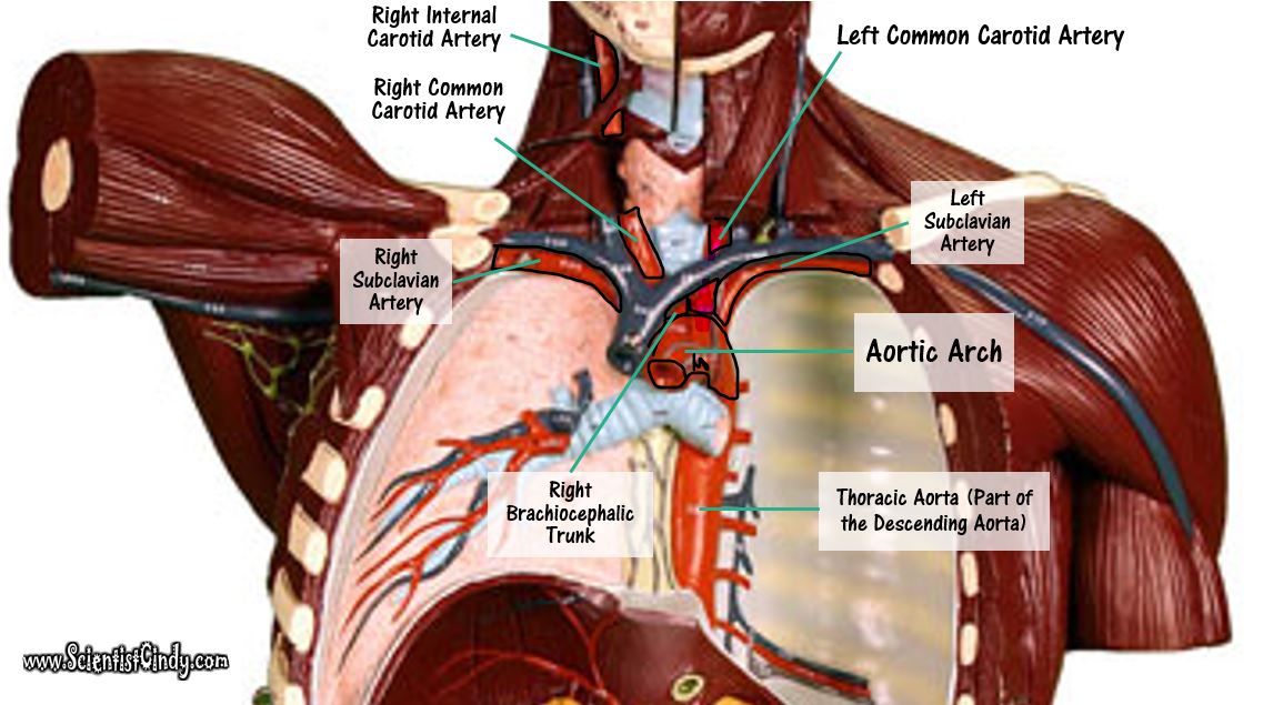
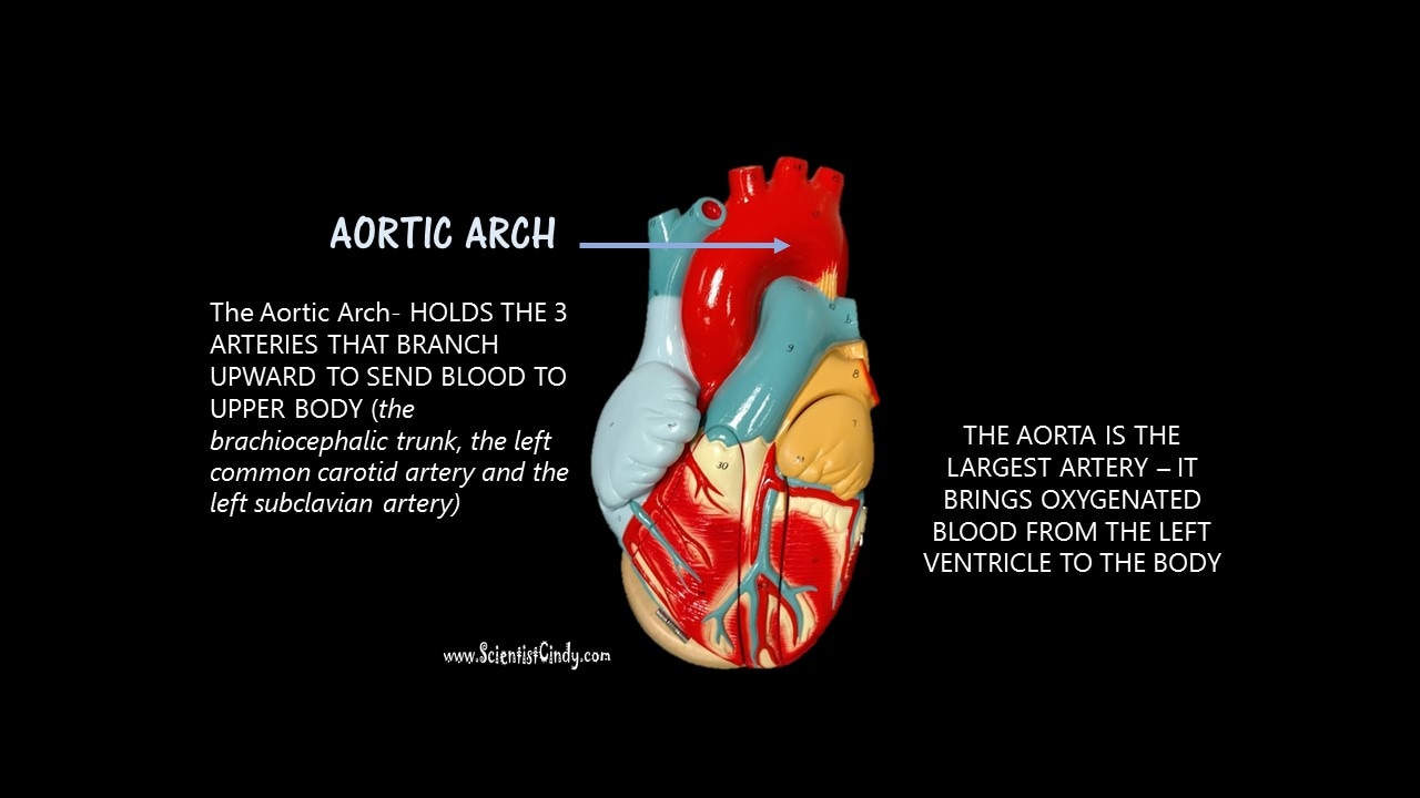
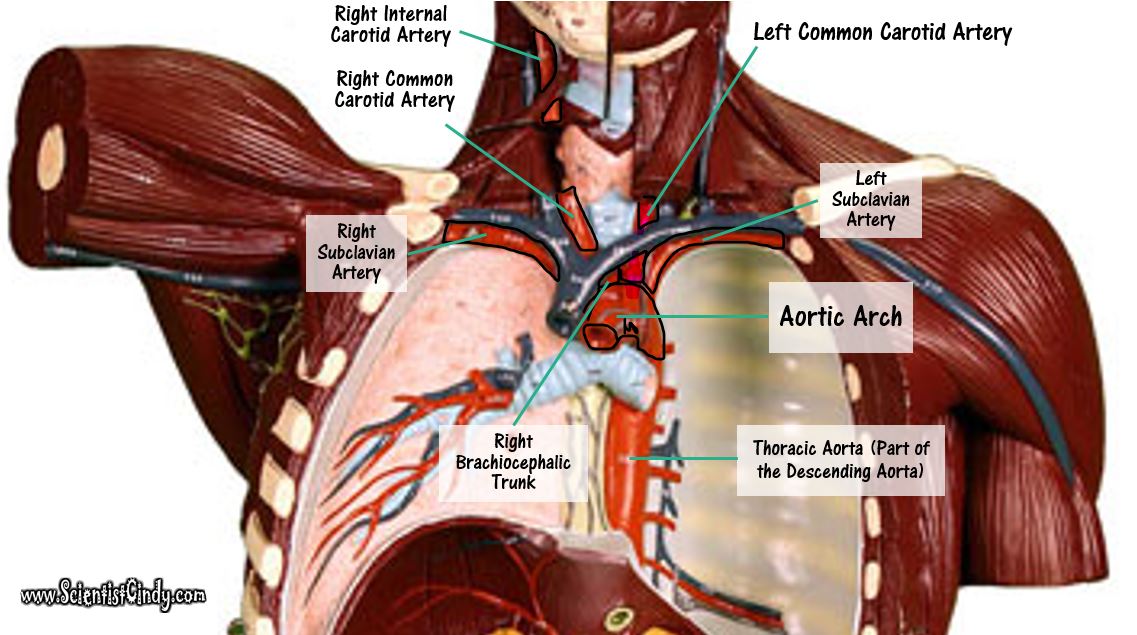
The aortic arch functions to send blood to the upper body via 3 branches.
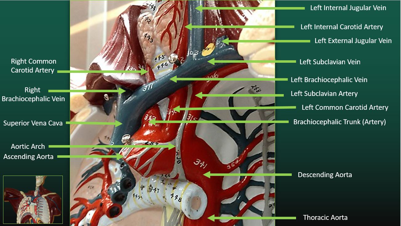
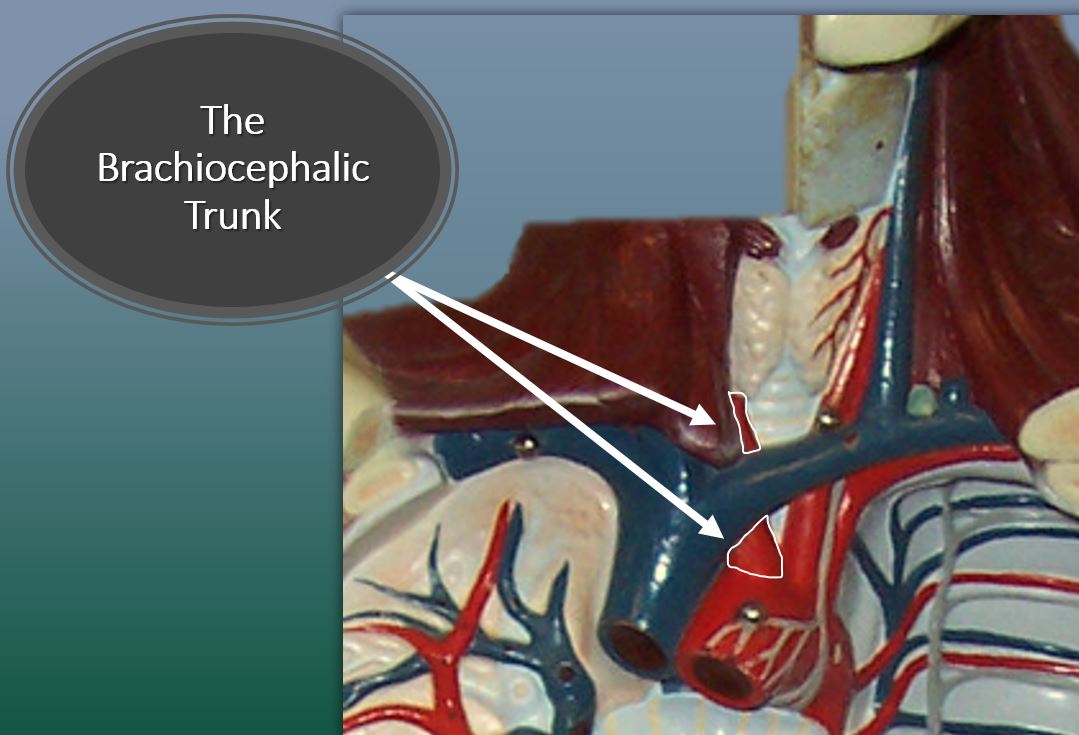
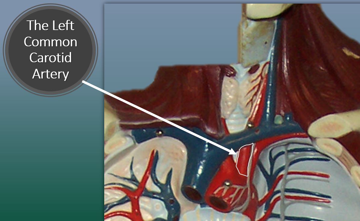
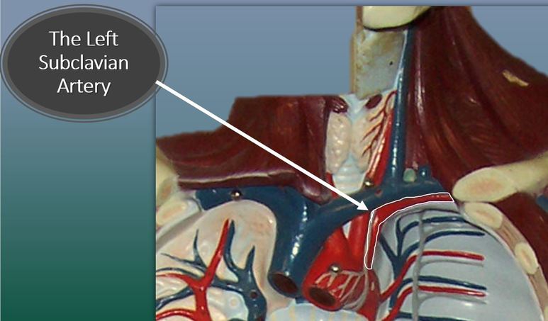
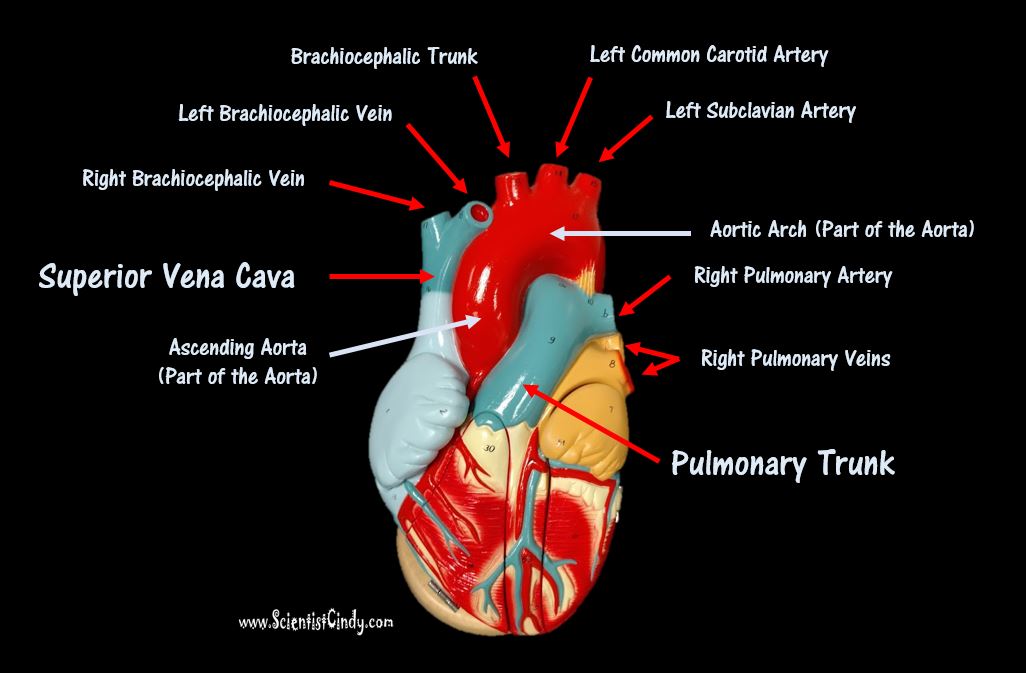
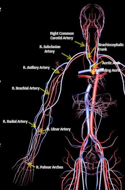
The 3 of the aortic arch are the 1) the brachiocephalic trunk 2) the left common carotid artery and 3) the left subclavian artery. The first branch is the brachiocephalic trunk (brachiocephalic artery). The brachiocephalic trunk sends blood to the right arm, and the head and neck. The brachiocephalic trunk branches off to form the right subclavian artery and the right common carotid artery. The right subclavian artery supplies blood to the right arm and the right common carotid artery supplies blood to the right side of the head and neck. The second branch is the left common carotid artery and it supplies blood to the left side of the head and neck. The third branch is the left subclavian artery and it supplies blood to the left arm. |
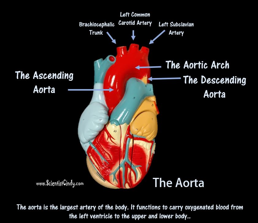
The ascending aorta brings oxygenated blood from the left ventricle (the blood passes through the aortic semilunar valve) to the portions of the aorta called the AORTIC ARCH and the DESCENDING AORTA. The aortic arch contains branches that send blood to the upper body. The descending aorta sends blood to the lower regions of the body.
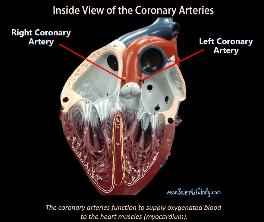
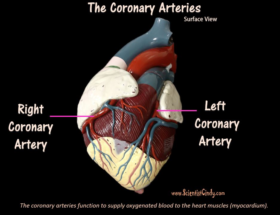
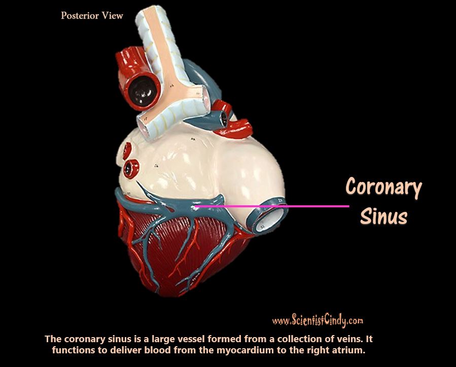
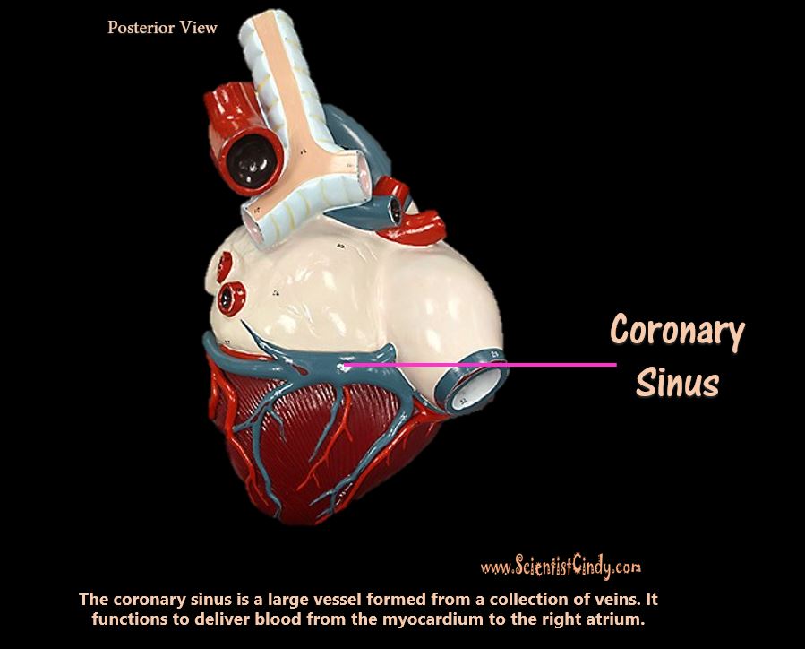
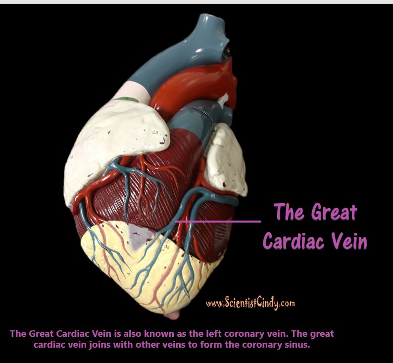
The heart itself needs a supply of blood. There are two arteries that begin at the base of the aorta called the right and left coronary arteries. The right coronary artery supplies oxygenated blood to the cardiac muscles on the right side of the heart and the left coronary artery supplies oxygenated blood to the cardiac muscles on the left side of the heart.
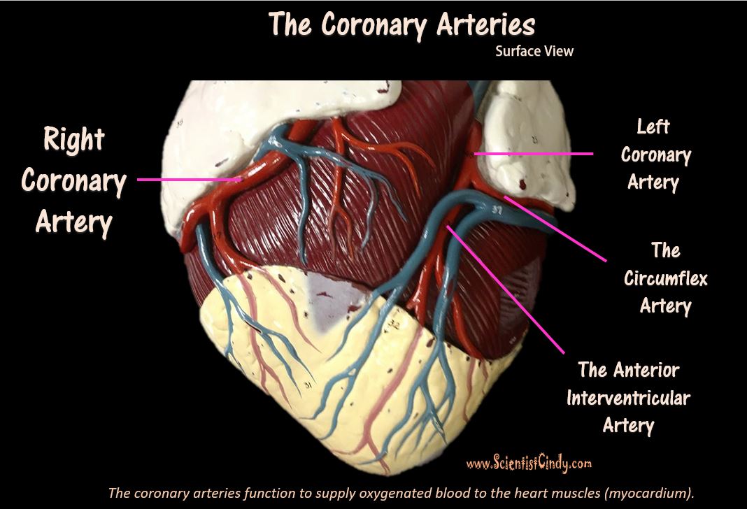
The right and left coronary arteries begin at the base of the aorta (the ascending aorta) and travel to the surface of the heart. The right coronary artery runs along the surface of the right atrium. The left coronary artery reaches the surface of the heart and quickly branches off to form two different arteries (see next image).
The left coronary artery branches off to form the circumflex artery and the anterior interventricular artery. The circumflex artery circles around the inferior border of the left atrium. The anterior interventricular artery continues downward lying in between the right and left ventricles.
Arteries of the Thoracic Region
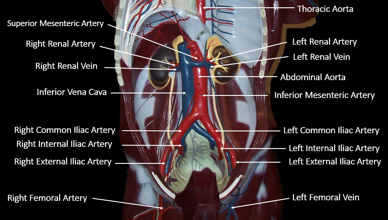
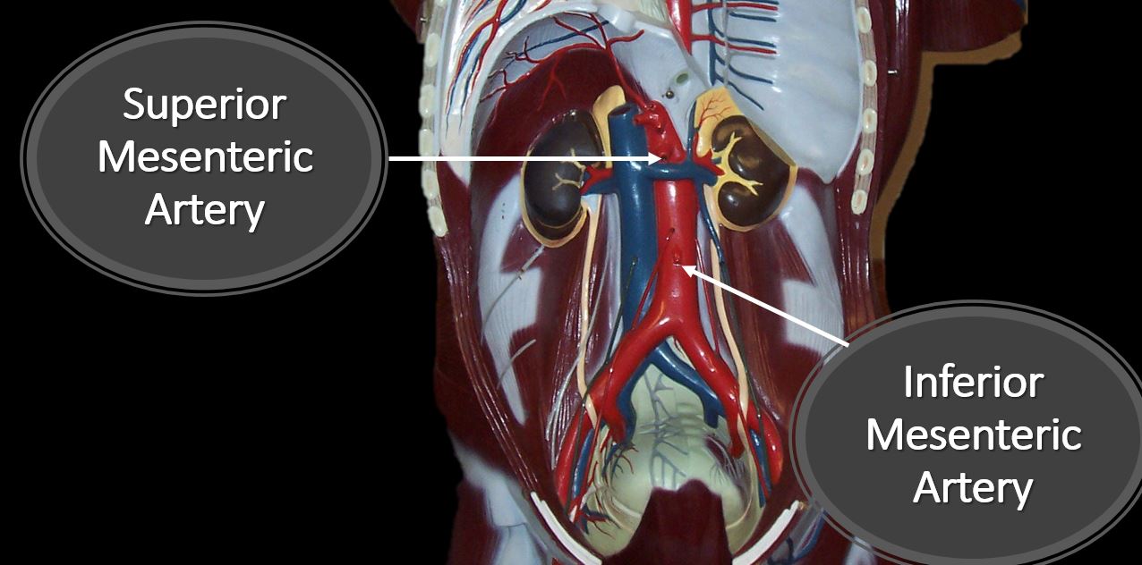
The descending aorta supplies blood to the lower body and has branches that travel to the abdomen, pelvis, perineum and the lower limbs.
The descending aorta is the portion of the aorta that comes just after the aortic arch. It turns downward and descends to deliver oxygenated blood to the lower portions of the body.
|
|
The Pulmonary Trunk and Its Branches
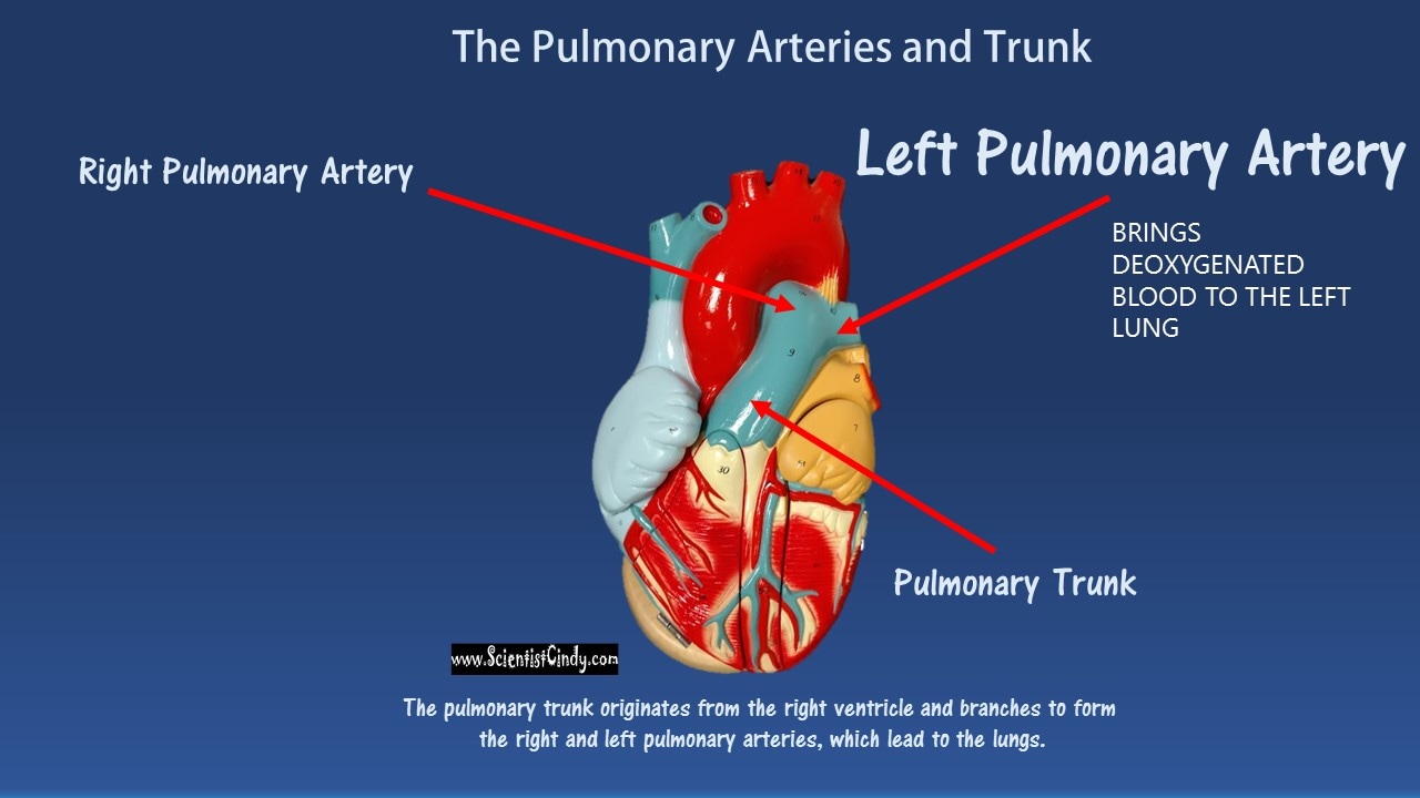
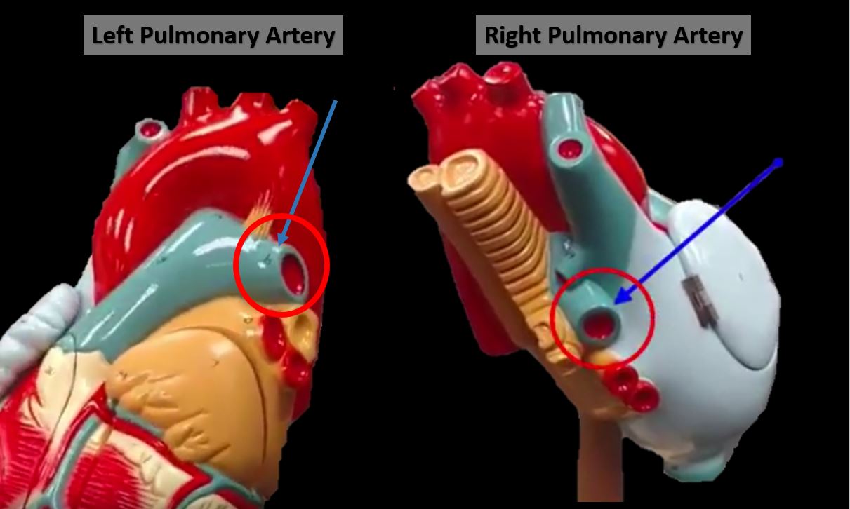
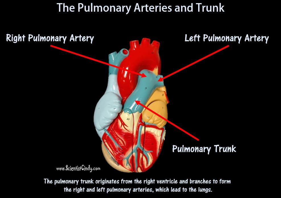
The next largest artery you see in the heart is colored BLUE because it is carrying deoxygenated blood from the right ventricle to the lungs. This artery is called the pulmonary trunk. The pulmonary trunk branches off to form the right and left pulmonary arteries. The right pulmonary artery carries the blood to the right lung and the left pulmonary artery carries blood to the left lung. |
THE VEINS
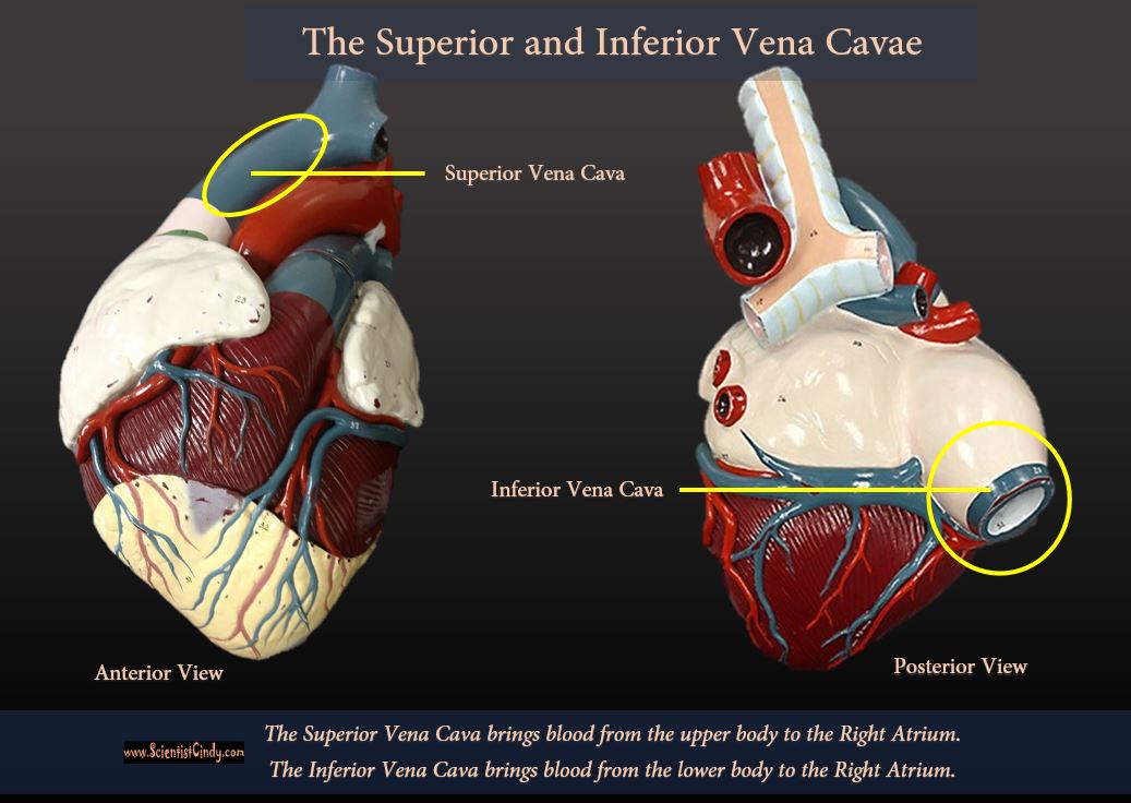
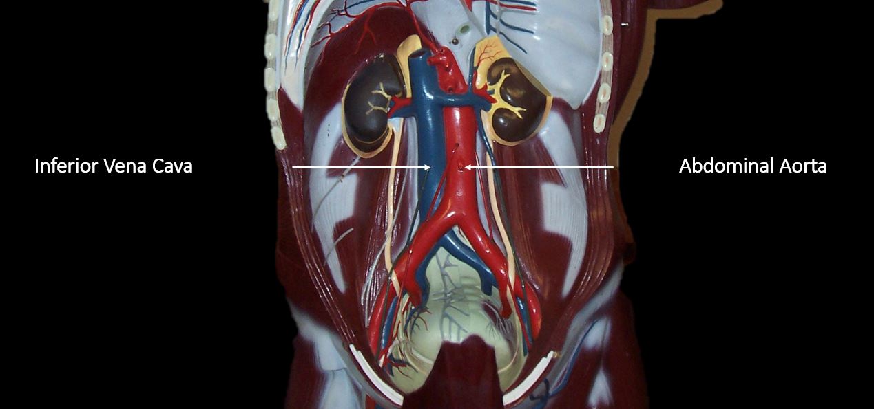
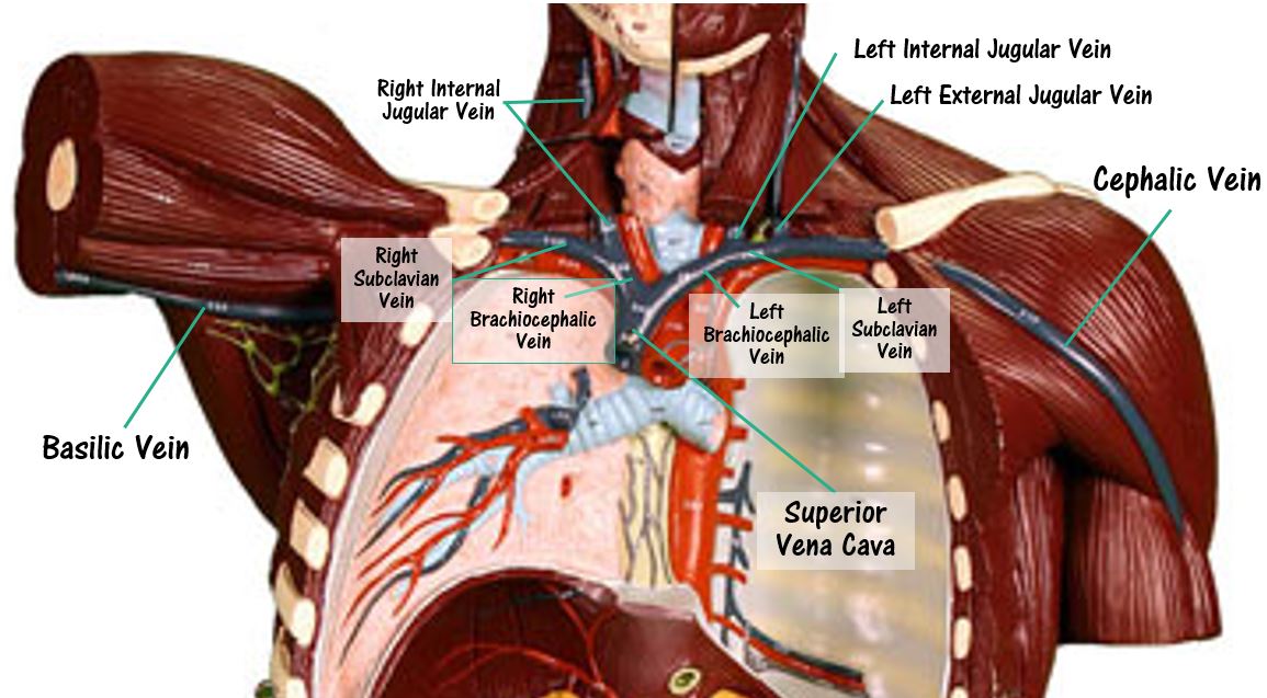
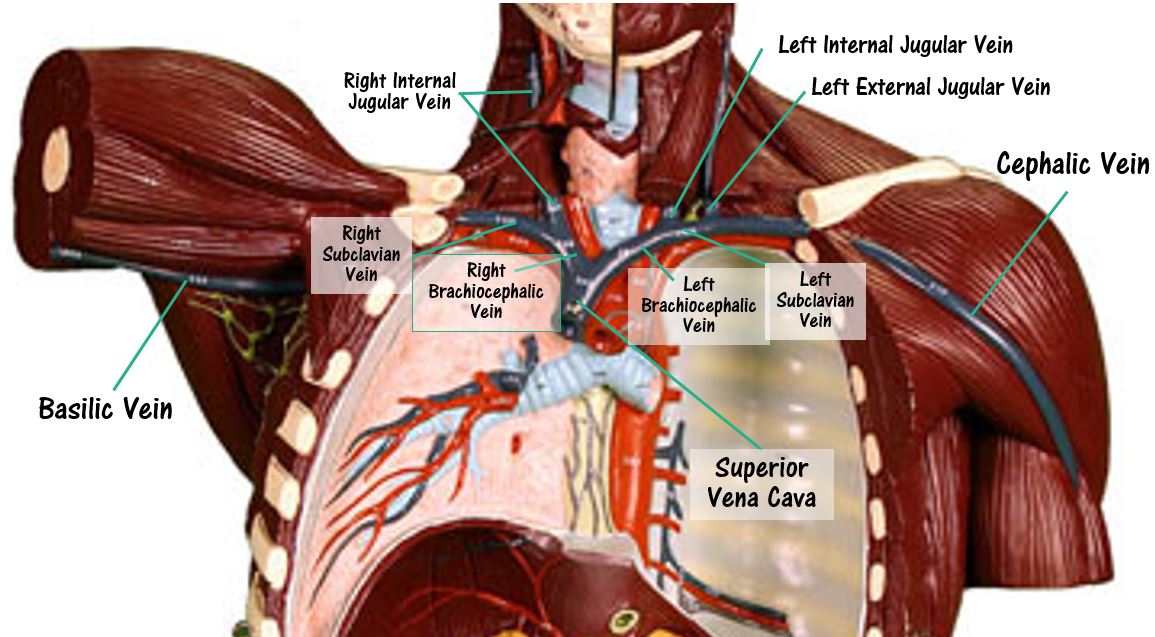
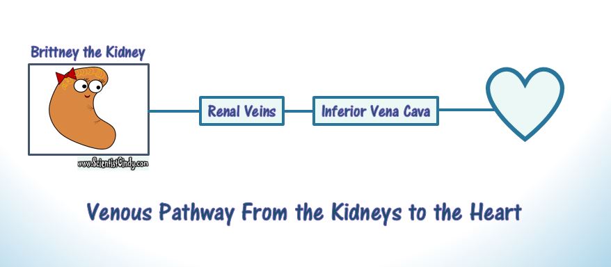
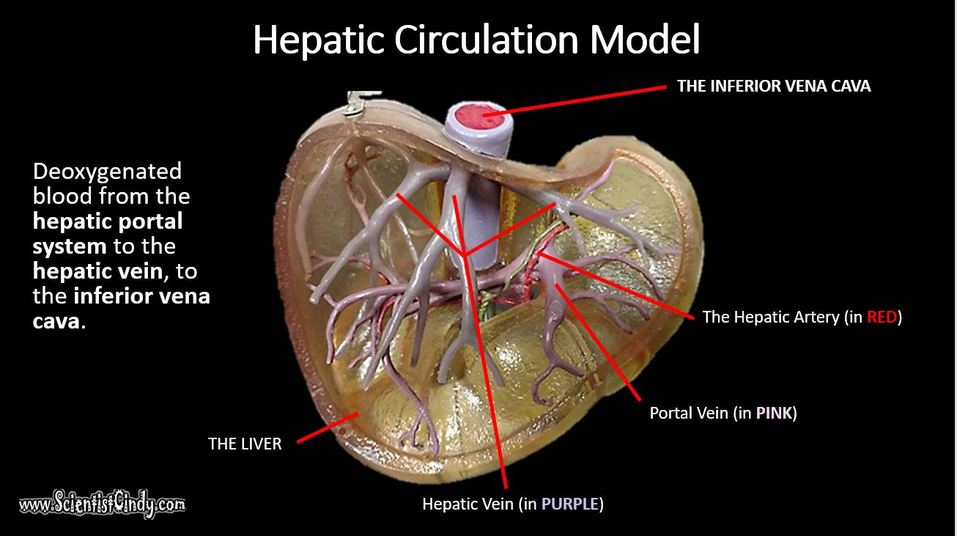
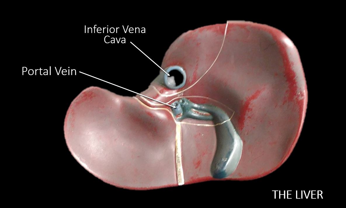
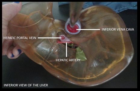
The largest veins of the body are the superior and inferior vena cava. The superior vena cava brings deoxygenated blood from the upper body to the right atrium. The inferior vena cava brings deoxygenated blood from the lower body to the right atrium.
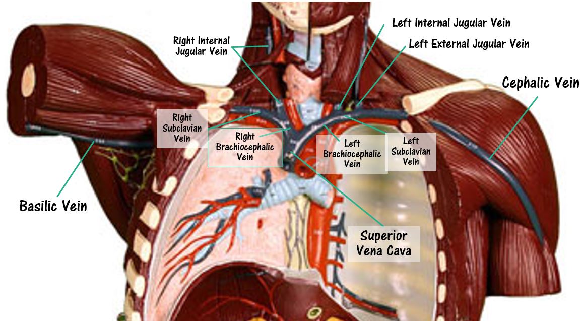
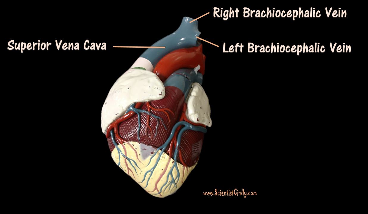
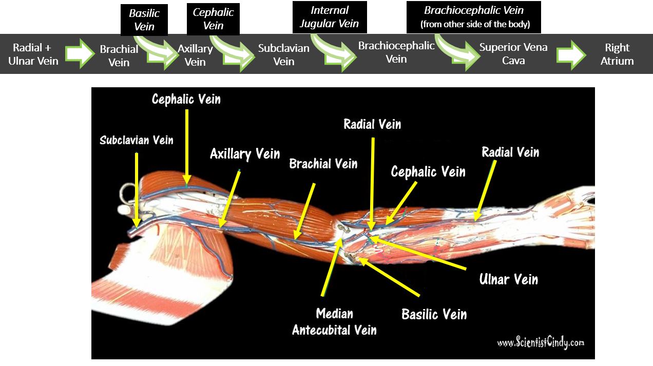
The right and left brachiocephalic veins come together to form the superior vena cava. The right brachiocephalic vein brings deoxygenated blood from the right side of the head and neck and the right arm, to the superior vena cava. The left brachiocephalic vein brings deoxygenated blood from the left side of the head and neck and the left arm, to the superior vena cava. |
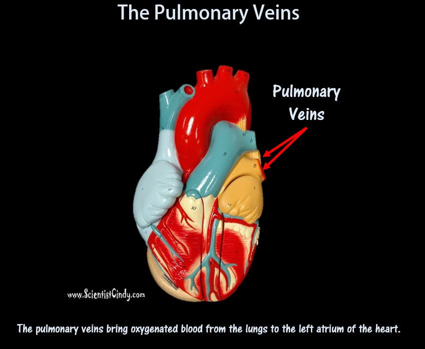
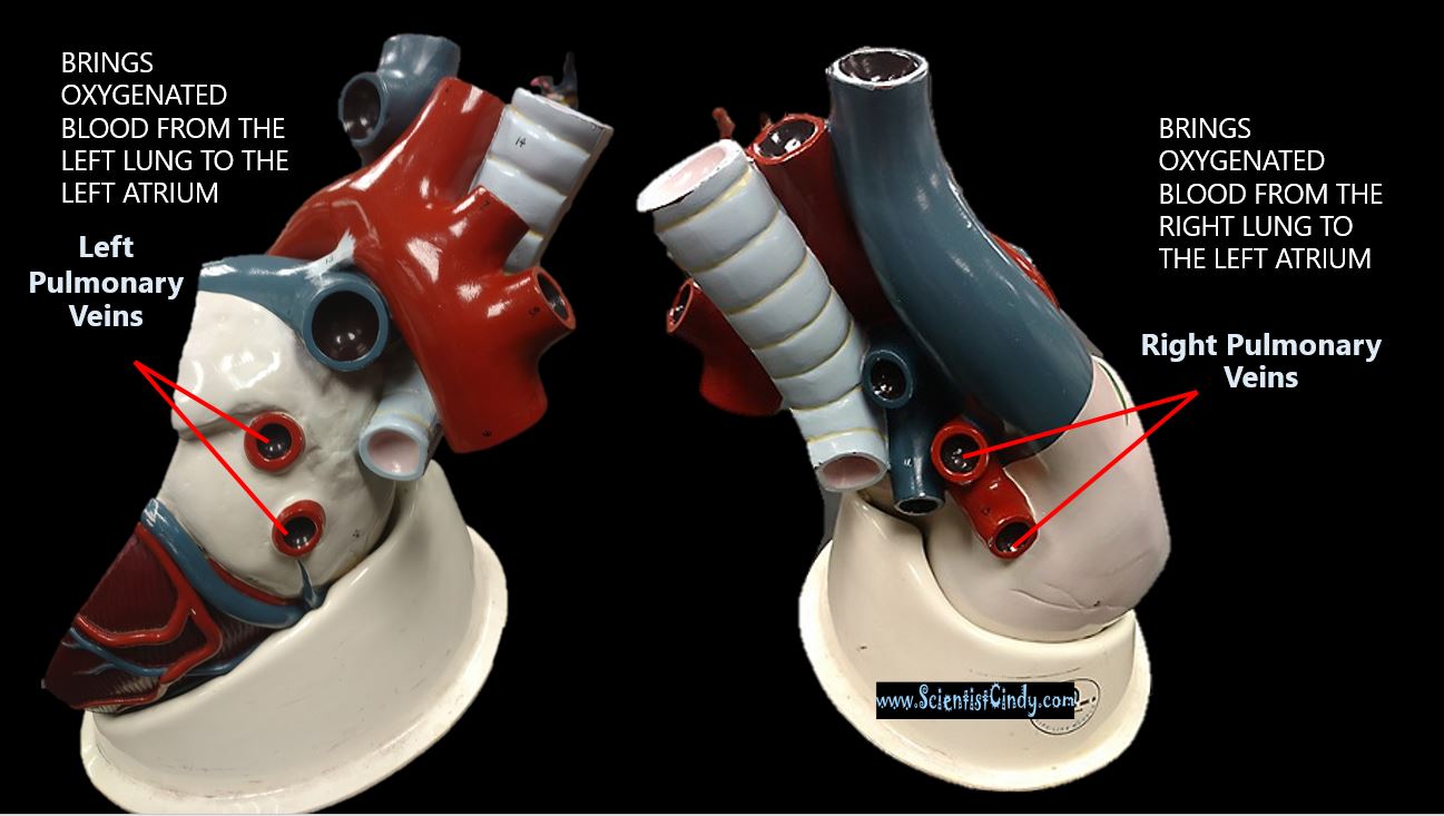
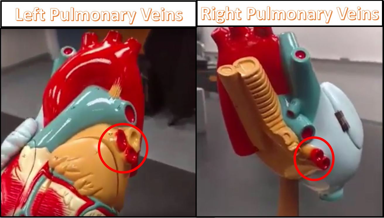
Oxygenated blood coming from the lungs returns to the heart through the right and left pulmonary veins. The right pulmonary veins bring oxygenated blood from the right lung to the left atrium of the heart. The left pulmonary veins bring oxygenated blood from the left lung to the left atrium of the heart.
THE CIRCUITS OF THE CIRCULATORY SYSTEM
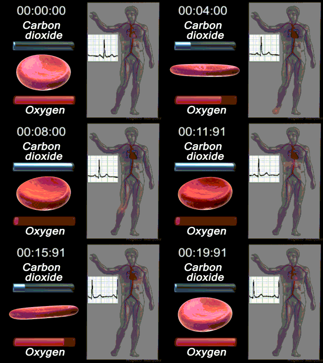
The circulatory system circulates blood around two major loops.
- 1) THE PULMONARY CIRCUIT
- - Functions to bring blood from the heart to the lungs and back to the heart
- 2) THE SYSTEMIC CIRCUIT
- - Functions to bring blood from the heart to all the parts of the body except the lungs and then back to the heart
The Arteries of the Head and Neck
Your brain uses a lot of blood for its size. About 20% of your blood supply is directed toward your brain to keep you reading this page! Let's look at the basics of how the blood gets there.
Arterial Pathways to the Brain
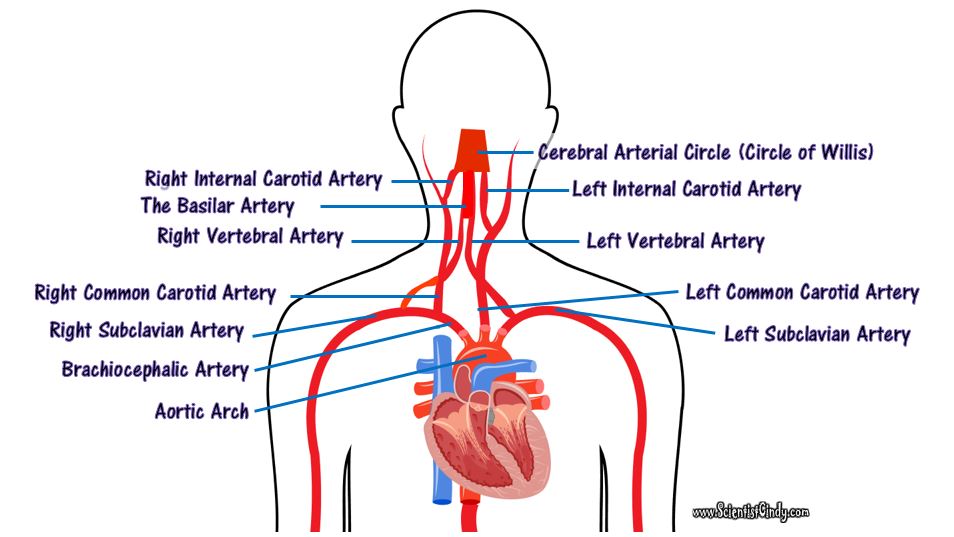
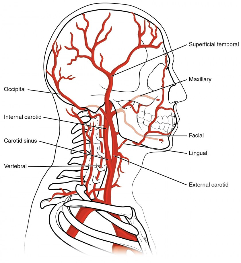
Oxygenated blood from the heart travels through he aorta. The aorta contains 3 branches; the brachiocephalic trunk (or artery), the left common carotid artery, and the left subclavian artery. All 3 of these branches carry blood from the aorta to the brain, head and neck.
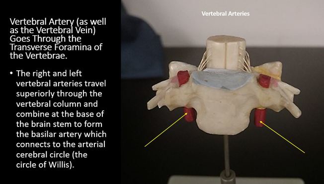
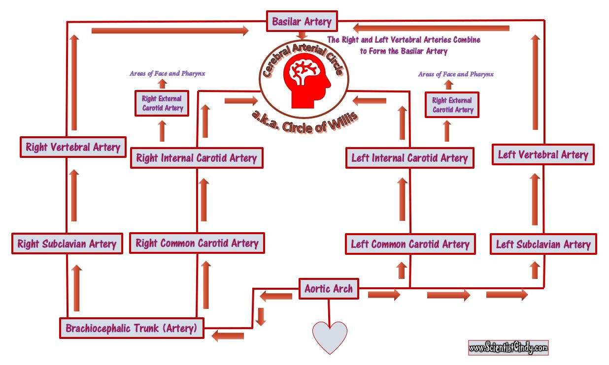
Oxygenated blood from the left ventricle leave the heart through the aorta. This blood travels through the ascending aorta to the aortic arch. The aortic arch has 3 branches that send blood to the upper portions of the body. Each of the 3 branches off the aorta have pathways that lead to the brain (the arterial cerebral circle).
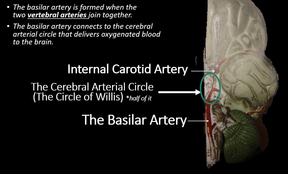
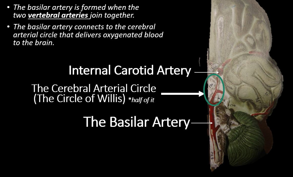
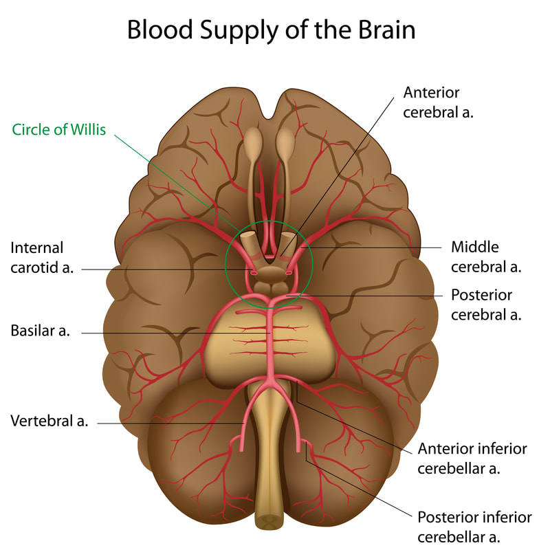
- The first branch off of the aortic arch is the brachiocephalic trunk. The brachiocephalic trunk (or artery) branches off to form the right subclavian artery and the right common carotid artery. The right subclavian artery brings blood to the right vertebral artery which joins with the basilar artery which lies at the base of the brain. The basilar artery connects to the cerebral arterial circle (a.k.a. the circle of Willis) which delivers oxygenated blood to the brain.
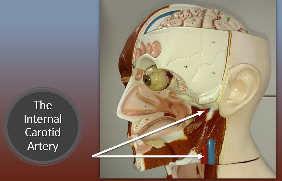
- The brachiocephalic trunk (the first branch off of the aortic arch) has another branch called the right common carotid artery. The right common carotid artery branches to form the right internal carotid artery which leads directly to the cerebral arterial circle (a.k.a. the circle of Willis) of the brain.
- The second branch coming off of the aortic arch is the left common carotid artery. The left common carotid artery ascends to form the internal carotid artery which leads directly to the cerebral arterial circle (a.k.a. the circle of Willis) of the brain.
- The third branch coming off of the aortic arch is the left subclavian artery. The left subclavian artery brings blood to the right vertebral artery which joins with the basilar artery which lies at the base of the brain. The basilar artery connects to the cerebral arterial circle (a.k.a. the circle of Willis) which delivers oxygenated blood to the brain.
The Veins from the Brain, Head and Neck Areas to the Heart.
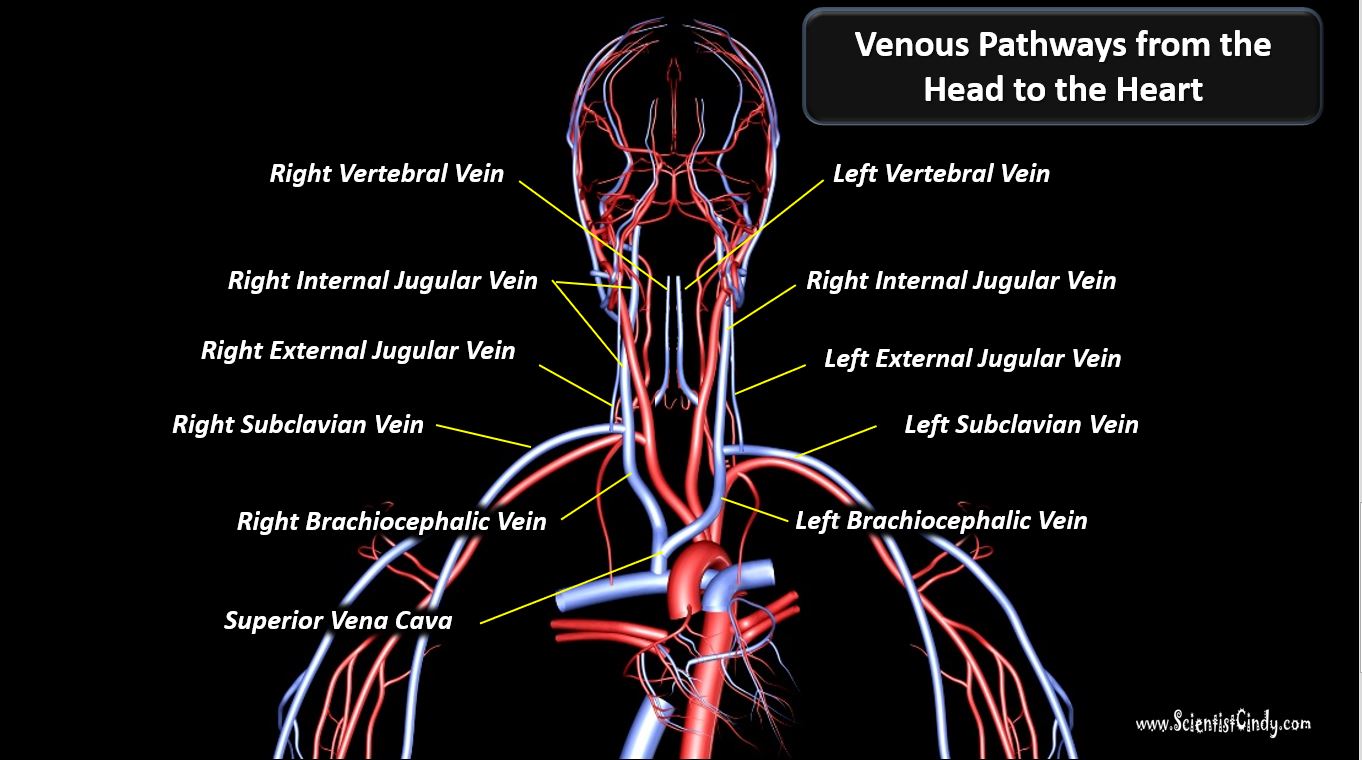
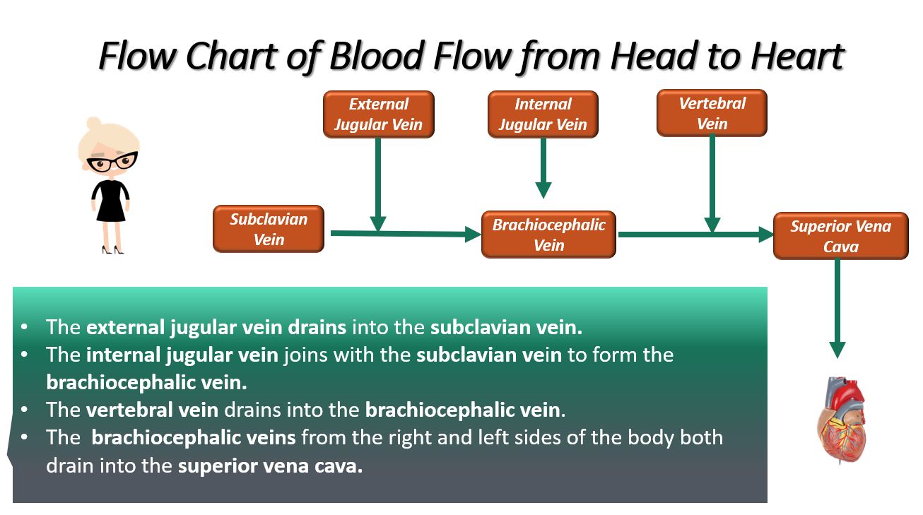
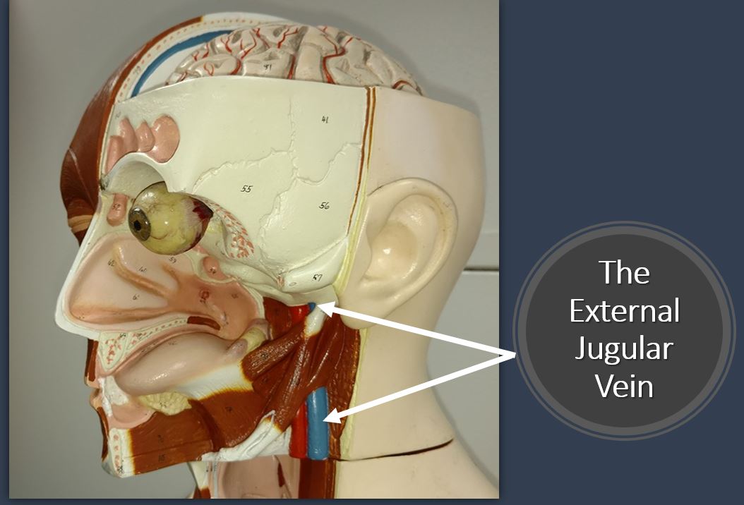
What goes up, must come down. Deoxygenated blood from the brain, head and neck to the heart. There are 6 major pathways that this blood from the brain, head and neck travels to the heart. These pathways begin at the right and left external jugular veins, the right and left vertebral veins and the right and left internal jugular veins.
The right and left external jugular veins travel to the right and left subclavian vein. From there the blood travels through the right and left brachiocephalic veins to the superior vena cava. The right and left vertebral veins connect to the right and left brachiocephalic veins which then travels to the superior vena cava. The right and left internal jugular veins also travel to the brachiocephalic veins which connects with the superior vena cava. After the deoxygenated blood reaches the superior vena cava, it flows into the right atrium of the heart.
The Blood Vessels of the Arm
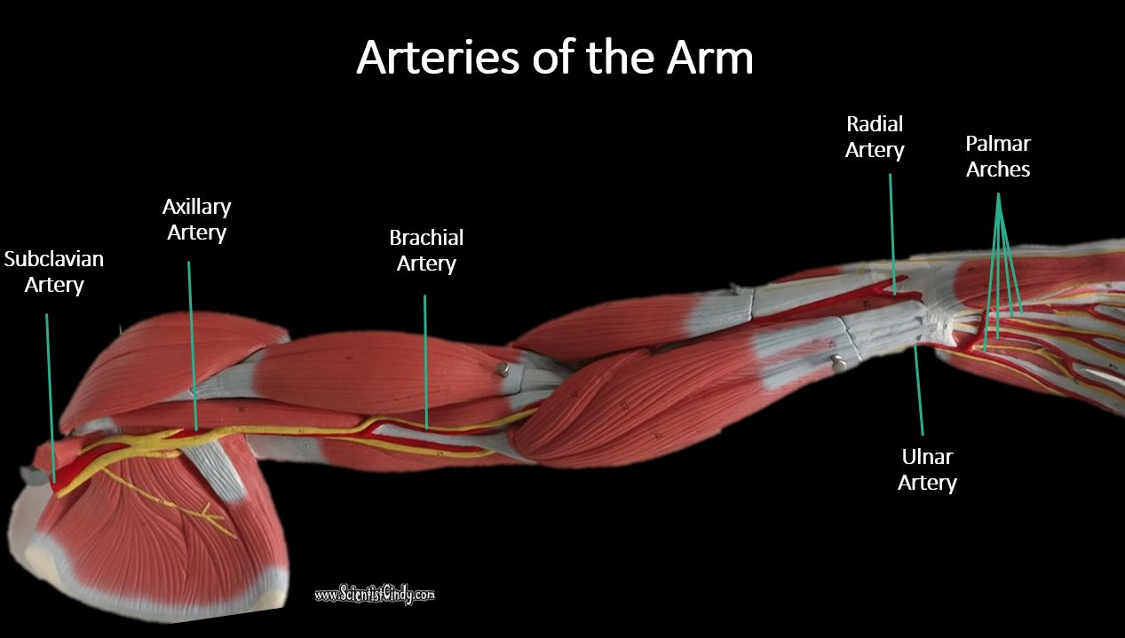
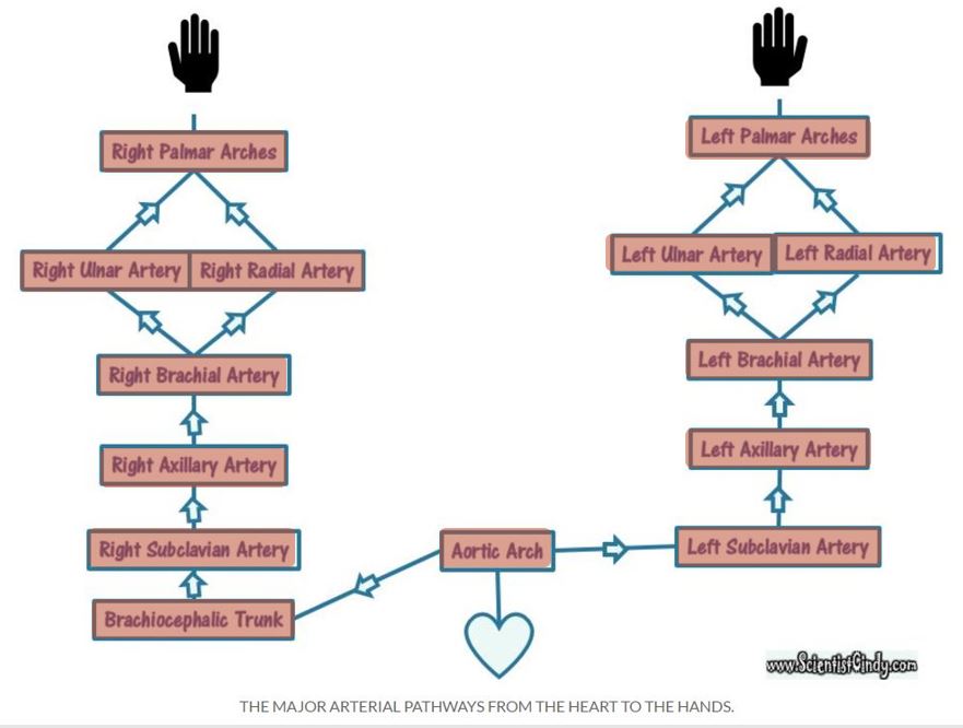
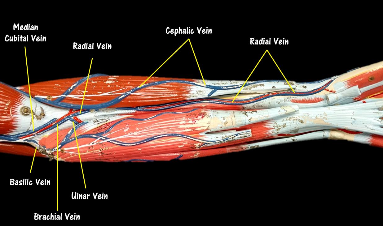
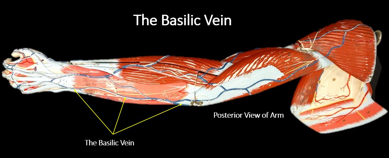
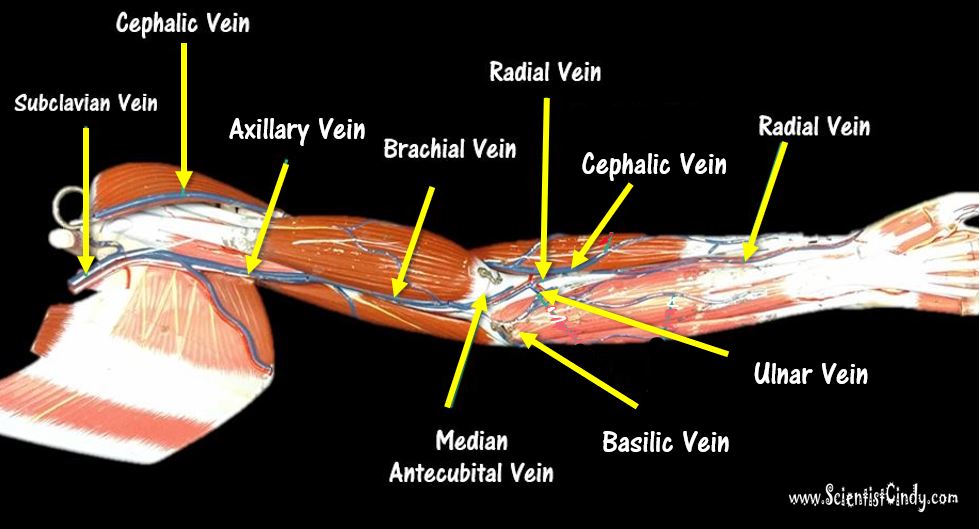
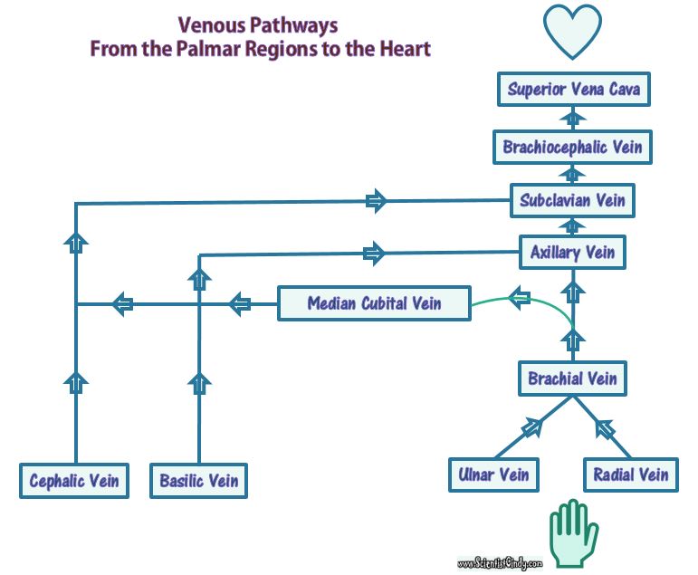
Oxygenated blood travels down the arms as well. Oxygenated blood leaves the left ventricle of the heart through the aortic semilunar valve and enters the ascending aorta. The blood travels superiorly to the aortic arch. Blood destined for the right arm enters the right brachiocephalic trunk (the first branch of the aortic arch) and then enters the right subclavian artery. Blood destined for the left arm travels though the 2nd branch of the aortic arch, which is the left subclavian artery. From this point, the arterial pathways from the right and left subclavian arteries to the palms (palmar arches) are identical. |

