The Appendicular Skeleton
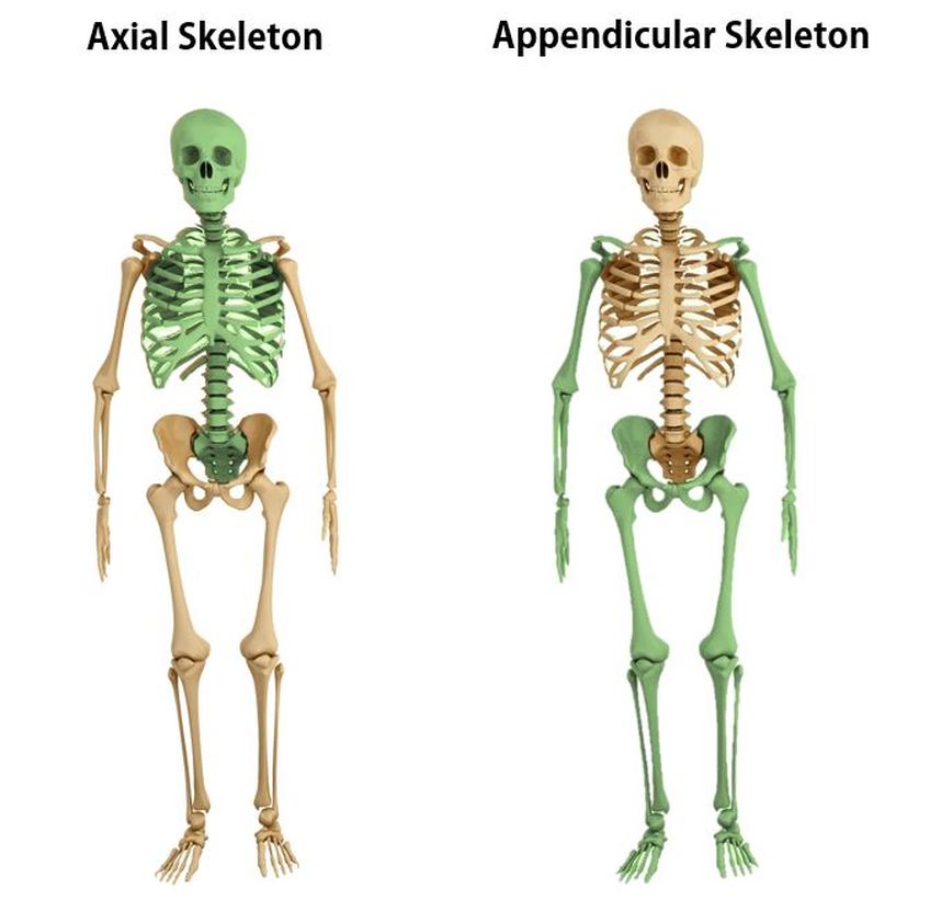
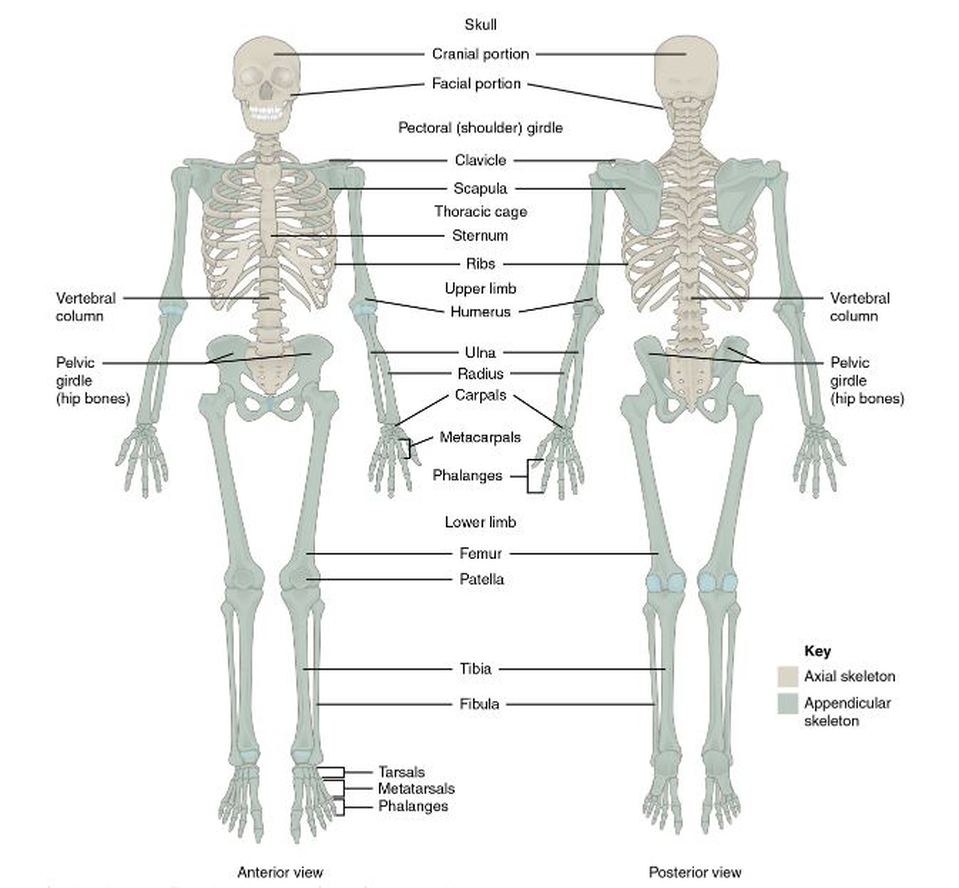
The human skeleton is divided into 2 categories; the axial skeleton and the appendicular skeleton. The axial skeleton forms the long axis of your body. The axial skeleton is divided into 3 categories; 1) the skull, 2) the vertebral column, and 3) the thoracic cage. The function of the axial skeleton is to support the head, neck, and trunk, and to protect the brain, spinal cord, and the thoracic organs. The bones of the appendicular skeleton make up the rest of the skeleton, and are so called because they are appendages of the axial skeleton. The appendicular skeleton includes the bones of the shoulder girdle, the upper limbs, the pelvic girdle, and the lower limbs. The appendicular skeleton has 126 bones. The axial skeleton and the appendicular skeleton make up the 206 named bones of your body!
THE ANATOMY OF THE PECTORAL GIRDLE
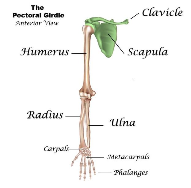
The pectoral girdle region of the skeleton consists of the following bones:
|
|
THE CLAVICLES (COLLAR BONES)
EDITED 2 MINUTE VIDEO YOU SHOULD WATCH!! Courtesy of Nicole Mashburn
|
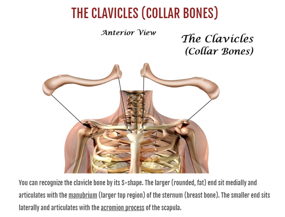
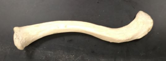
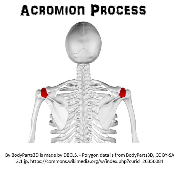
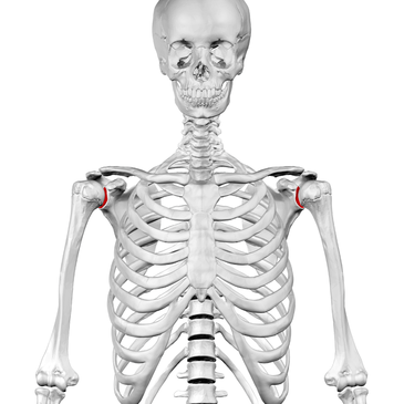
In this photo of an articulated skeleton, you can see how the medial aspect of the clavicle articulates with the manubrium of the sternum and how the lateral aspect articulates with the acromion process of the scapula.
|
|
THE SCAPULAE (SHOULDER BLADES)
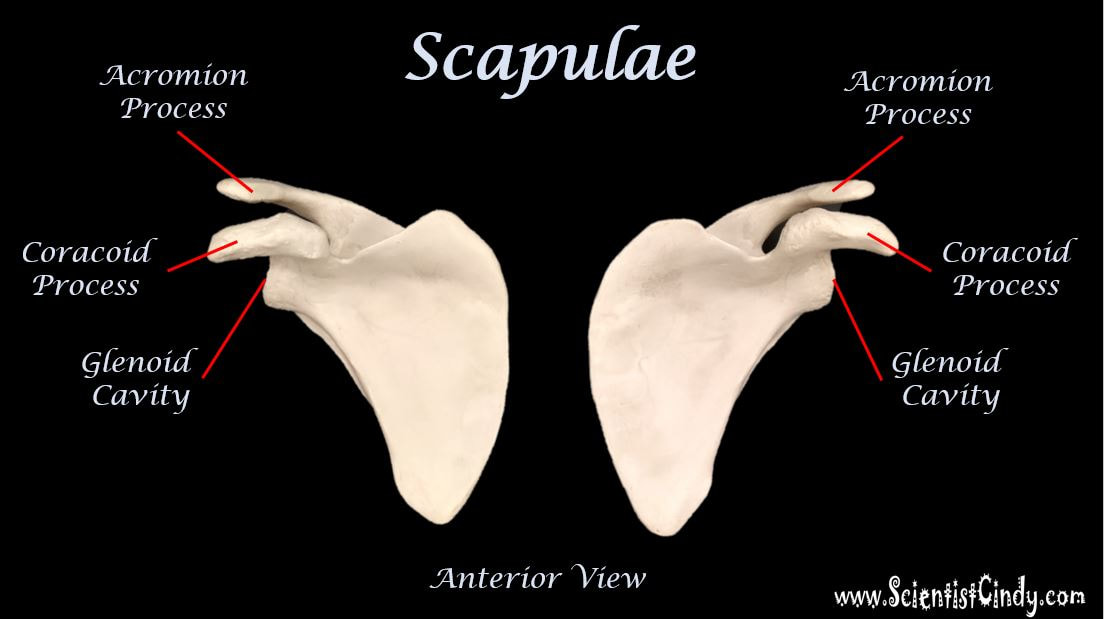
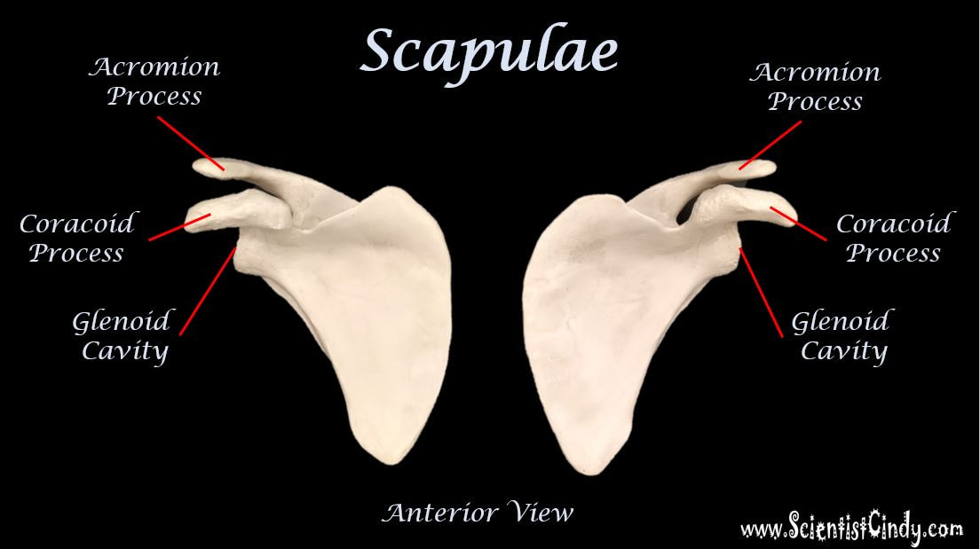
The Scapula has 4 Structures You Need to Know.
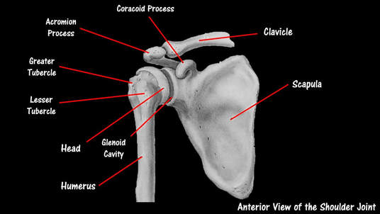
1) The Acromion Process 2) The Coracoid Process 3) The Glenoid Cavity 4) The Spine of the Scapula
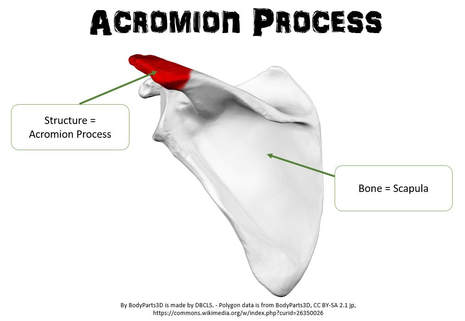
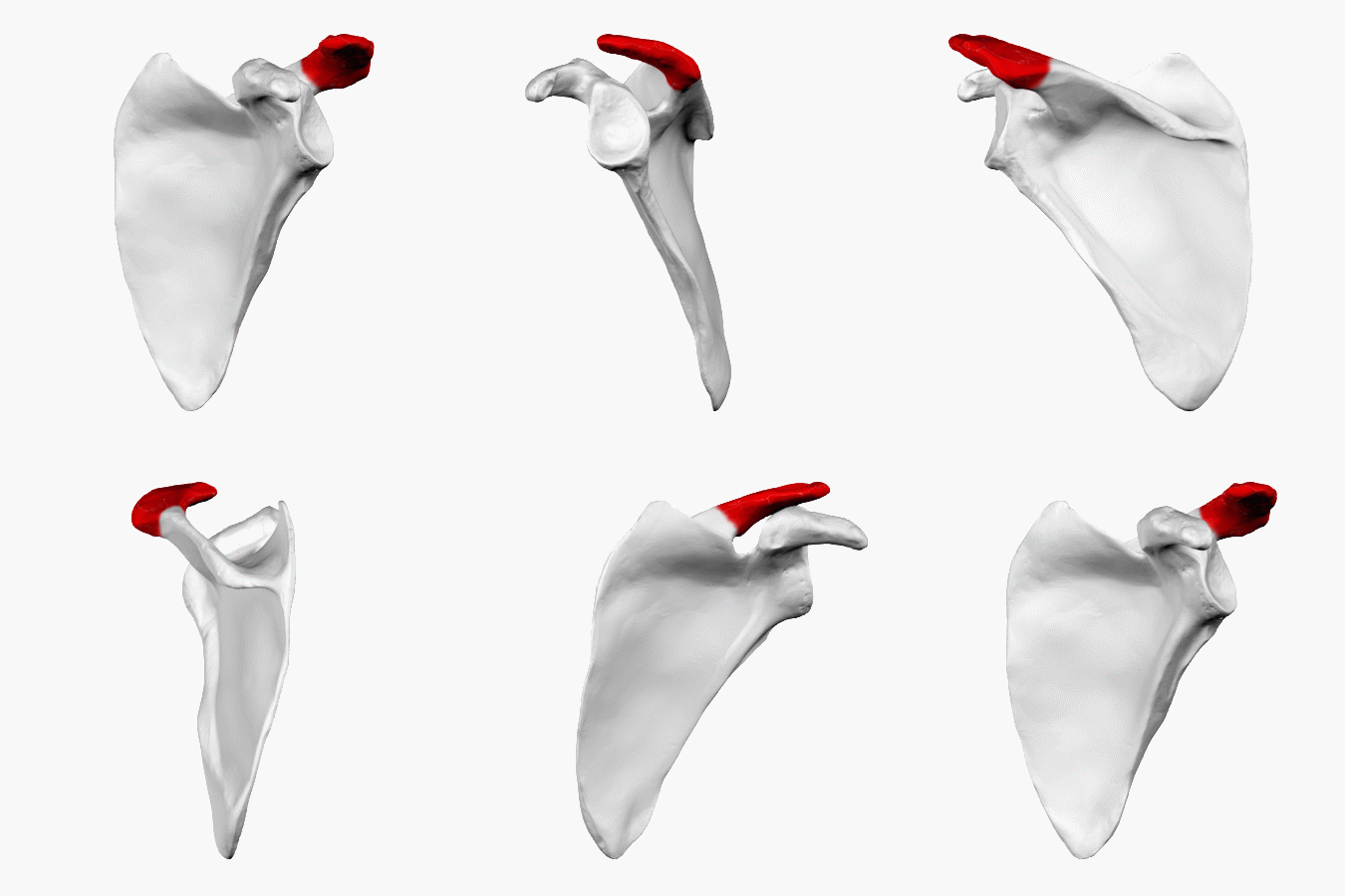
1) THE ACROMION PROCESS
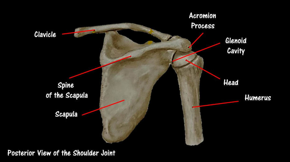
The Acromion Process - The acromion process articulates with the clavicle. Remember that the shoulder area is called the "acromial" region. The acromion process is an extension of the spine of the scapula. It extends from the posterior of the scapula the turns anterior at the acromial region to articulate with the clavicle.
2) CORACOID PROCESS
 Anterior View of the Shoulder Joint
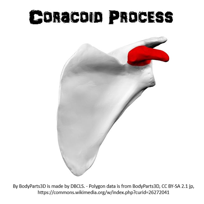
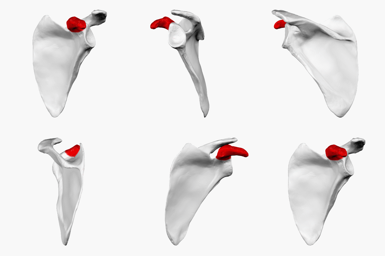
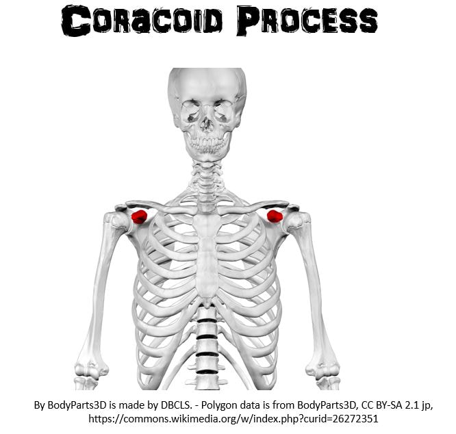
The Coracoid Process - The coracoid process looks like a raven's beak or a crooked finger sticking out. Its name comes from the Latin word for "raven's beak". This surface attaches to the biceps (upper arm muscles). The corocoid process extends from the anterior surface of the scapula, whereas the acromion process extends from the posterior aspect of the scapula.
The coracoid process lies on the lateral edge of the scapulae on the anterior aspect. The coracoid process does not articulate with any other bone. Instead, it acts as a surface for attachment of the bicep muscles (upper arm muscles).
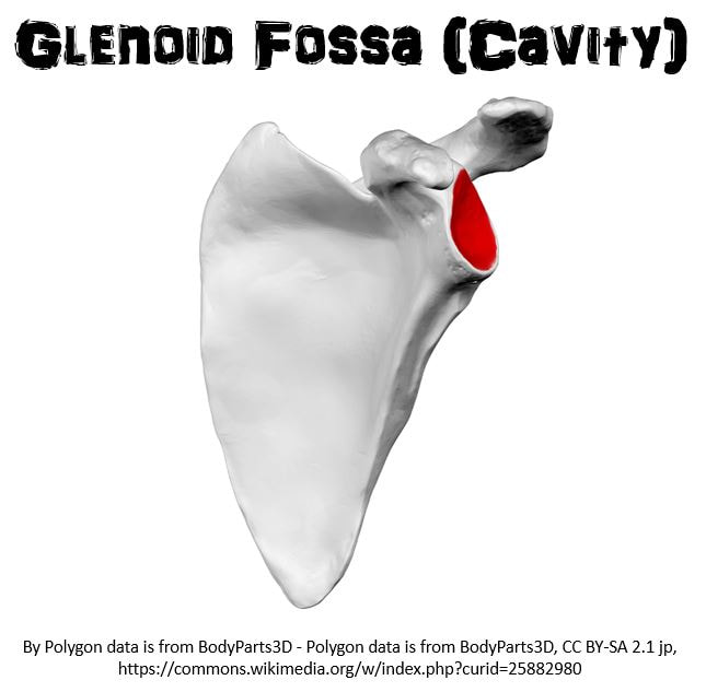
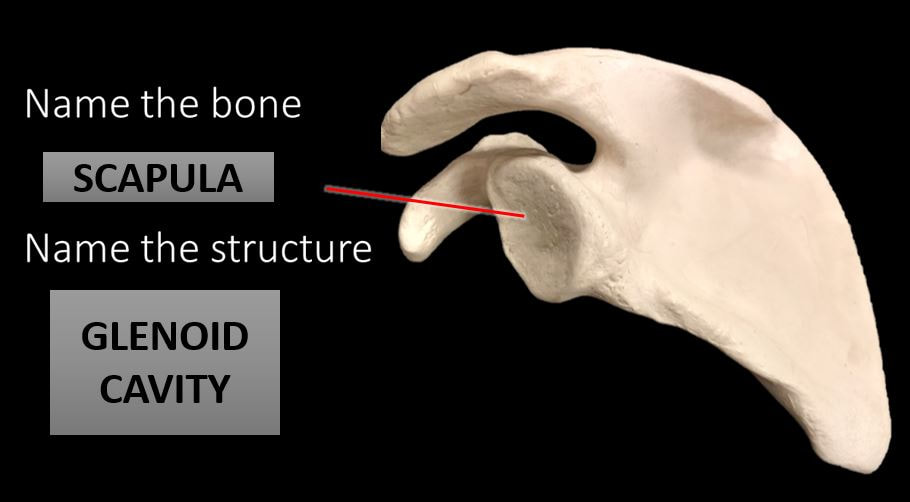
3) THE GLENOID CAVITY
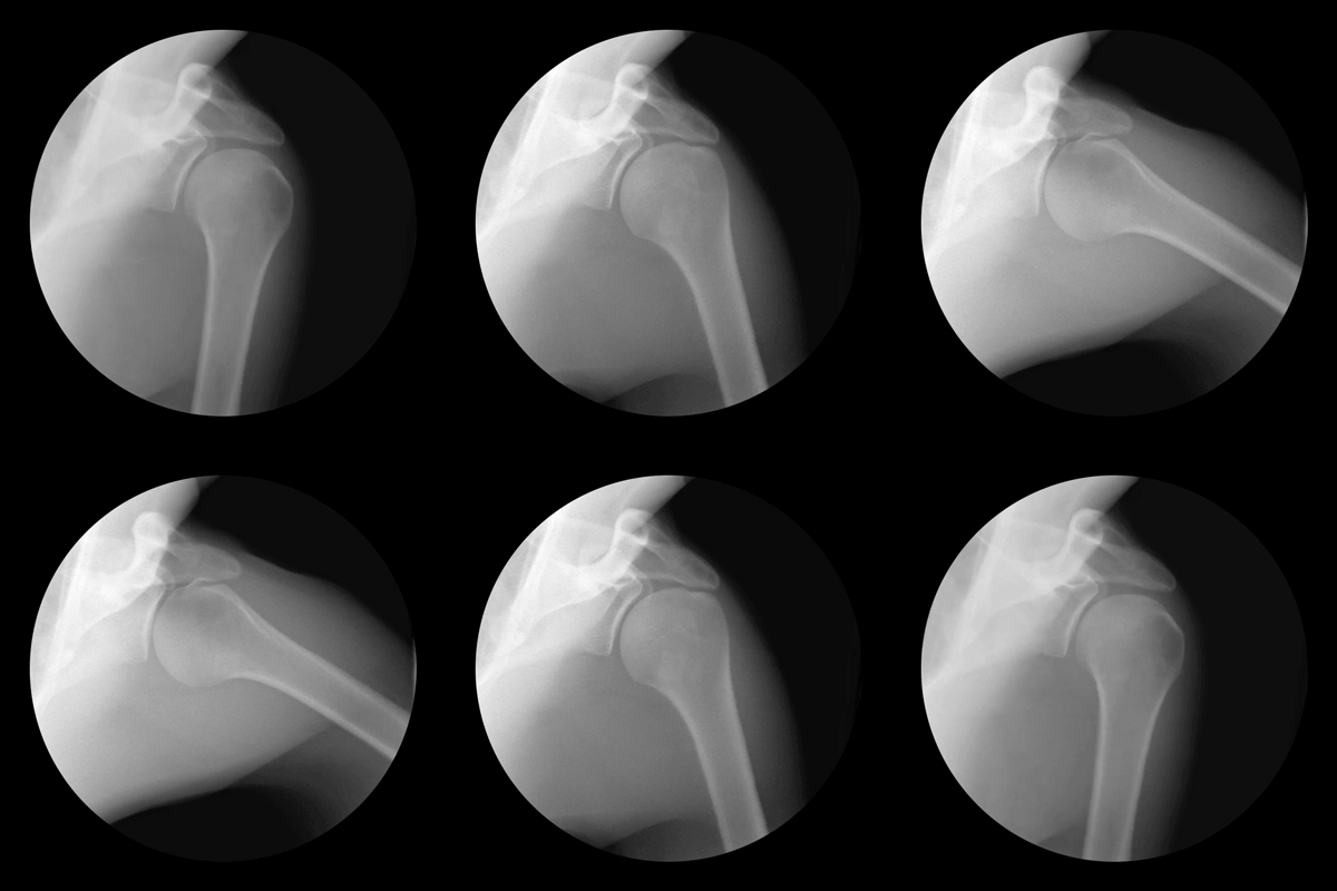
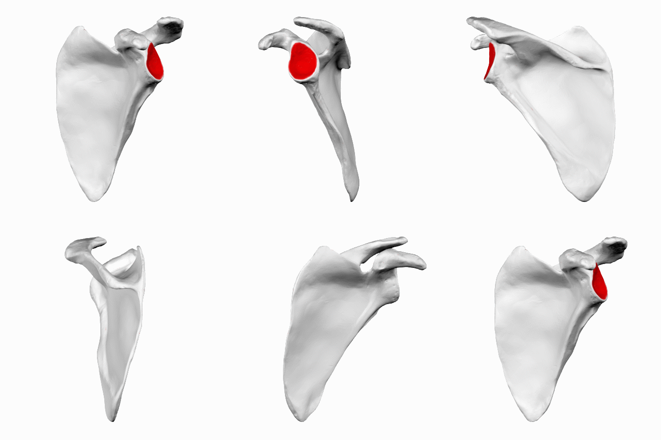
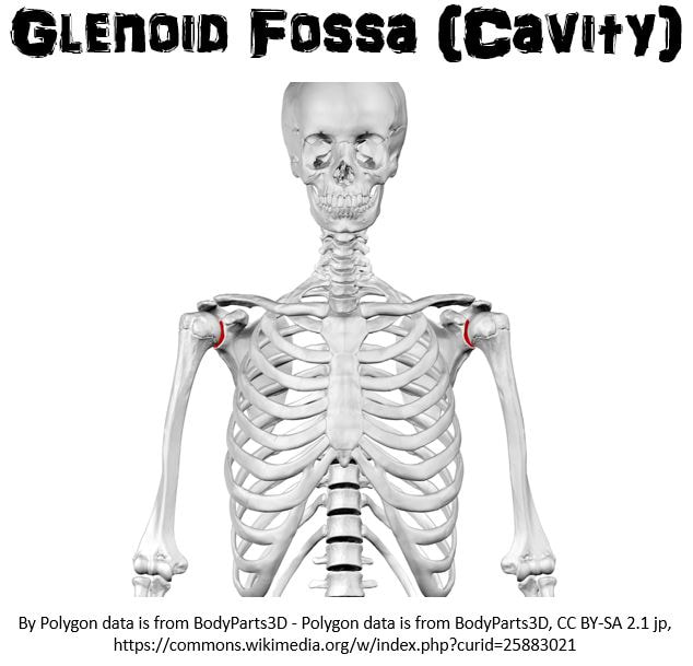
The Glenoid Cavity - The glenoid cavity is the socket or cavity that articulates with the head of the humerus bone to form the shoulder joint. Its name comes from the Greek word for "socket".
The shoulder joint is known as the glenohumeral joint. The glenohumeral joint is a ball-and-socket joint. The hip joint is also a ball-and-socket joint. These joints are specialized to allow for multi-directional and rotational motion. This high range of mobility, however, comes with a price. These joints are hot spots for pulled muscles.
The shoulder joint is known as the glenohumeral joint. The glenohumeral joint is a ball-and-socket joint. The hip joint is also a ball-and-socket joint. These joints are specialized to allow for multi-directional and rotational motion. This high range of mobility, however, comes with a price. These joints are hot spots for pulled muscles.
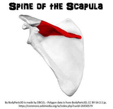
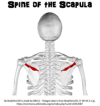
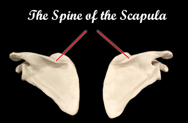
The Spine of the Scapula - The spine of the scapula is the raised portion that can be felt through the skin. This acts as a surface for back muscle attachment.
4) THE SPINE OF THE SCAPULA
EDITED 2 MINUTE VIDEO YOU SHOULD WATCH!! Courtesy of Nicole Mashburn
The Anatomy of the Upper Extremities
|
|
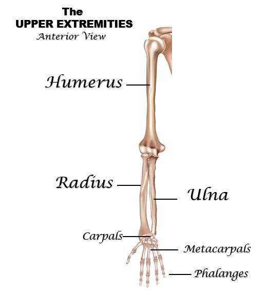
Bones of the Upper ExtremitiesThe area of the upper extremities includes the following bones:
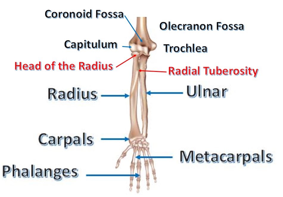
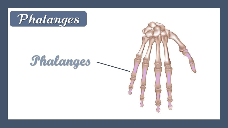
1) The Humerus (upper arm bone) 2) The Radius (lower arm bone on the "thumb side") 3) The Ulna (lower arm bone on the "pinky side") 4) The Carpals (wrist bones) 5) The Metacarpals (bones of hand/palm) 6) Phalanges (finger bones) |
THE HUMERUS (UPPER ARM BONE)
The Humerus has 7 Structures You Need to Know.
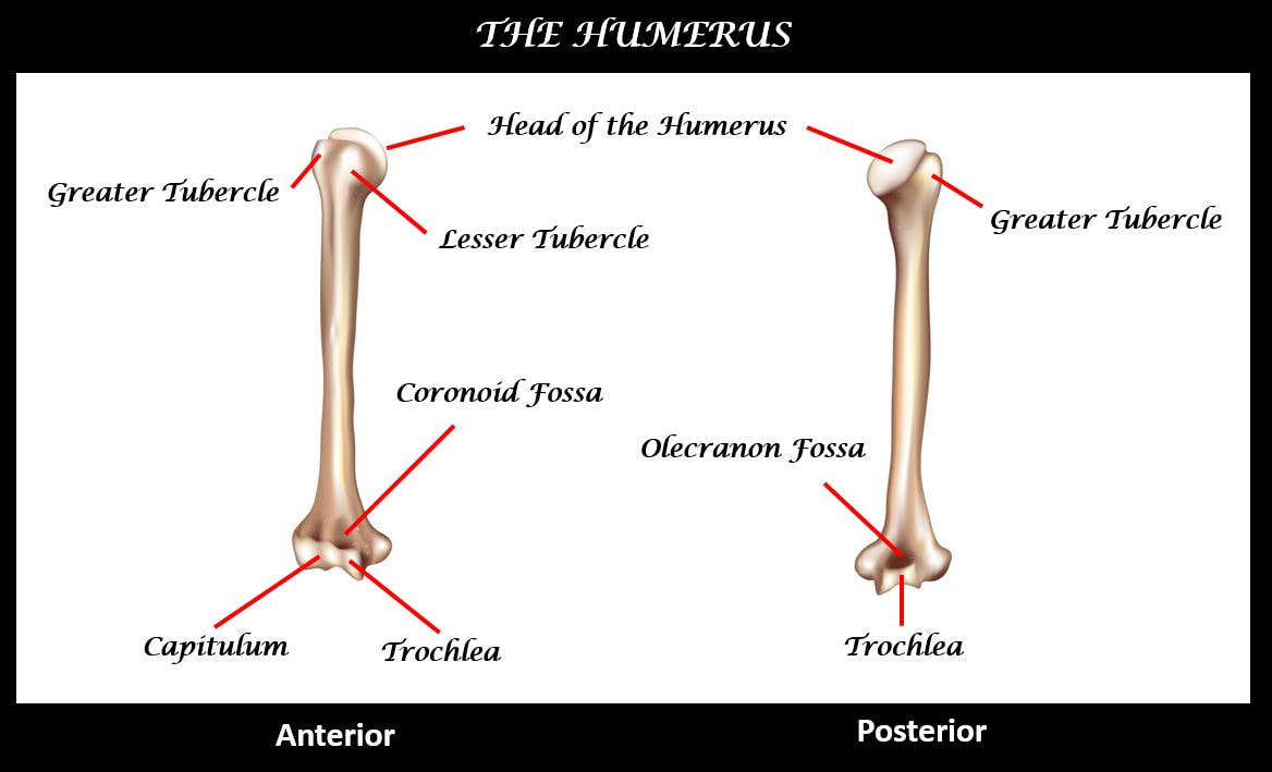
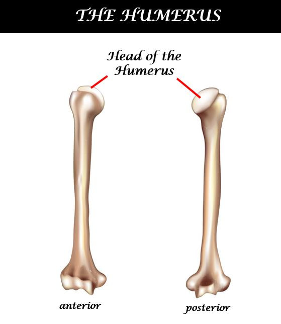
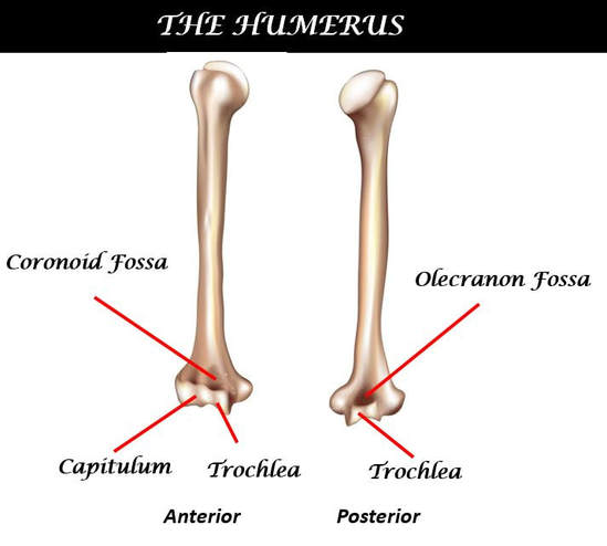
1) The Head 2) The Trochlea 3) The Capitulum 4) The Olecranon Fossa 5) Coronoid Fossa
6) The Greater Tubercle 7) The Lesser Tubercle
1) THE HEAD OF THE HUMERUS
2) THE GREATER TUBERCLE
|
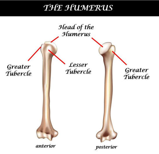
The Greater Tubercle - A tubercle is a general term for a medium-sized rounded protuberance. The greater tubercle lies at the posterior aspect of the superior humerus. It functions as an attachment site for the rotator cuff muscles (shoulder muscles).
3) THE LESSER TUBERCLE The Lesser Tubercle - The lesser tubercle is less pronounced than the greater tubercle and it lies at the anterior aspect of the superior humerus. It also functions as an attachment site for the rotator cuff muscles.
|
|
4) THE CAPITULUM
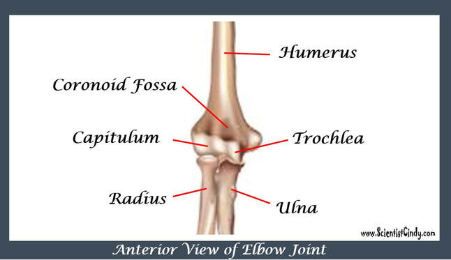
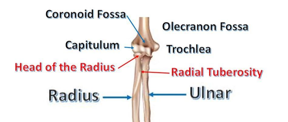
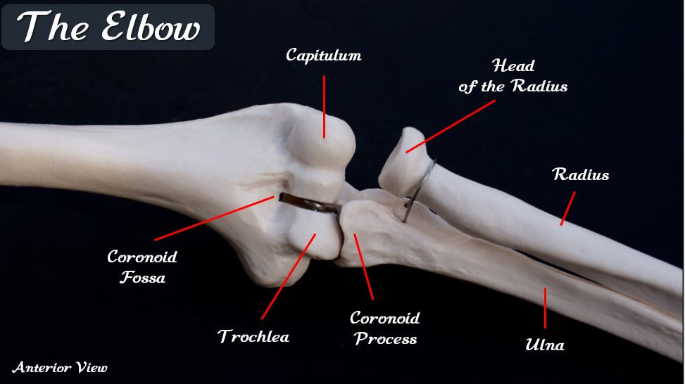
The capitulum lies on the "thumb side" of the humerus and articulates with the radius to form part of the elbow joint. The capitulum appears like a small "bald head". In fact, the name "capitulum" comes from the Latin word for "tiny head". For me, the word "decapitation" reminds me that the little bald head structure is called the capitulum.
5) THE TROCHLEA
|
|
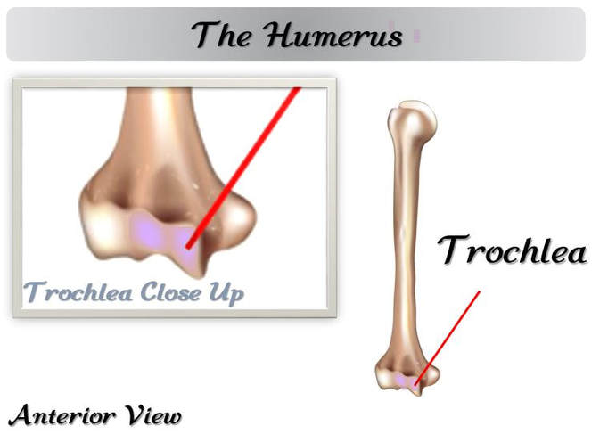
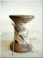
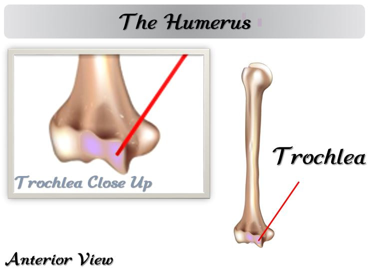
The trochlea is the point of articulation for the trochlear notch of the ulna. The trochlea lies on "pinky" side of the distal humerus and looks like a spool of thread.
The trochlea provides a surface for the trochlear notch of the ulna to 'rock back and forth, on to make up part of the elbow joint! |
6) THE CORANOID FOSSA
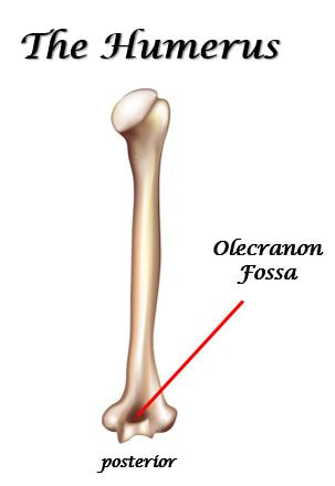
7) THE OLECRANON FOSSA
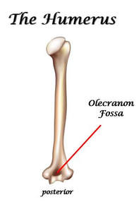
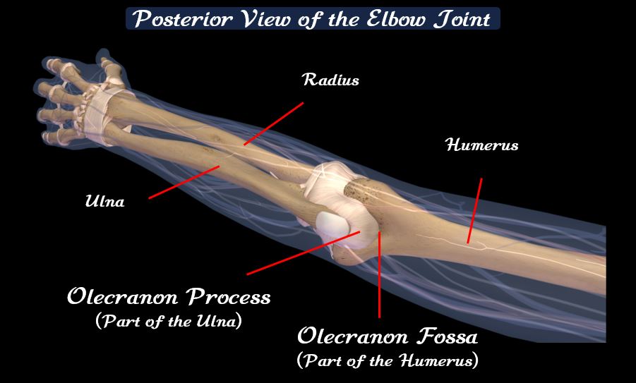
The Olecranon Fossa - The olecranon fossa is the deep triangular depression existing on the posterior aspect of the distal humerus. The function of the olecranon fossa is to receive the olecranon process of the ulna when the elbow joint is fully extended.
THE RADIUS
The Radius Has 2 Structures You Need to Know.
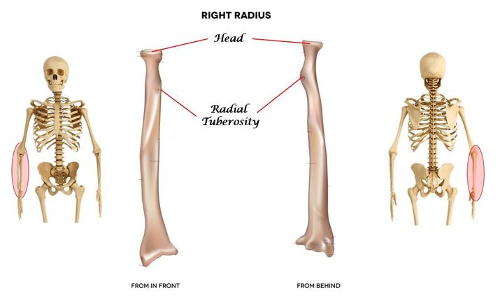
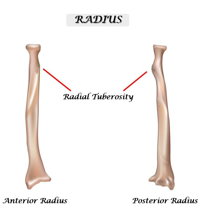
1) The Radial Tuberosity 2) The Head of the Radius
1) THE RADIAL TUBEROSITY
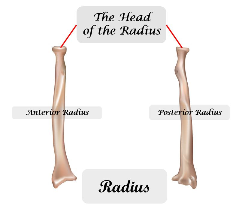
2) THE HEAD OF THE RADIUS
The head of the radius lies at the proximal end of the radius. It is disc-shaped with a slightly concave (inward curved) surface. The morphology (shape or appearance) of the head of the radius is very different from the more rounded head of the humerus. The head of the radius articulates with the capitulum of the radius to form part of the elbow joint. You may recall that the capitulum is named after the Latin word for "head". This round, convex (outwardly curved) surface articulates nicely with the head of the radius.
THE ULNA
The Ulna Has 3 structures That You Need to Know.
1) The Olecranon Process 2) The Coronoid Process 3) The Trochlear Notch
1) THE OLECRANON PROCESS
|
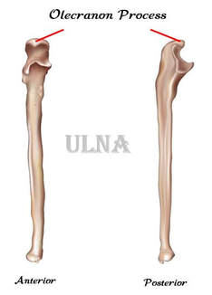
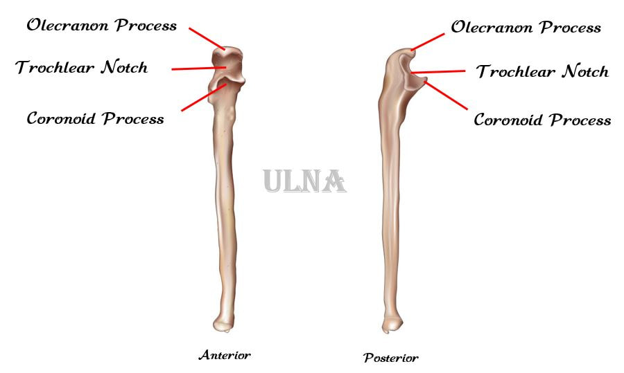
The pointy, bony protrusion located at the olecranal region is known as the olecranon or the olecranon process. This is the part of the body that is more commonly known as the "elbow". The olecranon process lies at the posterior aspect of the proximal ulna. When the elbow joint is extended, the olecranon process will articulate with the olecranon fossa on the posterior aspect of the distal humerus.
|
2) CORANOID PROCESS
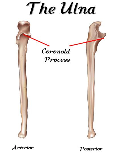
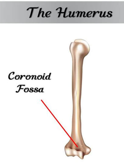
The coronoid process is the anterior portion of the proximal ulna. When the elbow joint is fully flexed, the coronoid process will articulate with the coronoid fossa at the anterior aspect of the distal humerus.
3) TROCHLEAR NOTCH
The deep concavity lying between the olecranon process and the coronoid process, is the trochlear notch of the ulna. The trochlear notch glides across the trochlea on the distal humerus when the elbow joint is moved.
Edited 3 minute video on the ulna and radius you should watch! Courtesy of Nicole Mashburn
THE HAND
The Hand Contains 3 Groups of Bones You Should Know.
1) The Carpals 2) The Metacarpals 3) The Phalanges
1) The Carpals
|
|
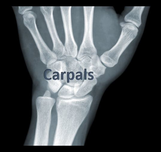
The carpals are the short, somewhat cube-shaped bones that collectively make up your wrist. You may have heard of a condition called "carpal runnel syndrome". The word "carpel" means wrist. Even though carpal tunnel syndrome is a nerve condition, the term can help you link the word "carpal" to the wrist.
The carpals are 8 separate bones that allow for multi-directional movement. |
2) The Metacarpals
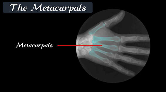
The metacarpals are the bones of your hand or palm area. Don't be deceived! The skeleton looks like it has really long fingers, BUT don't forget that metacarpals are making up part of what looks like those long fingers. The X-ray of the hand will help you visualize the location of the metacarpals. Another way to think about it is that the metacarpals will be the 1st set of long bones proximal to the carpals.
LOWER EXTREMITIES
THE PELVIC GIRDLE (PELVIS)
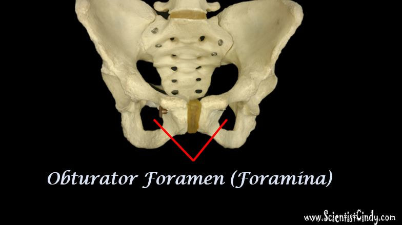
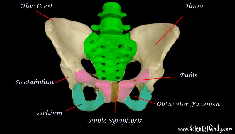
The pelvic girdle (also referred to as the pelvis) consists of the 2 coxal bones (also called the hip bones, the pelvic bones or the os coxae), the sacrum, and the coccyx (tailbone).
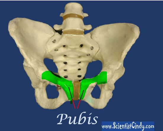
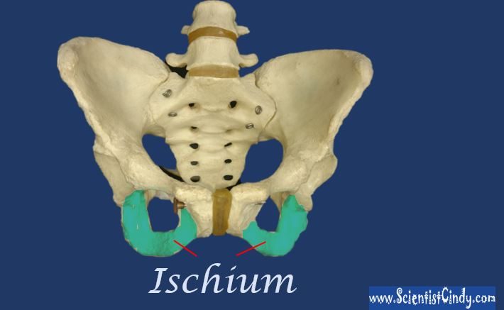
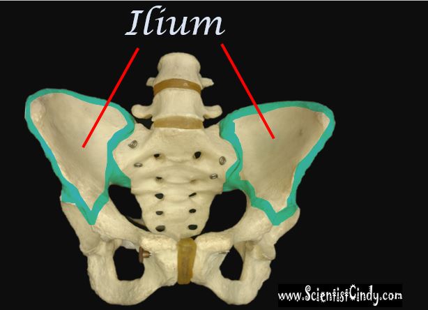
The coxal bone (or hip bone / pelvic bone / os coxae) is actually 1 bone that is formed from the fusion of 3 bones, called the illium (top / superior portion), the pubis (medial, anterior portion, and the ischium (lower / inferior portion).
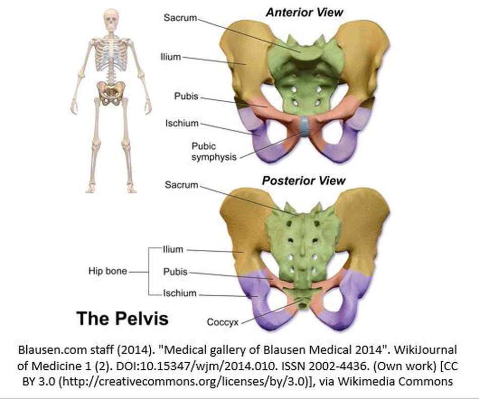
The hip bones are formed through the fusion of 3 plates during development. These form the 3 regions known as the ilium, the pubis and the ischium, which collectively form the hip bone.
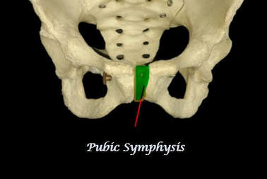
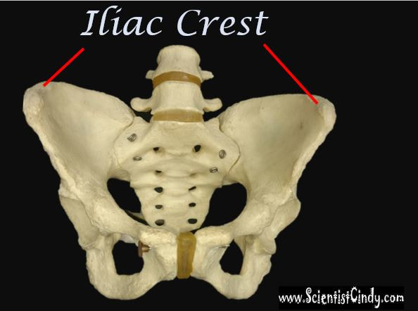
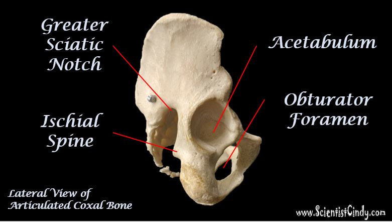
The Coxal Bones (aka Pelvic Bones / Hip bones / Os Coxae) have 9 structures you need to know.
|
1) The Pubic Symphysis -The structure of the joint that fuses the 2 pubic bones together anteriorly
3) Pubis - Medial portion of the hip bone
5) Iliac Crest - The part of your hips you can rest your hands on.
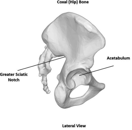
7) Greater Sciatic Notch - This lies at the posterior of the coxal bone opposite the ischial spine.
|
2) The Ishium - the lower portion of the hip bones
4) Ilium - Large Upper (Anterior) portion of the hip bone
6) Acetabulum - the socket for the femoral head (only viewable laterally)
8) Obturator Foramen - allows blood vessels and nerves to go through the bone.
|
VERY SHORT EDITED VIDEO SHOWING ANATOMY OF THE PELVIS ON A REAL BONE. HIGHLY RECOMMENDED!
LOWER LIMBS
THE FEMUR (UPPER LEG BONE)
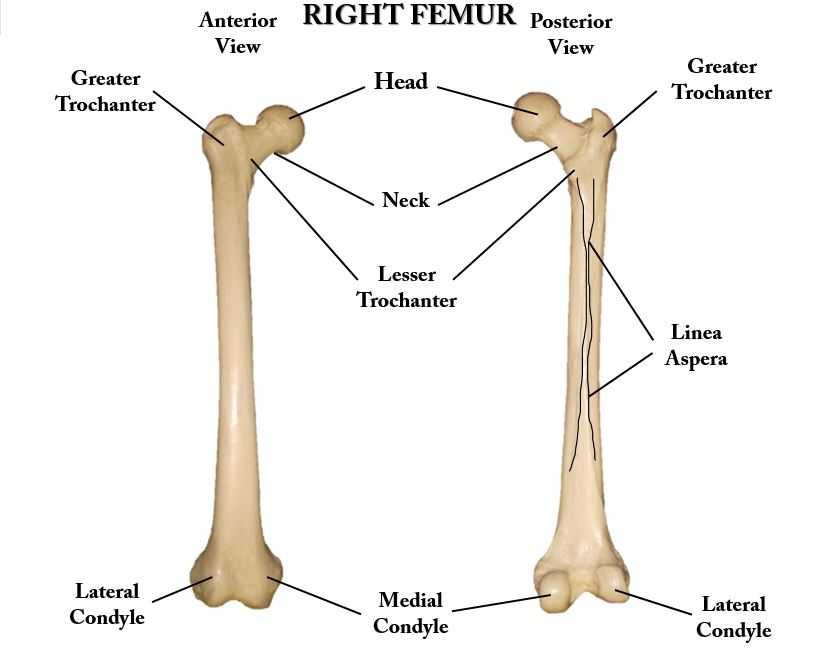
The FEMUR Has 7 Structures You Need To Know.
1) Head 2) Neck 3) Greater Trochanter 4) Lesser Trochanter
5) Medial Condyle 6) Lateral Condyle 7) Linea Aspera
|
|
The Structures of the Femur
TAKE A LOOK AT THIS VERY SHORT SUPER HELPFUL VIDEO I HAVE EDITED FOR YOU! |
THE PATELLA
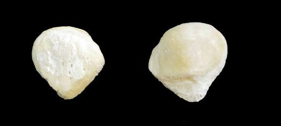
The patella is known as the kneecap. It is categorized as a sesamoid bone, because it is embedded in a tendon. The patella is a thick, circular-triangular bone which articulates with the femur (thigh bone) and covers and protects the knee joint.
THE TIBIA (SHIN BONE) AND FIBULA
The TIBIA has 3 structures that you need to know.
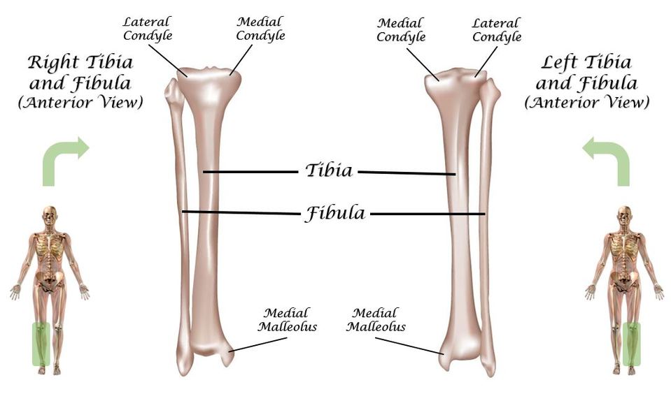
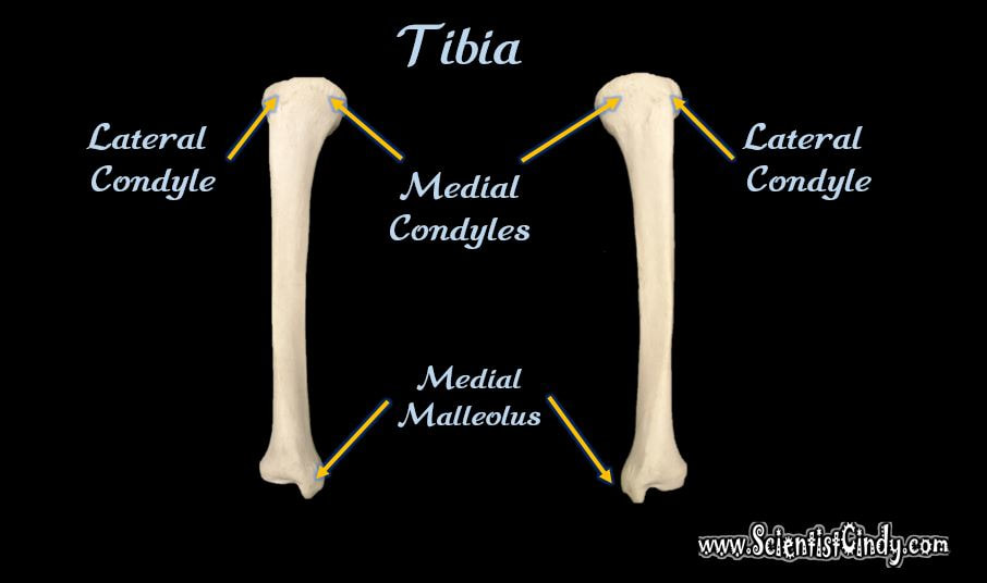
1) The Medial Condyle 2) The Lateral Condyle 3) Medial Malleolus
The TIBIA has 3 structures that you need to know.
1) The Medial Condyle 2) The Lateral Condyle 3) Medial Malleolus
1) The Medial Condyle 2) The Lateral Condyle 3) Medial Malleolus
|
|
The Medial and the Lateral Condyles lie at the proximal portion of the tibia and join with the femur to form part of the knee joint. The Medial Malleolus is the prominence on the inner side of the ankle. This can be felt through the skin. |
THE FOOT (TARSUS)
The Foot (Tarsus) has 5 Structures that You Need to Know.
1) The Tarsals 2) The Calcaneus 3) The Talus
4) The Metatarsals 5) The Phalanges
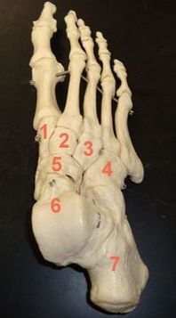
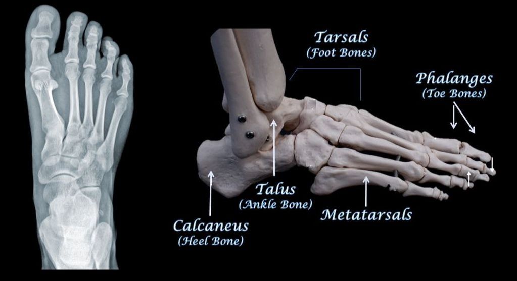
- The Tarsals (General Term for the 7 "Foot Bones") – The term tarsals refers to the 7 bones of the foot as a group. The tarsals are labeled numbers 1 - 7 in the photo. Each of these bones has a name. Don't worry though, you will only need to know the name of a 2 of these bones (the calcaneus (heel) bone and the talus (ankle).
- The Calcaneus (Heel Bone) - The calncaneus is the bone that form the heel portion of your foot. It is considered one of the tarsal bones of the foot and is labeled as #6 in the photo.
- The Talus (Ankle Bone) - The talus is the bone that form the ankle portion of your foot. It is considered one of the tarsal bones of the foot and is labeled as #7 in the photo.
|
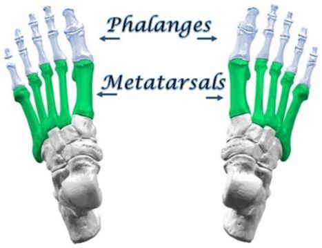
The Metatarsals - The metatarsals are the 5 long bones that make up the front (anterior) part of the foot. The metatarsals are anterior to the tarsals and posterior to the toe bones (phalanges). The Phalanges – The phalanges are the bones of the toes. The big toe has 2 phalanges, while the other toes have 3 phalanges. ALTERNATE VIEWS OF FOOT ANATOMY |

