Epithelial Cells and Tissue
|
Atoms come together to form molecules. There are about 100 trillion atoms in one cell. The human body contains probably somewhere around around 100 trillion cells as well! Depending in the source, estimates of the number of cell in the average human have ranged from as little as 5 trillion (OK, that's not little at all) to 200 trillion cells.
The cell is the fundamental unit of life. All living things are made up of one or more cells.
|
|
Tissues

|
|
Cells of that are of a similar type of are of the same embryonic origin, come together to form tissues of the same type.
- Neurons will come together to form nervous tissue.
- Muscle fibers will come together to form muscle tissue.
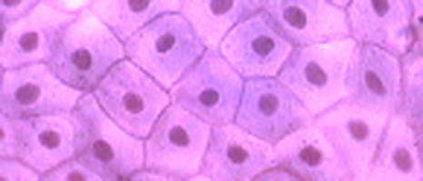
- Epithelial cells will come together to form epithelial tissue.
|
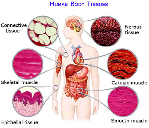
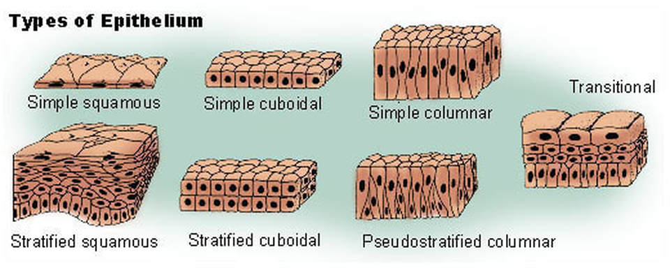
There are 4 tissue types.
Epithelial tissue, is more than just your skin. It is all of the inner and outer coverings of the body, including our the tracts and the cavities. A specialized form of epithelial tissue, called, glandular epithelial tissue (or simply glandular tissue) form the body's glands.
|
|
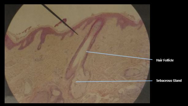
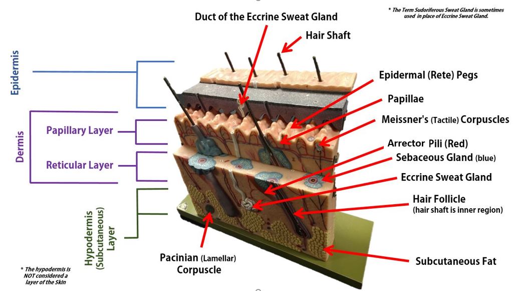
Epithelial tissue is a major component of the integumentary system. The integumentary system has 2 major components; 1) the cutaneous membrane (skin) and 2) the accessory structures. The accessory structures include the hair, nails, sebaceous (oil) glands, and sudoriferous (sweat) glands.
The word "cutaneous" simply means "skin" in Latin and is also called the integument which means “covering”. Different areas of the body will have various thickness between 1.5 to 4.0 millimeters (mm).
Characteristics of Epithelial Layers
Epithelial tissues have five distinguishing characteristics:
polarity,
supported by connective tissues,
being avascular but innervated, and
having the ability to regenerate.
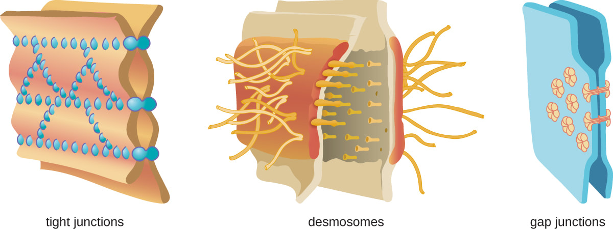
2) Specialized Contacts

Epithelial tissue is tightly woven together and is almost entirely composed of cells, with very little matrix. The individual epithelial cells are tightly linked to one another through specialized junctions, creating a water-tight barrier.
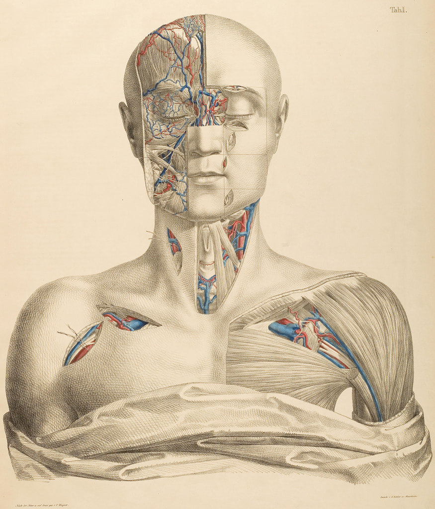
2) Epithelial Tissue is Avascular
Epithelial tissue is avascular, meaning that it does not contain blood vessels. The epithelial cells are nourished by substances diffusing from blood vessels in the underlying connective tissue.
3) Epithelial Tissue is Innervated
4) Polarity
Epithelial tissue has a very distict orientation. The top and bottom portions of the cells have specialized features that allow for specific functions. The portion of the cell or tissue that is more superficial, is called the APICAL side or APICAL surface. The deeper portion of the cell or layer of tissue is called the BASAL or BASALAR side or surface.
One side of the epithelial cell is oriented towards the surface of the tissue, body cavity, or external environment and the other surface is joined to a basement membrane. The basement layer is non-cellular in nature and helps to cement the epithelial tissue to the underlying structures.
One side of the epithelial cell is oriented towards the surface of the tissue, body cavity, or external environment and the other surface is joined to a basement membrane. The basement layer is non-cellular in nature and helps to cement the epithelial tissue to the underlying structures.
Epithelial tissue lines every surface of our bodies, inside and out. Our glands are also made up of epithelial tissue.
Special Characteristics of Epithelial Tissue
Cellularity – composed almost entirely of cells.
Small amount of extracellular matrix. Narrow spaces between cells.
Small amount of extracellular matrix. Narrow spaces between cells.
Functions of the Epithelium
(1) protection, (2) absorption, (3) filtration, (4) excretion, (5) secretion, and (6) sensory reception,
The skin performs a variety of functions. We may spend a lot of time preparing our dead skin cells to look their best for the world, but your skin does a lot more than just give you an attractive face! Let's take a look at the ways your skin is hard at work to keep you movin' and groovin' throughout your day!
(1.) Protection
|
The primary function of your skin is to provide a physical protective barrier against harmful environmental elements, dehydration and physical forces. The epithelium allows your internal organs and soft tissues to be somewhat shielded from the outside world. It provides a barrier that is able to withstand some wear and tear. It can get bumped, bruised or scraped and it is able to heal relatively quickly.
This is because the epidermis is composed of stratified squamous tissue which provides layers of small, squamous cells which are rapidly and easily replaced. The barrier of your skin also protects you from harmful elements like bacteria, viruses, chemicals and even contains melanin (within specialized cells called melanocytes) which provides protection from harmful UV radiation from the sun. |
|
YOU SHALL NOT PASS!
Epithelial tissue is selectively permeable, allowing only certain substances to enter the body at any time. The skin is our first line of defense against harmful elements in our environment, such as disease-causing microbes, reactive substances and irritants. Your skin functions to prevent excess water loss from the body.
(2) absorption
Excretion - Small quantities of nitrogenous wastes, such as ammonia, uric acid, and urea are removed through sweat.
(3) filtration
 Gland Gland
Epithelial cells that produce secretions are called gland cells or glandular epithelial cells. Like any good cell, the gland cells will come together to form glandular epithelial tissues.
Epithelial tissue assists in maintaining homeostasis. For example, sweat glands respond to heat by emitting secretions that reach the outer surface of the skin to keep us cool. The secretion of sweat, mucus, enzymes, and other products that are delivered by ducts come from the glandular epithelium. 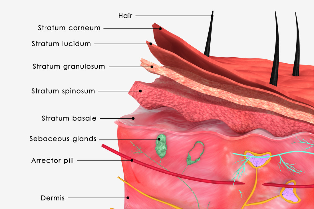
|
|
(3) filtration
Vitamin D is a steroid that gets converted into calcitriol in the body, calcitriol is a hormone that assists in the metabolism of calcium. this is why you will sometimes see "with vitamin D" marked on products containing calcium.
(4) excretion
Removing unwanted substances from the body
(5) secretion
4. Sensory Reception.
Our epithelium is our interface will the outside world. Our physical sensation of touch, pressure, pain and temperature all come from specialized sensory receptor cells in our epithelium. Our skin gathers sensory information about the outside world that gets relayed to the brain. Your skin has sensory receptors for temperature, touch and pain.
Our epithelium is our interface will the outside world. Our physical sensation of touch, pressure, pain and temperature all come from specialized sensory receptor cells in our epithelium. Our skin gathers sensory information about the outside world that gets relayed to the brain. Your skin has sensory receptors for temperature, touch and pain.
(6) sensory reception
SKIN
The beauty of the skin is more then SKIN DEEP!
|
Properties of epithelial tissue
|
Your skin's thickness varies in different areas of your body. Skin can be categorized as either thick skin or thin skin. Your thick skin is located in regions of your body that has a lot of physical contact with the outside world. These areas include your fingertips, your palms and the soles of your feet. These extra layers help to protect the soft tissues from abrasions. Thick skin differs from thin skin in its structure. While the epidermis is thicker in thick skin than in thin skin, but the layer of the dermis turns out to be thinner. Also, thick skin does not contain hairs, sebaceous glands, or sweat glands. The epithelium of thick skin contains 5 epidermal layers, whereas thin skin contains only 4 layers.
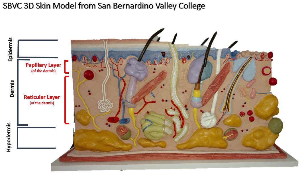
The Layers of the Skin
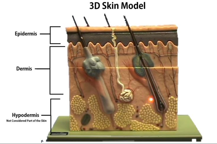
The outermost surface of your skin (the superficial region) is made of a thick epithelial tissue called epidermis. The word "derm" means "skin". The prefix "epi-" means "above". Beneath the epidermis, (deep to the epidermis) is the layer of the dermis. The dermis is composed of connective tissue. The skin is composed of the epidermis and the dermis. Below the skin (deep to the skin) there is another layer called the hypodermis. "Hypo-" means "below". This layer is composed of areolar connective tissue and adipose tissue. The hypodermis is NOT considered to be part of the skin itself.
The skin is composed of 2 primary layers:
1) the epidermis
2) the dermis
The apical (outer most) layer is called the epidermis. The prefix "epi-" means "above". Lying underneath the epidermis is the dermis. Each of these primary layers is broken down into subsequent layers.
Epdermal tissue is categorized as a keratinized stratified squamous epithelial tissue. The most abundant type of cell found in the epidermis are keratinocytes. Keratinocytes produce keratin which is a fibrous protein that gives strength to epidermis. The keratin protects the deeper tissue layers not only by providing a physical barrier to the outside world, but they provide antibiotics. and enzymes that act to detoxify the harmful chemicals to which our skin is exposed. Keratinocytes form in the deepest portion of the epithelium (the stratum basale) and then are continously pushed outward until they die and eventually get sloughed off of the body. We loose millions of these dead skin cells every day! your epidermis is completely replaced every 35-45 days.
The keratinocytes undergo physical changes as they mature and move closer to the surace of your skin. The keratinocytes are created from stem cells that extist in the stratum basale. As these cells are pushed up by the production of new cells beneath them, they make theor way to higher (more apical) epithelial layers. When the keratinocytes reach the stratum spinosum (the layer superficial to the stratum basale) they begin to produce keratin. Once they reach the stratum granulosum, these cells are busy making lots of keratin which fills their cytoplasm. The keratinocytes begin to die as they become part of the stratum lucidum, becoming smaller and clear. They begin to loose their organelles as they die. By the time these dead keratinized squamous epithelial cells reach the skin's surface, they are completely dead and are characterized as "corneocytes". The corneocytes are basically dead, flat sacs completely filled with fibrous keratin.
There are no blood vessels in the epidermis, so the epidermis does not have its own blood supply.The epidermis get nutrients and gasses , oxygen, and vitamins travel to the epidermis through the rete pegs, which are made up of a network of very small blood vessels that project down to the dermis layer.
The Epidermis
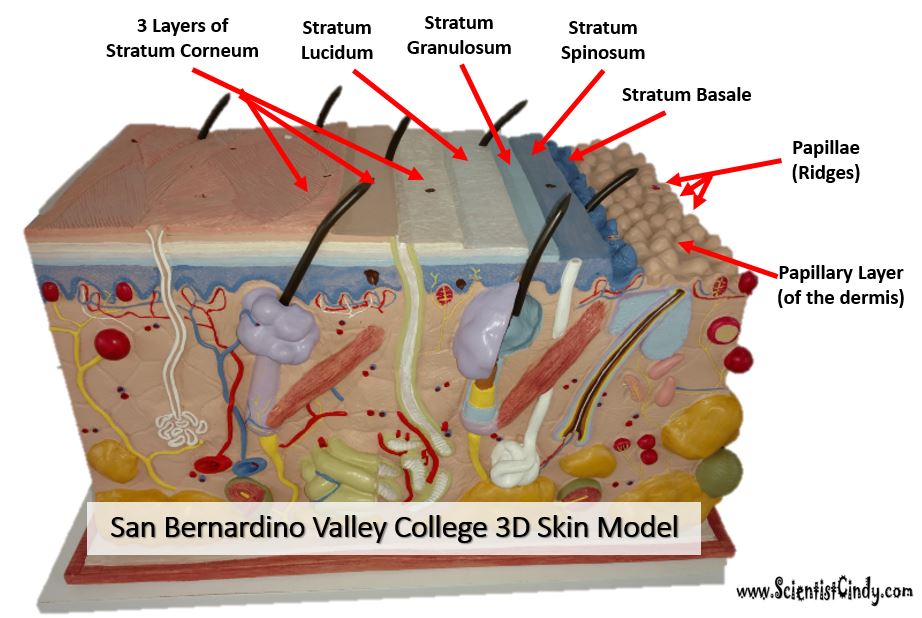
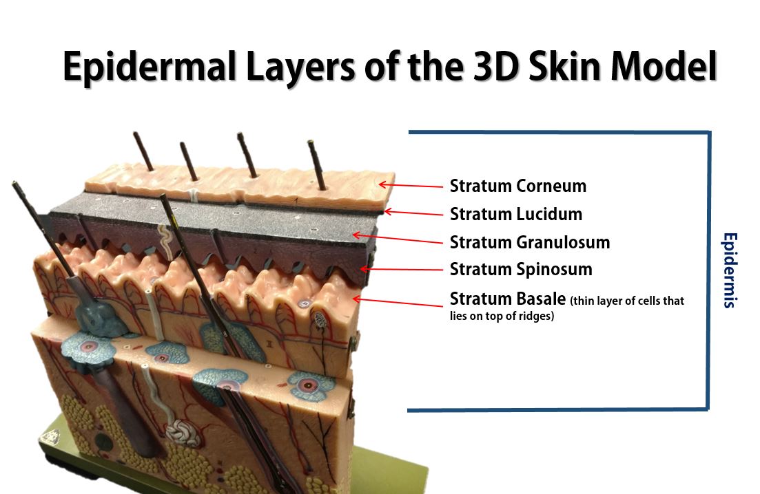
The Layers of the Epidermis
The epidermis consists of 5 layers in thick skin / 4 layers in thin skin.
- Stratum Corneum - Translates as "horny layer".
- Stratum Lucidem - Translates as "clear layer". ONLY EXISTS IN THICK SKIN
- Stratum Granulosum - Translates as "granular layer".
- Stratum Spinosum - Translates as "spinous layer/prickle cell layer".
- Stratum Basale - Translate as "basal layer".
Need a Mnemonic?? Try, Come, Let's Get SunBurned!
C = Stratum Corneum
L = Stratum Lucidem
G = Stratum Granulosum
S = Stratum Spinosum
B = Stratum Basale

The Layers of the Epidermis
(HINT: they go in alphabetical order).
Starting at the apical (top) end...
1. The Stratum Corneum (Horny Layer)
The stratum corneum is a keratinized
stratified squamous epithelium that contains four
types of cells. The four cells types cells that make up the stratum conrneum are 1) keratinocytes, 2) melanocytes, 3) tactile epithelial
cells, and 4) dendritic cells
The stratum corneum is Latin for 'horny layer'. Its odd name comes from the dead squamous cells called corneocytes that make up the apical surface of the tissue. The corneocytes form several layers of flattened (squamous) cells with no nuclei and no organelles. This is the layer that includes the final keratin product, which is a combination of cytokeratin and keratohyaline.
2. *The Stratum Lucidum
The stratum lucidum is Latin for "clear layer". This epidermal layer gets its name from translucent appearance of the dead skin cells that make it up. *This layer only exists in the thick skin of the soles of your feet, your palms and your fingertips.
3. Stratum Granulosum (Granular Layer)
The stratum granulosum is Latin for granular layer. In this layer, the keratinocytes have become squamous cells that contain granules of keratohyaline, a precursor to the extracellular keratin that protects the skin tissue from abrasion.
4. Stratum Spinosum (Prickle Cell Layer)
The stratum spinosum is Latin for "spiny". After forming in the basal cell layer, keratinocytes migrate upwards to form the stratum spinosum. In this layer, they develop short projections that attach via desmosomes to adjacent cells. The stratum spinosum is also known as the "prickly layer" because of these characteristic spines. The cells in this layer produce cytokeratin, an intermediate filament precursor to keratin.The cells in this layer experiences shrinking of its microfilaments during the slide staining process.
5. Stratum Basale (Basal Layer)
The stratum basale is the deepest of the 5 epidermal layers. The primary cell of the epidermis is the keratinocyte stem cells which give a continuous supply of new cells to replace old dead ones. The stratum basale also contain sensory nerves called Meissner's (Tactile) Corpuscles that are sensitive to light tactile sensations (light touch). This layer also contains melaocytes which produce the pigment melanin which acts to filter out harmful UV radiation.
Video of Skin Model for San Bernardino Valley College
Video of 3D Skin Model - Layers of the Epidermis
For Rio Hondo
This 3D skin model is identical to the one we use for our practical exam. The video below can be found at youtu.be/o3so9gKQItE
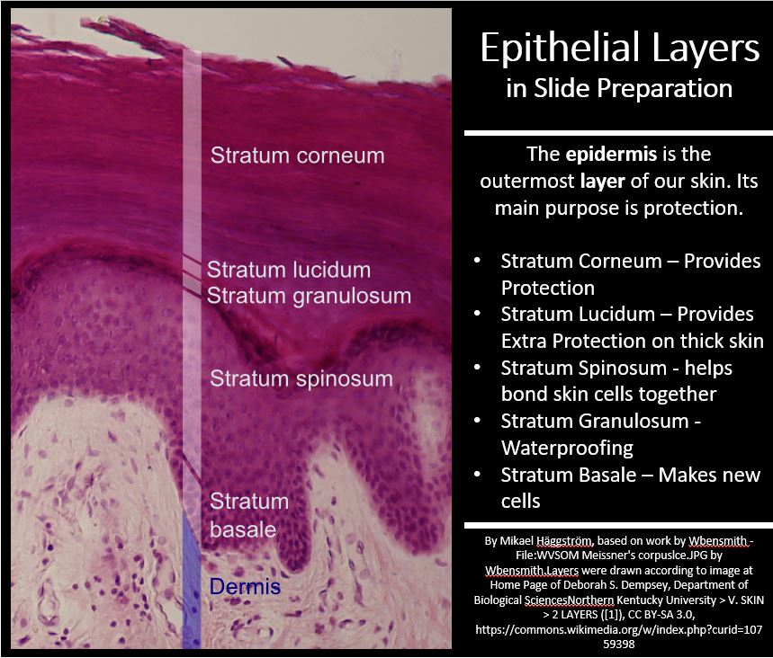
The epidermis is characterized as keratinized stratified squamous epithelium.
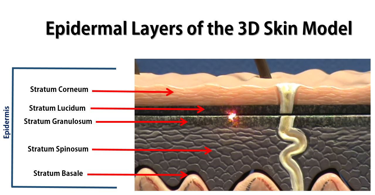
The picture below is a close up of the epidermal region of our 3D skin model.
The stratum basale in our 3D model appears at the interface between the wavy beige region and the gray region. It is a single-cell layer that has stem cells
The picture below is a close up of the epidermal region of our 3D skin model.
The stratum basale in our 3D model appears at the interface between the wavy beige region and the gray region. It is a single-cell layer that has stem cells
3D Skin Model from San Bernardino Valley College
The epidermis consists of 4 cell types.
Touch receptors of the skin.
Your skin has specialized sensory receptors called touch (tactile) receptors or mechanoreceptors which responds to mechanical pressure or distortion by generating a signal that will ultimately reach the brain.
There are four main types of mechanoreceptors in your skin
- Keratinocytes - The most abundant cell type in the epidermis. They are created in the stratum basale and pushed upward toward the skin's surface. These cells make keratin and act to protect deeper layers of soft tissue. These cells die as they approach the surface of the skin. Once they are dead and shriveled up, they are called corneocytes.
- Melanocytes - Pigmented cells of the stratum basale region that produce melanin which protects from UV radiation.
- Merkel Cells - function as touch receptors in association with sensory nerve endings (Merkel disc); located at epidermal-dermal junction
- Langerhans’ Cells - epidermal macrophage-like cells that help activate the immune system via receptor-mediated endocytosis; arise from bone marrow;
Touch receptors of the skin.
Your skin has specialized sensory receptors called touch (tactile) receptors or mechanoreceptors which responds to mechanical pressure or distortion by generating a signal that will ultimately reach the brain.
There are four main types of mechanoreceptors in your skin
- Pacinian corpuscles
- Meissner's corpuscles
- Merkel's discs
- Ruffini endings
The Dermis and Hypodermis
Video of 3D Skin Model - Dermis and Hypodermis
This 3D skin model is identical to the one we use for our practical exam. The video below can be found here: youtu.be/o3so9gKQItE

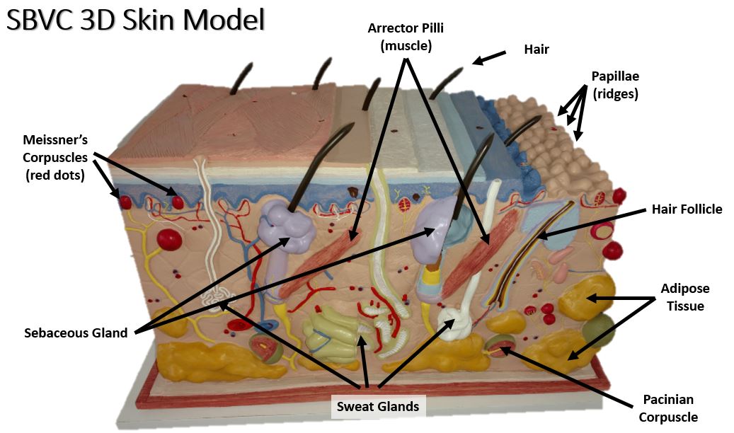
3D Skin and Hair Follicle Model
Dermis and Hypodermis
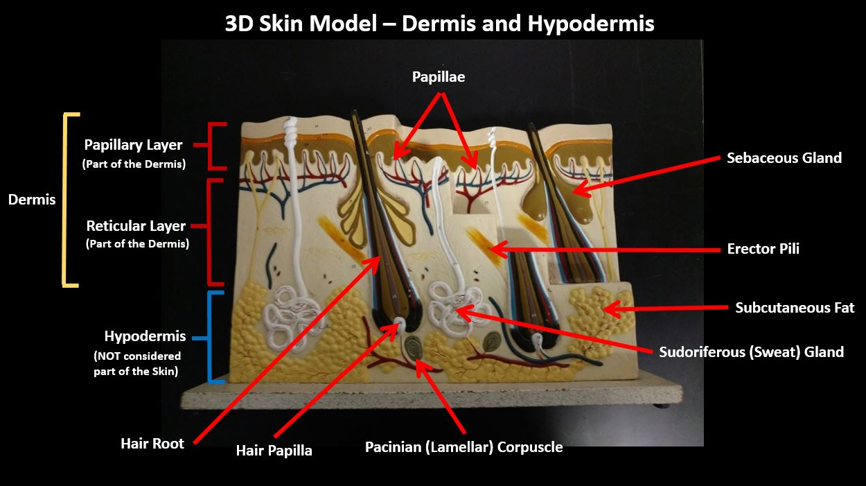
Rio Hondo Model #2
VIDEO - Glands and Sensory Structures of the Skin
Melanocytes are specialized cells that lie within the skin. They produce a brown pigment (melanin) that functions to provide protection from damaging UV radiation from the sun. The pigment filters out the UV light. When your skin is exposed to more sunlight, your melanocyte darken the skin by increasing the production of melanin.
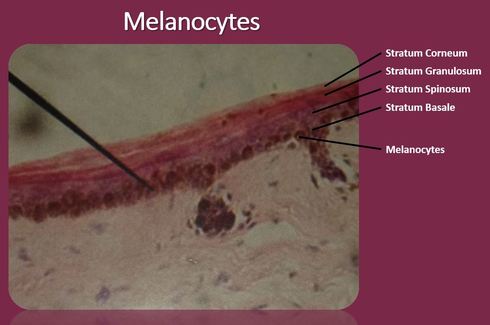
Melanocytes - make melanin - makes skin dark. Protects from UV Light. UV light is carcinogenic. This is why getting too much sun can cause skin cancer.
Slides of Hair Follicle