THE MUSCULAR SYSTEM
Muscles of the Head and Neck
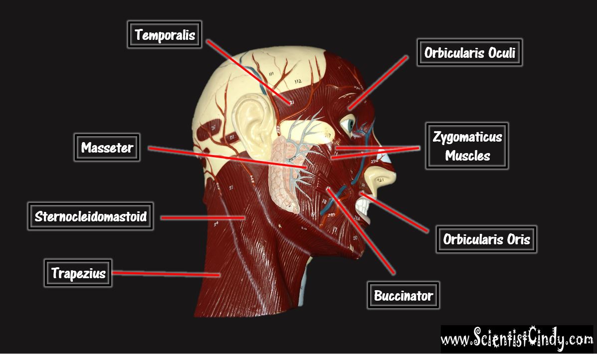
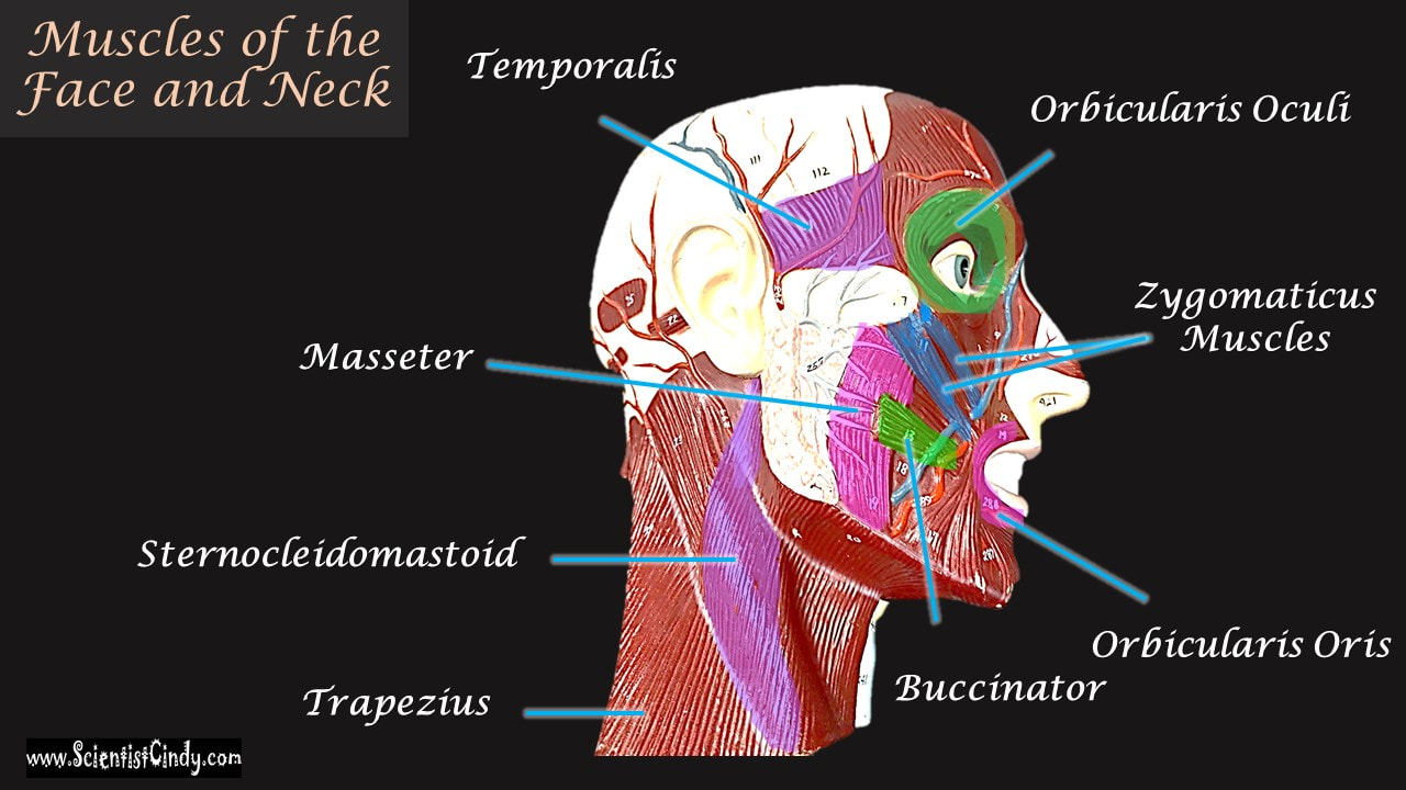
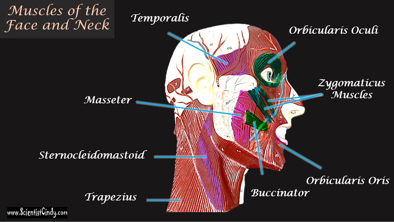
MUSCLES OF THE HEAD AND FACE
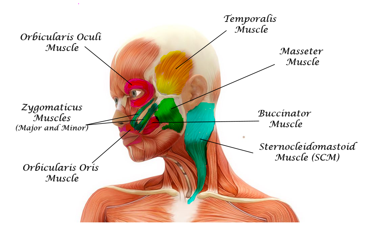
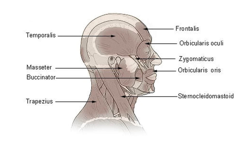
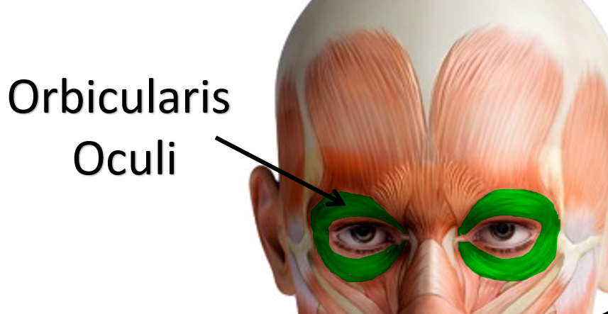
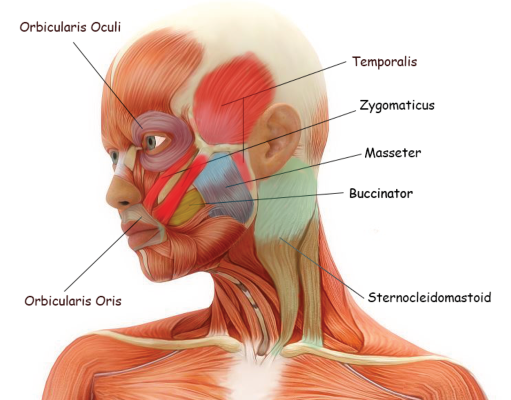
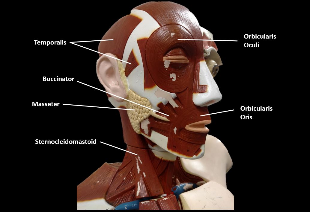
Orbicularis Oculi Muscles
Function: Closes the Eyes
The orbicularis oculi muscle is in charge of your "blinking" motion and being able to squint or close your eye tightly. It appears as a ringlike band of muscle, called a sphincter muscle, that surrounds the eye. Sphincter muscles are arranged in a circular pattern.
The orbicularis oculi muscle is in charge of your "blinking" motion and being able to squint or close your eye tightly. It appears as a ringlike band of muscle, called a sphincter muscle, that surrounds the eye. Sphincter muscles are arranged in a circular pattern.
|
Click to set custom HTML

|
|
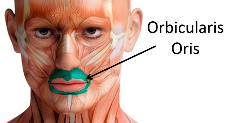
Orbicularis Oris Muscles
Function: Puckers (purses) Lips
The orbicularis oris muscle is a sphincter muscle that encircles the mouth. It is sometimes called the kissing muscle because it causes the lips to close and pucker. |

|

|
|
Muscles of Mastication (Temporalis and Masseter)
|
Temporalis Muscles
Function: Elevates Mandible
The temporalis muscle (or temporal muscle) is one of the muscles you use for chewing (mastication). When you clinch your jaw, you see a couple of muscles contract at the jaw joint. If you clench your jaw, you can see and feel it contracting at the temples on both sides of your head. It's attached to the mandible (jaw) and to the skull's temporal bone, or temporal fossa.
When the jaw is clinched, the muscles of mastication are visible. The upper muscle is the temporalis and the lower, more noticeable muscle is the masseter.
TEMPORALIS MUSCLE
By Anatomography - Anatomography( configuration page of this image[1], CC BY-SA 2.1 jp, https://commons.wikimedia.org/w/index.php?curid=20910061
When the jaw is clinched, the muscles of mastication are visible. The upper muscle is the temporalis and the lower, more noticeable muscle is the masseter. 
|
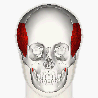
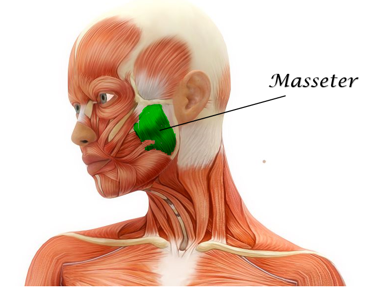
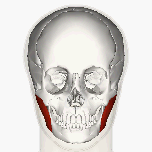
Masseter Muscles
Function: Closes Jaw
The most visible muscle of mastication is the masseter muscle. It is a strong, superficial muscle that function to close the jaw.
The name "masseter" comes from a Greek meaning "one who chews".
The masseter is the major muscle of the jaw. It's principle action is to close the jaw, but it also acts in the side-to-side and forward and back (i.e. protraction and retraction) movement of the jaw.
The most visible muscle of mastication is the masseter muscle. It is a strong, superficial muscle that function to close the jaw.
https://upload.wikimedia.org/wikipedia/commons/6/62/Masseter_muscle_animation.gif
|
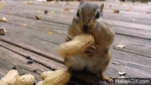
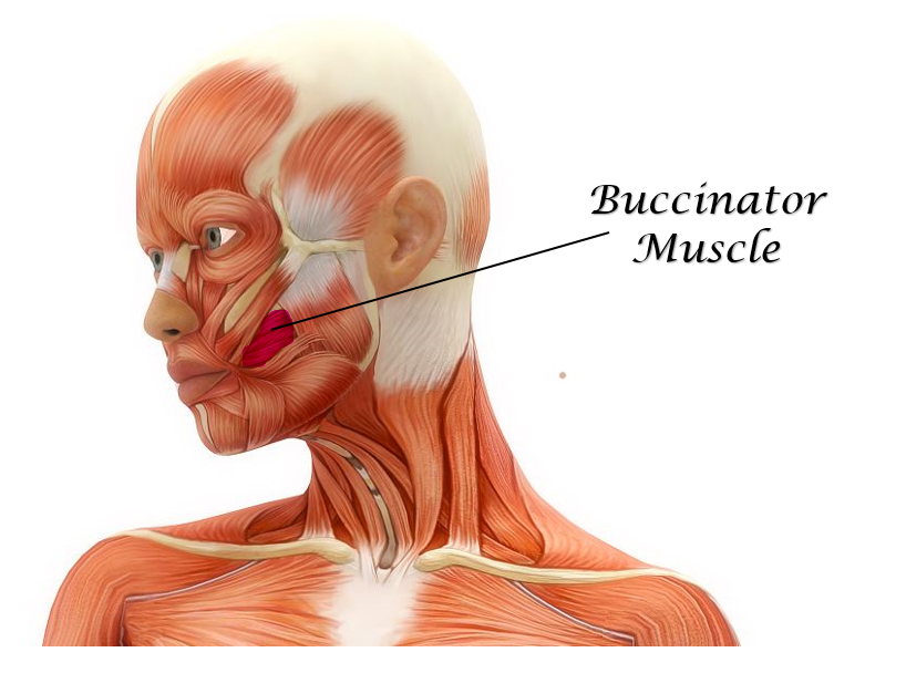
Buccinator Muscles
Function: Holds Food in Mouth (Keeps Food Between Grinding Teeth)
|
Your buccinators are the muscles of your cheeks. You may recall that the cheek is considered the "buccal" region. These anatomical terms come from the Greek word, "bucca".
The buccinator functions to flatten the cheek area and help hold food in the right place so it can get chomped by your teeth, instead of falling into the pockets of your cheeks between the cheek and gums. |
|
|
|
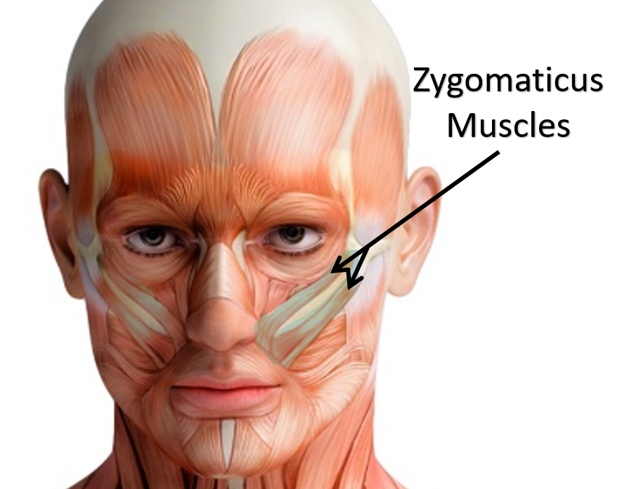
Zygomaticus Muscles
|
Zygomaticus Major Muscles of the face.
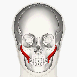
By Anatomography - Anatomography (setteing page of this image file), CC BY-SA 2.1 jp, https://commons.wikimedia.org/w/index.php?curid=20936738
|
Function: Smiling
The zygomaticus muscles are the muscles of your face that help you show emotion by smiling.
You have a zygomaticus major and a zygomaticus minor. These muscles act together to form the smile on your face by pulling up the corners of your mouth. This also allows the poofing out your 'happy' cheeks. Zygomaticus Minor Muscles of the face.
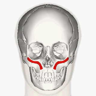
By Anatomography - Anatomography (setteing page of this image file), CC BY-SA 2.1 jp, https://commons.wikimedia.org/w/index.php?curid=21148978
|
The zygomaticus major is larger than the minor and sits lower on the face, extending down to the mouth. The zygomaticus minor is higher and closer to the nose and upper cheek. Both of the zygomaticus muscles also function to keep food in the mouth when chewing.
|
|
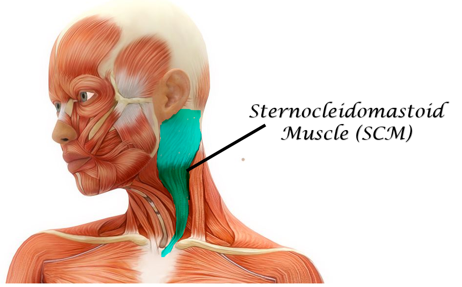
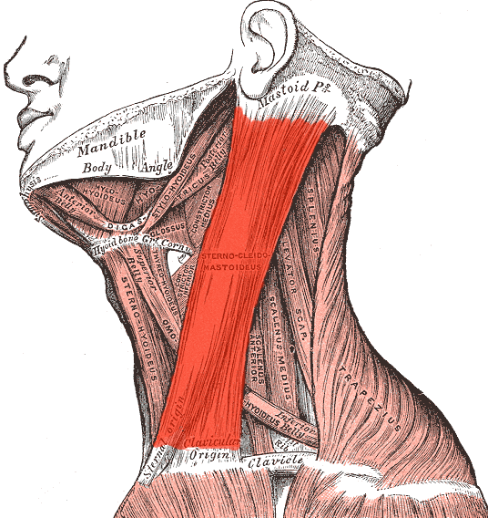
Sternocleidomastoid Muscles
|
Function: Flexes the Head
The sternocleidomastoid muscles (sternomastoid or SCM) are the muscles of your neck responsible for rotation of your head and flexion (tilting head on one shoulder). It is one of the largest cervical muscles and is easy visible when a person turns their head from side to side.
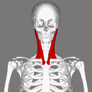
The 'sterno-' portion of its name comes from that fact that one of its origination points is at the sternum (more specifically at the manubrium which is the upper portion of the sternum). It also originates at the clavicle (collar bone), which is where the 'cleido-' portion of its name comes from.
|
|
The name ends with "mastoid", because the sternocleidomastoid muscle ends with an attachment at the mastoid process of the temporal bone The name. sternocleidomastoid, describes the muscles' points of connectivity within the body.
Summary of the Muscles of Head and Neck
MUSCLES OF THE TORSO (trunk)
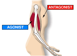
How can I tell the function of muscle? The function of a muscle can be inferred by the position of the muscle relative to the joint it crosses.
|
Remember, a muscle attaches to at least 2 bones and spans at least 1 joint. In order to determine the function of the muscle, you need to see if that muscle passes in front or behind the joint it spans.
Remember that muscles only have 2 actions; they can either contract (shorten) or relax. This means that muscles will essentially 'pull' on the bone it attaches to. |
|
Rules for determining muscle function:
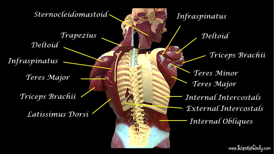
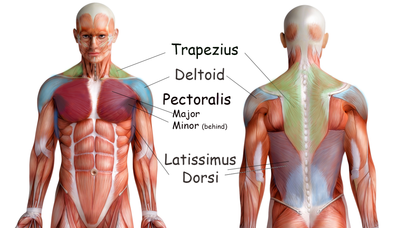
The Superficial Muscles of the Thorax Include the Trapezius, the Pectoralis (Major and Minor), the Deltoid and the Latissimus Dorsi
|
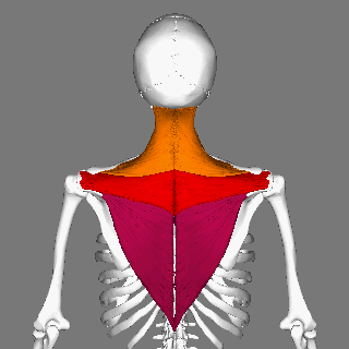
Trapezius Muscles
|
|
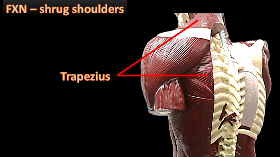
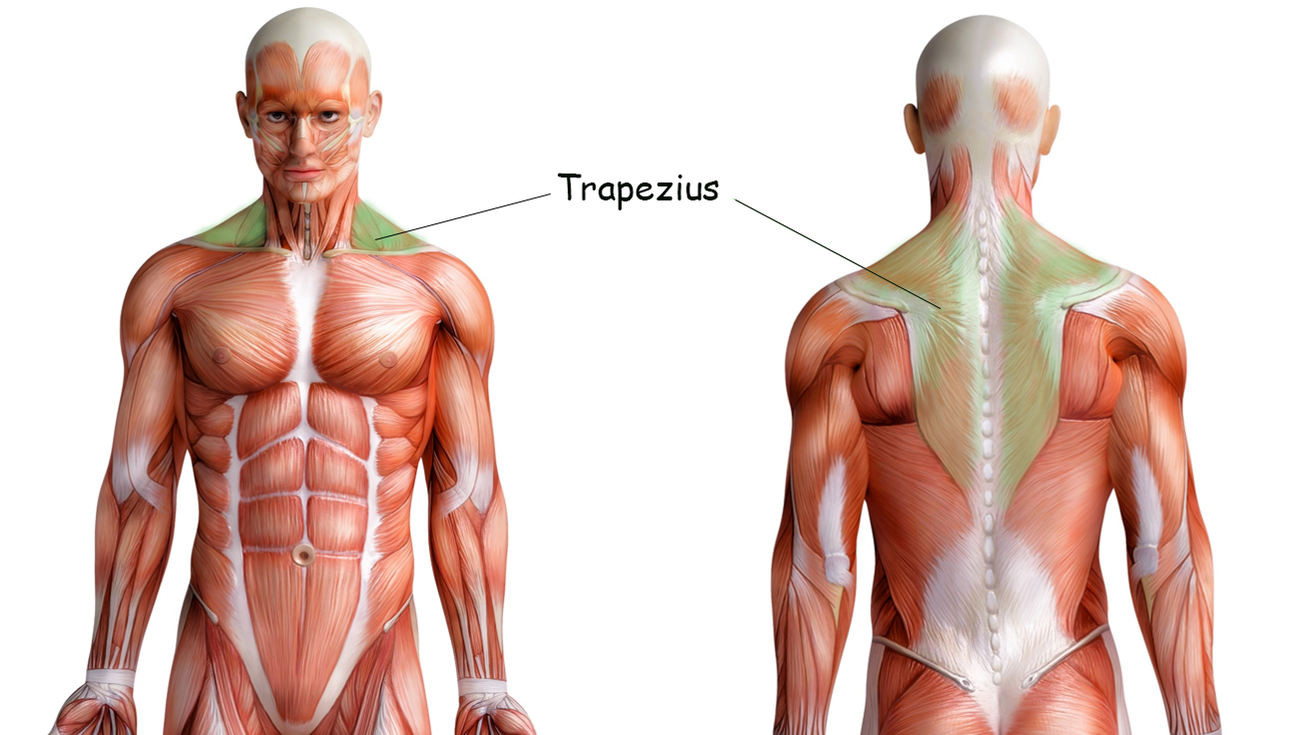
Function: Shrugs Shoulders
The trapezius muscles are the large major muscles of the upper back. The trapezius muscles function to move, rotate and stabilize the scapula (shoulder blade).
By Anatomography - en:Anatomography (setting page of this image)., CC BY-SA 2.1 jp, https://commons.wikimedia.org/w/index.php?curid=22182550
|
|
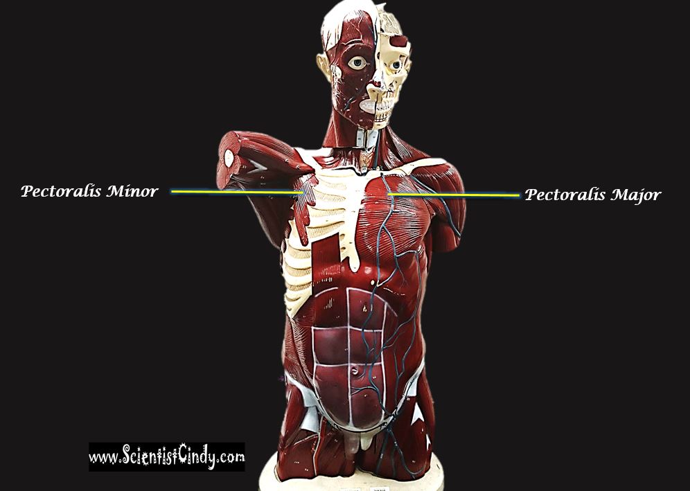
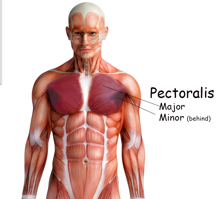
Pectoralis Major and Minor Muscles
Function: Flexion, rotation and adduction of the arm.
The pectoralis major is a large muscle of the upper chest region (thoracic region). It connects the bones of the chest to the shoulders and upper arms. It originates at the sternum and the costal cartilage (from the costal cartilage of the 2nd to the 5th pair of ribs). The pectoralis major attaches to the clavicle and to the humerus, just below the shoulder. The pectoralis major muscle allows you to move your arm across the body.
The pectoralis minor muscles lie underneath the pectoralis major muscles closer to the axillary region (region of the armpit). The pectoralis minor and are considerably smaller. It is smaller of the two sets of muscles that connect the bones of the chest to the shoulder and upper arm. Sitting in the axilla and largely below the pectoralis major, the thin pectoralis minor muscle fastens to the shoulder blade (scapula) at the coracoid process and connects it to the center of each side of the chest near the cartilage of the middle ribs (anterior, sternal surfaces generally of the third through fifth ribs).
By Anatomography - Anatomography (setteing page of this image.),
CC BY-SA 2.1 jp, https://commons.wikimedia.org/w/index.php?curid=21204069
By Anatomography - Anatomography (setteing page of this image.),
CC BY-SA 2.1 jp, https://commons.wikimedia.org/w/index.php?curid=21204069
|
|
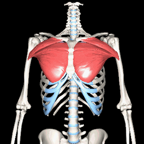
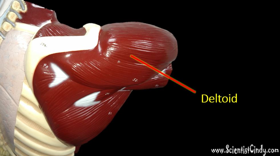
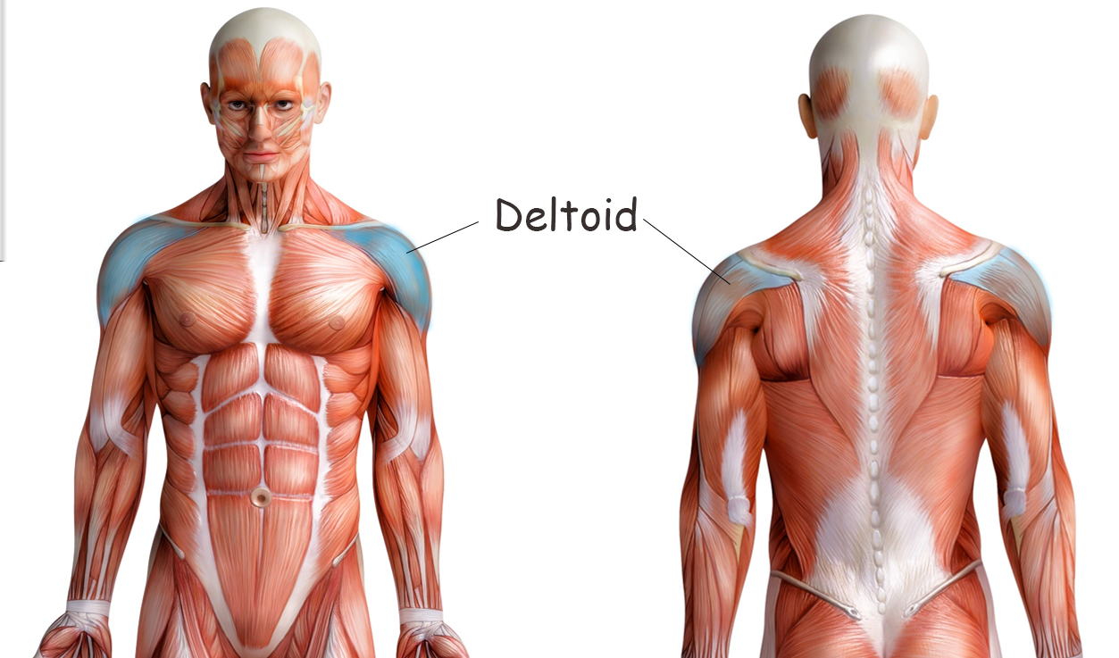
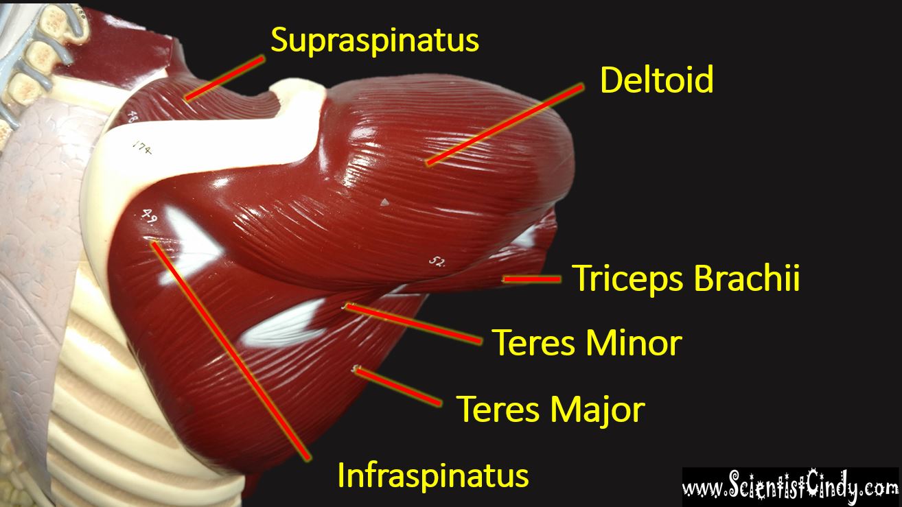
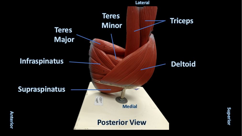
Deltoid Muscles
The deltoid muscle is the rounded muscle of the shoulder and upper arm. The deltoid muscle is named after the Greek letter "delta", because it has a similar triangular shape. The deltoid is attaches to clavicle (collarbone), the scapula (shoulder blade), and the humerus (upper arm bone).
Contraction of the deltoid muscle results in a wide range of movement of the arm at the shoulder due to its location and the wide separation of its muscle fibers.
Contraction of the deltoid muscle results in a wide range of movement of the arm at the shoulder due to its location and the wide separation of its muscle fibers.
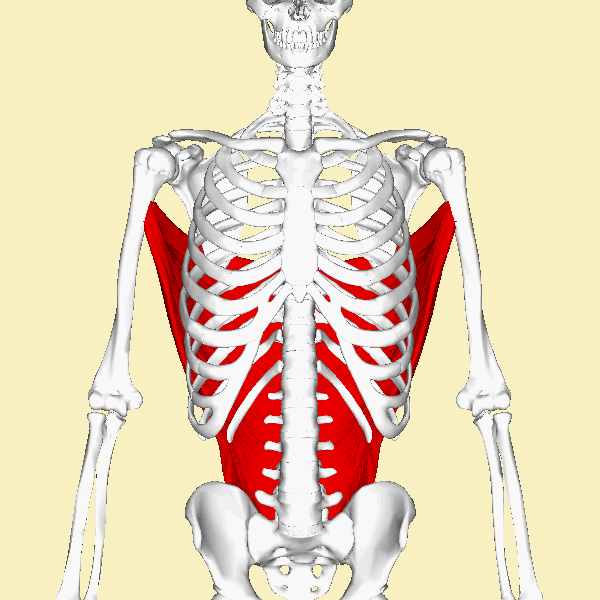
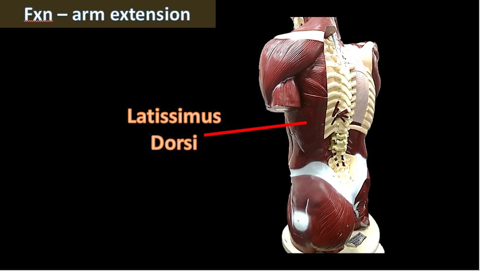
Latissimus Dorsi
Latissimus dorsi comes from the words ‘latissimus’ = widest and ‘dorsi’ = back.
|
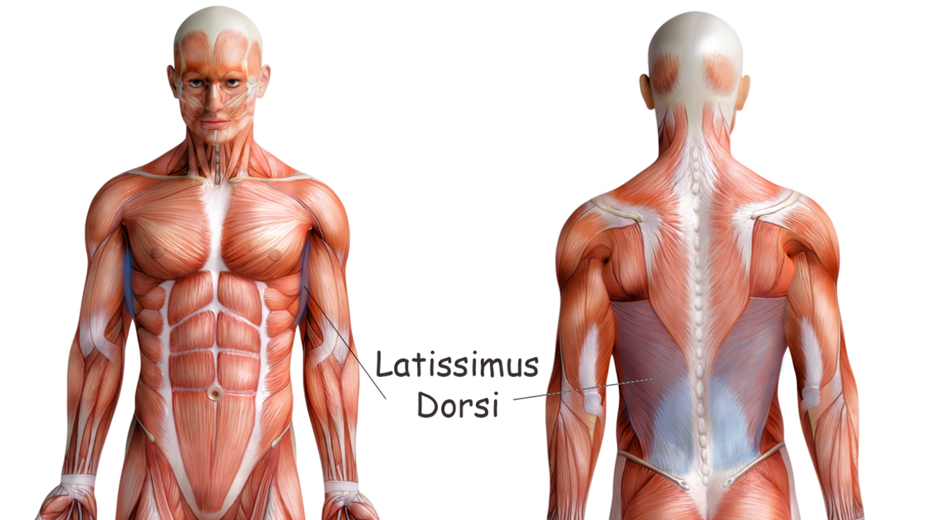
FUNCTION : Major muscle for arm extension, adduction and medial rotation of arm.
Other functions – Bringing the arm down forcefully (hammering) Swimming Reaching overhead Chin-ups |
Summary of the Superficial Muscles of the Torso
The Deep Muscles of the Torso Include the Internal and External Intercostal Muscles and the Muscles of the Rotator Cuff (Supraspinatus, Infraspinatus and the Teres Major)
Internal and External Intercostal Muscles
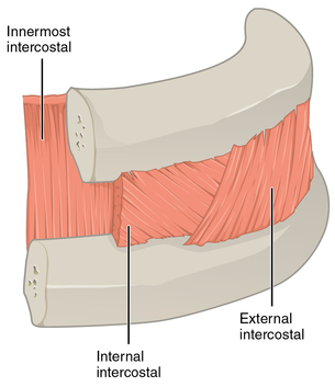
IMAGE Courtesy of By CFCF - Own work, CC BY-SA 4.0, https://commons.wikimedia.org/w/index.php?curid=44308826
The internal intercostals form the intermediate intercostal muscle layer. The internal intercostal muscles pull the rib cage down to allow for expiration(or exhalation).
Internal Intercostals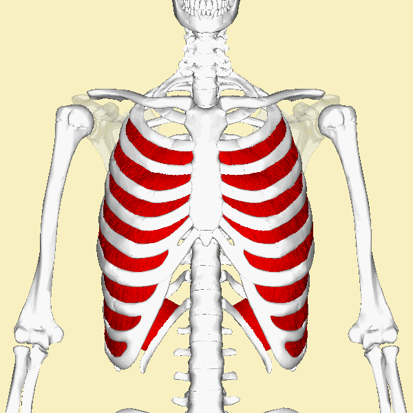
|
The function of the deep muscles of the thorax is to assist in respiration (breathing). The medical term for inhalation (or breathing inwardly) is inspiration and the medical term for exhalation (or breathing outwardly) is expiration. In order for inspiration to occur, the thoracic cavity (which includes the pleural cavities) must expand (or increase in volume). Conversely, in order for expiration (exhalation) to occur, the thoracic cavity must collapse (or decrease in volume). Just FYI - The deepest muscle layer of the thoracic wall attaches to the internal surfaces of the ribs. The function of the deepest layer is unknown. The external intercostal muscles function to expand the thoracic cavity upon inspiration (inhalation) by acting to lift the rib cage. The external intercostal muscles make up the most superficial of the 3 layers of intercostals.
External Intercostals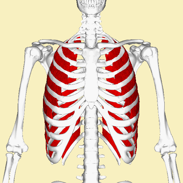
|
|
IMAGE and GIFa By Anatomography - en:Anatomography (setting page of this image), CC BY-SA 2.1 jp, https://commons.wikimedia.org/w/index.php?curid=22763359
|
GIFa By Anatomography - en:Anatomography (setting page of this image), CC BY-SA 2.1 jp, https://commons.wikimedia.org/w/index.php?curid=22763359
|
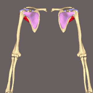
Four muscles make up the rotator cuff that encircles the shoulder joint.
|
|
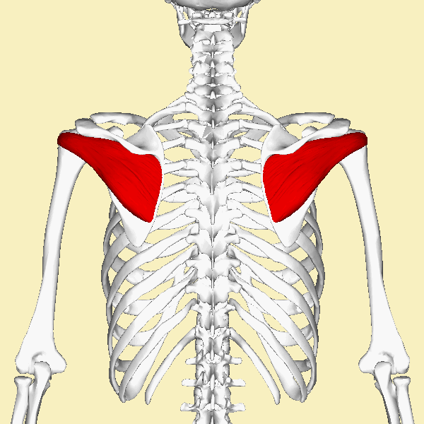
Supraspinatus
|
Function = initiates abduction of arm
The supraspinatus muscles originate at the supraspinatus fossa (which lies just above the spinous process of the scapulae and inserts at the greater tubercle of the humerus. The supraspinatus muscles function to abduct the arm and to stabilize the shoulder. It is located superiorly at the posterior aspect of the scapulae.
GIF Courtesy of Young Lae Moon, CC BY 3.0, https://commons.wikimedia.org/w/index.php?curid=23337523
|
GIF Courtesy of Anatomography - en:Anatomography (setting page of this image), CC BY-SA 2.1 jp, https://commons.wikimedia.org/w/index.php?curid=22799710
|
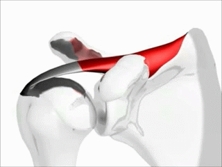
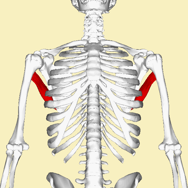
Infraspinatus
|
Function = laterally rotates the arm; helps stabilize the shoulder
The infraspinatus muscles connect the scapulae and the humerus as one of the rotator cuff muscles. It lies at the posterior aspect of the scapulae and is below (inferior to) the supraspinatus. The infraspinatus muscle originates at the infraspinous fossa of the scapula, which lies just below the spine of the scapulae. The literal translation for infraspinatus in "below spine" which refers to its origin beneath the spine of the scapulae. . This muscle inserts at the middle part of the greater tubercle of the humerus. The infraspinatus functions to laterally rotate the arm and it helps stabilize the shoulder by drawing the humerus toward the glenoid fossa of the scapula.
|
GIF Courtesy of Anatomography - en:Anatomography (setting page of this image), CC BY-SA 2.1 jp, https://commons.wikimedia.org/w/index.php?curid=22787515
|
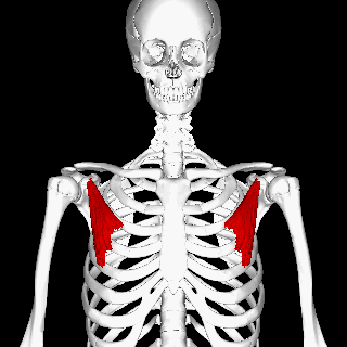
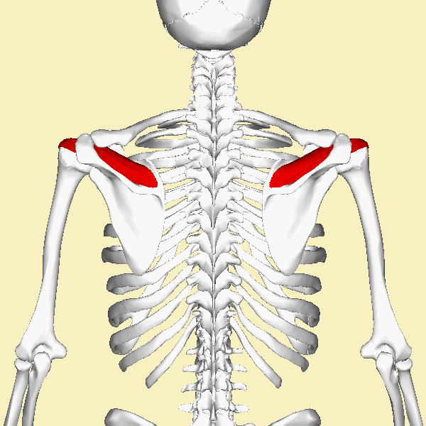
Teres MajorFunction: Adduction of the arm; Extends arms, Medially Rotates arm.
The word 'teres' comes from a Latin word meaning 'rounded'. The teres major muscle does not live up to its name very well, because it looks more like a thick flattened rectangle. The teres major muscle originates at the inferior aspect of the scapulae and inserts into the humerus.
By Anatomography - en:Anatomography (setting page of this image), CC BY-SA 2.1 jp, https://commons.wikimedia.org/w/index.php?curid=22783829
|
|
Muscles of the Abdomen
Muscles of the Abdomen
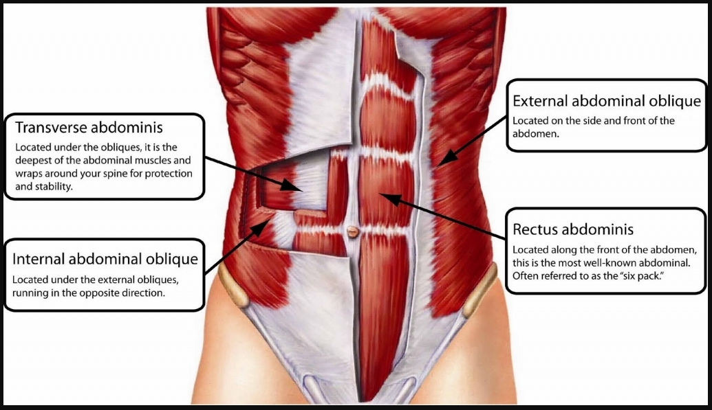
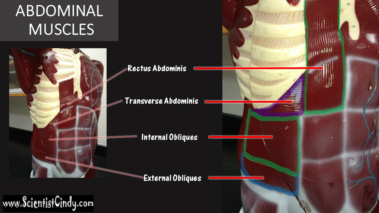
The abdominal muscles include the following:
• External oblique • Internal oblique • Transversus abdominus • Rectus abdominis
• External oblique • Internal oblique • Transversus abdominus • Rectus abdominis
The walls of the abdomen do not have bones protecting it. This allows for the abdomen to be more flexible, but it leaves the area more vulnerable to injury. For this reason, your abdominal wall contains strong, broad, sheet-like muscles. The anterior and lateral portions of the abdominal wall are composed of three broad, flat sheets of muscle. These are the external obliques, the internal obliques, and the transverse abdominis.
HOW THE STRIATIONS OF THE MUSCLE YOU ARE LOOKING AT, WILL AN IMPORTANT CLUE THAT TELLS YOU WHAT ABDOMINAL MUSCLE IT IS.
RECTUS ABDOMINIS - striations are oriented VERTICALLY (up and down)
TRANSVERSE ABDOMINIS - striations are oriented HORIZONTALLY (on the transverse plane)
EXTERNAL OBLIQUES - striations are oriented DIAGONALlY DOWNWARD
INTERNAL OBLIQUES - striations are oriented DIAGONALLY UPWARD
|
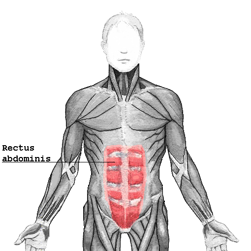
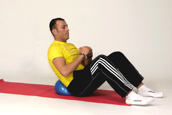
Rectus AbdominisFunction = Flexing the lumbar spine, placing the ribs closer to the pelvis, like when you are doing a "crunch" sit up.
The rectus abdominis muscles are what is commonly referred to as your "abs" or your "six-pack". These are the muscles that run vertically on each side of the anterior wall of the abdomen. There two parallel muscles are separated by the linea alba, which is a vertical band of connective tissue that runs down the midline. The rectus abdominis runs from the pevis to the anterior portions of the lower ribs and the xiphoid process of the sternum.
The "six pack" appearance of the rectus abdominis muscles is due to bands of connective tissue, called the tendinous intersections. |
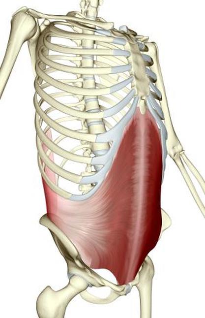
External Obliques
|
Function = Assists is trunk rotation and flexion of the vertebral column.
The external oblique muscles are named according to their location. The word 'oblique' means 'to run at an angle'. The external obliques originate by the lower ribs and insert at a few locations within the pelvis. The external obliques function to flex the vertebral column and to compress the abdominal wall. The external obliques lie on either side of the rectus abdominis muscles. The function of the external oblique muscles is to allow the trunk to twist to the opposite side of whichever external oblique is contracting. For example, the right external oblique contracts to turn the body to the left. They also function in trunk rotation and lateral flexion.
|
external oblique
|
Internal Obliques
|
The rectus abdominis connects the lower ribs to the pubic bone which is located at the front of the pelvis. The main function of the rectus abdominis is to move the body between the ribcage and the pelvis
Function = Assists is trunk rotation and flexion of the vertebral column.
The internal oblique muscles lie underneath (deep) to the external obliques. These muscles attach to the the area of the rectus abdominis muscles that is located just inside of the hip (coxal) bones. These muscles operate in the opposite direction to the external oblique muscles. For example, twisting the trunk to the left requires the left side internal oblique and the right side external oblique to contract together.
Transverse AbdominisFunction = Compresses abdomen inwards
|
|
The word 'transverse' means 'horizontal' and 'abdominis' means 'of the abdomen'. Therefore the 'transverse abdominis' translates literally to mean 'horizontal muscles of the abdomen'. The traverse abdominis muscles are the deepest (innermost) muscles of the abdominal wall. The fibers of this muscle run horizontally. It functions to compress the abdomen and assists in child delivery in females.