Skeletal Muscle, Electromyography and Stretch Reflexes
|
We use our skeletal muscles to move our bodies, maintain posture, stabilize joints, and generate heat. While many skeletal muscles are under conscious control, stretch reflexes are controlled unconsciously. Skeletal muscles respond to stimuli in response to signals it receives from the nervous system. Therefore, the nervous system and skeletal muscles function as a unit. Reflexes function to prevent bodily injury.
LAB EXERCISES:
|
|
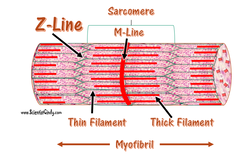
Anatomy of Skeletal Muscle
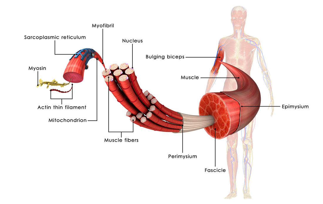
Skeletal muscle is highly organized and follows the following morphology:
1) Skeletal muscle is striated and is composed of muscle fibers (cells)
2) These muscle fibers contain myofibrils
3) Muscle fibers are grouped together to form muscle fascicles
4) Muscle fascicles are grouped together for form muscle bundles
5) The functional unit is the sarcomere
6) myosin (thick) and actin (thin) filaments are highly organized which results in striations.
The neuromuscular junction
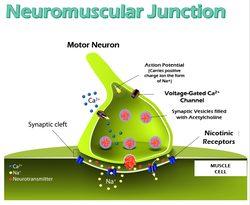
The neuromuscular junction is the site where a motor neuron communicates with a skeletal muscle fiber. The central nervous system (CNS) and skeletal muscle communicate to cause skeletal muscles to contract. Motor neurons receive an action potential from the CNS. When the action potential (AP) reaches the synaptic bulb, voltage gated calcium channels open and calcium ions diffuse into the synaptic bulb to trigger exocytosis of the neurotransmitter acetylcholine (ACh). ACh diffuses across the synaptic cleft and binds to the ACh-gated channels on the motor end plate (MEP) of the sarcolemma (plasma membrane of the muscle fiber). The ACh-gated channels open allowing sodium ions and potassium ions to diffuse across the MEP. A rush of sodium ions crosses into the muscle fiber and depolarizes the membrane resulting in an end-plate potential (EPP). If the EPP reaches threshold, then the action potential travels across the sarcolemma and into the transverse tubules (t-tubules). The voltage-sensing receptors (L-type calcium channels called dihydropyridine receptor, DHP) on the t-tubule change conformation and open the calcium ion release channels (ryanodine receptors, RyR) that are on the sarcoplasmic reticulum (SR; the smooth ER of the muscle cell). Calcium ions diffuse into the cytosol of the muscle fiber and bind to troponin. Troponin changes conformation and moves tropomyosin away from the myosin-binding site on actin. Myosin and actin can now interact to form cross-bridges.
The Cross-Bridge Cycle
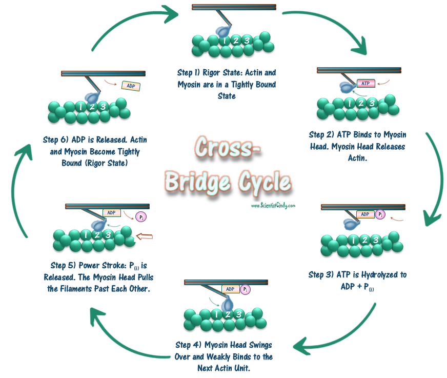
Cross-bridge cycling will continue until ACh is removed from the synaptic cleft by acetylcholine esterase (AChE) or via diffusion. Once ACh is removed, ACh-gated channels on the MEP close resulting in a loss of the AP on the muscle fiber. The SR then closes its calcium ion channels and the calcium ion pumps return the calcium into the SR. Force production is dictated by many factors and processes. Motor units are composed of somatic motor neuron(s) and all the muscle fibers they innervate. Motor units range from small to large. The smaller motor units have a lower threshold while the larger motor units have a higher threshold.
The Muscle Twitch
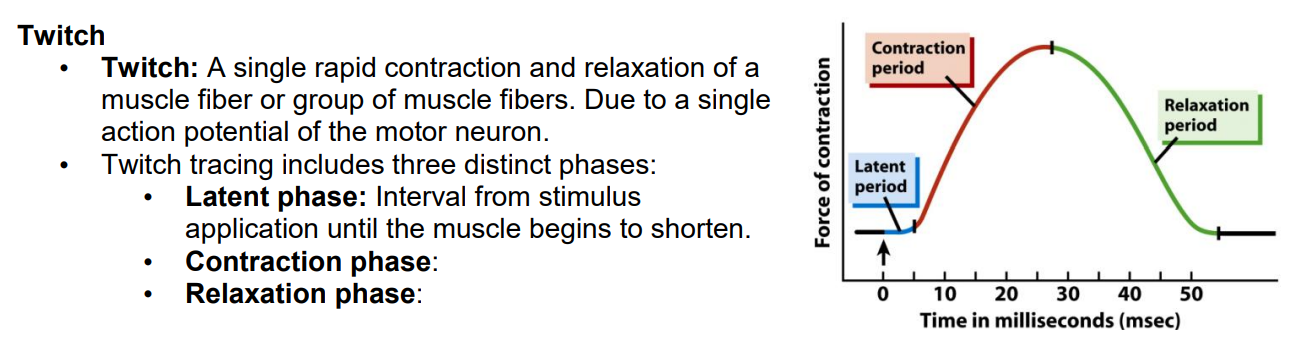
The TWITCH proceeds in 3 distinct phases:
- THE LATENT PERIOD (excitation-contraction coupling) - The LATENT PERIOD is the time between the application of the stimulus and the first sign of muscle contraction, when excitation-contraction coupling takes place.
- THE CONTRACTION PERIOD (tension generation) - The CONTRACTION PERIOD is the time period starting at the onset of contraction and ending at the time in which peak tension occurs.
- THE RELAXATION PERIOD (muscle relaxation) - The RELAXATION PERIOD is the period of time that occurs from the time of peak tension to the point when tension returns to baseline level. During this part of the phase, Ca2+ is being pumped back into the sarcoplasmic reticulum, the cross-bridge cycle stops and the muscle is stretching back to its original length.
Muscle Fiber Recruitment and Tetanus
You can recruit different motor units to produce the force that you require. A muscle fiber twitch is affected by the length of the muscle. Muscles at optimal length produce optimal force because there is an optimal amount of cross bridges being formed. Muscles that are shortened produce less than optimal force because the myosin filaments run into the Z disks inhibiting them from sliding and produce less tension. Muscles that are too long produce less than optimal force because there is not enough overlap for optimal cross bridging resulting in less sliding and less force production.
A tetanic contraction can be either unfused (incomplete) or fused (complete).
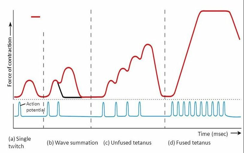
Unfused Tetanus
An unfused tetanus is when the muscle fibers do not completely relax before the next stimulus because they are being stimulated at a fast rate; however there is a partial relaxation of the muscle fibers between the twitches.
Fused Tetanus
Fused tetanus is when there is no relaxation of the muscle fibers between stimuli and it occurs during a high rate of stimulation. A fused tetanic contraction is the strongest single-unit twitch in contraction. It occurs when the contracting tension in the muscle remains constant in a steady state. This is the maximal possible contraction. During tetanic contractions, muscles can shorten, lengthen or remain constant length.
A tetanic contraction (also called tetanized state, tetanus, or physiologic tetanus, the latter to differentiate from the disease called tetanus) is a sustained muscle contraction evoked when the motor nerve that innervates a skeletal muscle emits action potentials at a very high rate. During this state, a motor unit has been maximally stimulated by its motor neuron and remains that way for some time. This occurs when a muscle's motor unit is stimulated by multiple impulses at a sufficiently high frequency. Each stimulus causes a twitch. If stimuli are delivered slowly enough, the tension in the muscle will relax between successive twitches. If stimuli are delivered at high frequency, the twitches will overlap, resulting in tetanic contraction.
Stretch reflexes
|
Stretch reflexes involve:
|
|
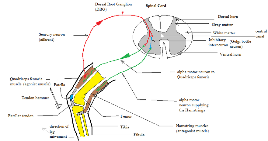
A good example of a stretch reflex is the patellar tendon reflex (knee jerk). When you tap the patellar tendon, you rapidly stretch the muscle causing the quadriceps and therefore the muscle spindles within the quadriceps to stretch. The muscle spindles sense the stretching of the muscle and fire more rapidly.
The sensory neuron sends action potentials to the spinal cord and results in two responses:
1) the sensory neuron synapses with somatic motor neuron within the ventral horn of the spinal cord causing the somatic motor neuron to send action potentials to the quadriceps resulting in contraction
2) the sensory neuron synapses with an interneuron in the grey matter of the spinal cord which results in inhibiting the somatic motor neuron of the hamstrings causing them to relax.
When the quadriceps contract and the hamstrings relax, the knee will extend (knee jerk). Today you will study your own stretch reflexes.
Lab Exercise A. - Stretch Reflexes
|
Equipment • Textbook/resources • Reflex hammer (quickly and firmly strike with either the blunt or proper end) • Relaxed student (have them close their eyes and count down from 100 for all jerk reflexes)
Protocol 1. Draw a representative stretch reflex (feedback loop). Look up the knee jerk in your textbook/resource and draw the feedback loop. Label the stimulus, sensor, afferent pathway, controller, efferent pathways, and responses. |
|
Jaw Jerk Reflex
2. Jaw Jerk Exercise
- a. Tell the volunteer to relax their jaw and open slightly.
- b. Place your pointer and middle finger in front of the mandible and place your ring finger below the chin to support the jaw.
- c. Firmly strike your pointer finger in a downward motion using the wide end of the reflex hammer.
- Normal Jaw Jerk Reflex: Slight or Absent Elevation of the Mandible
- Abnormal Jaw Jerk Reflex: Exaggerated or pronounced elevation of the Mandible
Normal Jaw Jerk Reflex |
Abormal Jaw Jerk Reflex |
The jaw jerk reflex or the masseter reflex is a stretch reflex used to test the status of a patient's trigeminal nerve (cranial nerve V) and to help distinguish an upper cervical cord compression from lesions that are above the foramen magnum. The mandible—or lower jaw—is tapped at a downward angle just below the lips at the chin while the mouth is held slightly open. In response, the masseter muscles will jerk the mandible upwards. Normally this reflex is absent or very slight. However, in individuals with upper motor neuron lesions the jaw jerk reflex can be quite pronounced.
Biceps Jerk Reflex
3. Biceps Jerk Reflex Exercise
- a. Face the volunteer and cradle their right arm by placing their right elbow in your right palm and placing their right hand on top of your forearm. Their elbow should be slightly bent.
- b. Jiggle their arm to make sure they are relaxed.
- c. Use your fingers to firmly press in the cubital fossa to look for the tendon.
- d. Firmly strike the tendon using the pointed end of the reflex hammer.
- e. Repeat steps 1-4 with the left arm.
- Normal Biceps Jerk Reflex: The biceps should twitch, and the elbow should flex.
- Abnormal Biceps Jerk Reflex: Exaggerated or absent reflexes are used as clues to the location of neurological disease.
Normal Bicep Jerk ReflexBiceps reflex is a reflex test that examines the function of the C5 reflex arc and the C6 reflex arc. Exaggerated or absent reflexes are used as clues to the location of neurological disease
|
Abnormal Bicep Jerk Reflex - Hyperflexia |
Triceps Jerk Reflex
4. Triceps Jerk Reflex Exercise
- a. Stand next to your patient and cradle their elbow. Their humerus should almost be parallel to the floor.
- b. Jiggle their arm to make sure they are relaxed.
- c. Use your fingers to firmly press to look for the triceps tendon.
- d. Firmly strike the tendon using the wide end of the reflex hammer.
- e. Repeat steps 1-4 with the left arm.
- f. Normal response: The triceps should twitch, and the elbow should extend.
Normal Triceps Jerk Reflex |
Abnormal Triceps Jerk Reflex |
Knee Jerk Reflex
5. Knee Jerk Reflex Exercise
- a. Ask the volunteer to sit on the table. Make sure their feet dangle from the side of the table.
- b. Use your fingers to firmly press under the right patella and look for the patellar ligament.
- c. Firmly strike the ligament using the wide end of the reflex hammer.
- d. Repeat steps 1-3 for the left side.
- Normal Knee Jerk Reflex: The quadriceps should twitch, and the knee should extend (kick forward)
- Abnormal Knee Jerk Reflex: Exaggerated or absent reflexes are used as clues to the location of neurological disease.
Ankle (Achilles) Jerk Reflex
Ankle (Achilles) Jerk Reflex
- a. Ask the volunteer to sit on the table. Make sure their feet dangle from the side of the table.
- b. Support the right heel of the foot by cupping it in your hand.
- c. Locate the calcaneal (Achilles’) tendon.
- d. Firmly strike the tendon using the wide end of the reflex hammer.
- e. Repeat steps 1-3 for the left side.
- Normal Ankle (Achilles) Jerk - Plantar flexion
- Abnormal Ankle (Achilles) Jerk - Exaggerated or absent reflexes are used as clues to the location of neurological disease.
Normal Ankle (Achilles) Jerk |
Abnormal Ankle (Achilles) Jerk Reflex - Hyperflexia |
7. Plantar (Babinski) Reflex Exercise
- a. Ask the volunteer to sit on the table. Make sure their feet dangle from the side of the table.
- b. Using the metal handle, firmly and swiftly trace the right foot in the following pattern. Begin by tracing the lateral side of the plantar surface from the heel towards the toes and then continue medially across the ball of the foot.
- c. Repeat steps 1-2 for the left side.
- Normal Plantar (Babinski) Reflex IN ADULTS - plantar flexion, curling of the toes. Recorded as a negative Babinski sign.
- Abormal Plantar (Babinski) Reflex IN ADULTS - dorsiflexion of the great toe and other toes fan outward. Recorded as a positive Babinski sign. If this is seen in an adult, then they may have damage in the corticospinal tract.
- Normal Plantar (Babinski) Reflex IN INFANT - dorsiflexion of the great toe and other toes fan outward. Recorded as a positive Babinski sign.
- Abnormal Plantar (Babinski) Reflex IN INFANT - plantar flexion, curling of the toes. Recorded as a negative Babinski sign.
Normal Plantar (Babinski) Reflex in ADULT |
Abnormal Plantar (Babinski) Reflex in ADULT |
Abnormal Plantar (Babinski) Reflex in INFANT
|
Babinski reflex is one of the normal reflexes in infants. Reflexes are responses that occur when the body receives a certain stimulus.
The Babinski reflex occurs after the sole of the foot has been firmly stroked. The big toe then moves upward or toward the top surface of the foot. The other toes fan out. This reflex is normal in children up to 2 years old. It disappears as the child gets older. It may disappear as early as 12 months. |
|
Lab Exercise B - Stimulator Experiment (Electrode Stimulation Experiment)
Equipment • Electrodes • Electrolyte gel • Elastic bands • Stimulator with variable stimulation settings (duration, voltage, and frequency) • Alcohol swabs • Follow directions and ask for help in necessary.
Protocol:
1. Draw a figure displaying a twitch, summation, incomplete (unfused) tetanus, and complete (fused) tetanus. Label the axes and the stimuli.
2. Locate the motor points on the forearm (finger flexor muscle group).
3. Place a droplet of electrolyte gel on each electrode.
4. Place the electrodes on the motor points and secure them with the elastic bands.

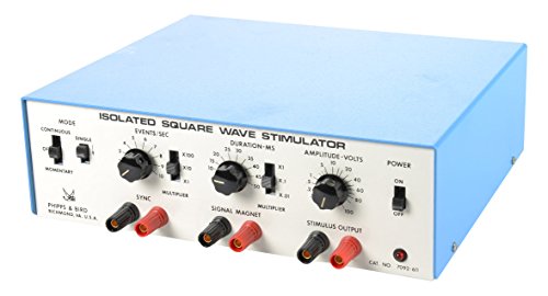
5. Initial Twitch experiment
- a. INITIAL STIMULATOR SETTINGS:
- i. Duration of the stimulus = 2 msec
- ii. Voltage = 20V
- iii. Frequency = single pulse
- b. Look at the fingers and deliver a stimulus.
- c. Determine if a twitch was seen. If so, then this is your threshold setting. If not, then continue to the next step.
- d. Change the Voltage to 30V and deliver a stimulus. Determine if a twitch was seen. If so, then move on to the next experiment. If not, adjust the electrode placement and repeat this experiment until you see a twitch.
- a. Begin by using the settings that you determined in the Initial Twitch experiment.
- b. For each test, lower the VOLTAGE in a stepwise manner until you determine threshold. The threshold is the minimum voltage needed to cause a twitch to occur.
- c. Determine the threshold (lowest voltage) needed to stimulate a twitch.
- a. Begin by using the settings that you determined in the Initial Threshold experiment.
- b. Lower the DURATION of the stimulus to 0.1 msec.
- c. Determine how the twitch results change as duration is varied.
- a. Begin by using the settings that you determined in the Initial Threshold experiment.
- b. Increase the VOLTAGE by 5V and deliver a stimulus.
- c. Repeat step 2 however you should not surpass 30V. If your volunteer does not want to proceed to a higher voltage, then you should move on to the next experiment.
- d. Determine how the twitch results change as voltage is varied.
- a. Begin by using the settings that you determined in the Initial Threshold experiment.
- b. Increase the Frequency in a stepwise manner until summation occurs. Record these settings.
- c. Continue to increase the Frequency in a stepwise manner until incomplete (unfused) tetanus occurs. Record these settings.
- d. Continue to increase the Frequency in a stepwise manner until complete (fused) tetanus occurs. Record these settings.
EXAMPLE OF EXPERIMENTAL RESULTS
Initial Twitch Experiment
EXPERIMENT |
DURATION OF STIMULUS |
VOLTAGE |
FREQUENCY - events per second |
DESCRIPTION |
INITIAL TWITCH EXP. |
2 ms |
20 mV |
1 per second |
It was found that an initial stimulus of 20mV with a duration of 2ms given at a frequency of 1 event per second, yielded a twitch response. |
THRESHOLD EXP. |
2 ms |
15 mV |
1 per second |
It was found that a minimum stimulation of 15mV was needed to cause a muscle twitch. Therefore the threshold voltage was found to be 15mV. |
DURATION EXP. |
0.1 ms |
15 mV |
1 per second |
Decreasing the duration of the stimulus to 0.1ms was insufficient to cause a twitch response. |
VOLTAGE EXP. |
2 ms |
various |
1 per second |
The strength of the twitch response grew stronger as voltage was increased. |
FREQUENCY EXPERIMENT - SUMMATION |
2 ms |
15 mV |
15 per second |
Summation of the twitch response (muscle contraction lasting a bit longer than a single twitch) was experienced when using a stimulus frequency of 15 events per second. |
FREQUENCY EXPERIMENT - INCOMPLETE (UNFUSED) TETANUS |
2 ms |
15 mV |
20 per second |
Incomplete or unfused tetanus (steady contraction with shaking) response was experienced when using a stimulus frequency of 20 events per second. |
FREQUENCY EXPERIMENT - COMPLETE (FUSED) TETANUS |
2 ms |
15 mV |
25 per second |
Complete or fused tetanus (steady contraction without shaking) response was experienced when using a stimulus frequency of 25 events per second. |
THRESHOLD - When identifying the threshold of a twitch, the amps of the stimulator are adjusted. You can think of amps as the strength of a stimulus. The threshold of a twitch is defined as the smallest current required to produce any recognizable sign of muscle contraction. This may appear as small jerking motion of a muscle (twitch). As the Amps of the stimulator are increased, the strength of the response also increases. This increase is due to the recruitment of additional muscle fibers to contract.
- There can be no measurable response at 0 mA. At 0 mA no muscle fibers are contracting.
- Usually the first twitch begins around 4–6 mA. , and the maximum contraction force begins around the 12–15 mA range.
- At threshold just a few muscle fibers are being recruited.
- the maximum contraction (or “maximal stimulus” - the current at which the response no longer increases) - the maximum contraction force begins around the 12–15 mA range.
- By definition, the maximal stimulus is seen when 100% of recruitable fibers are contracting.
- Above maximal stimulus, the number of contracting fibers cannot increase.