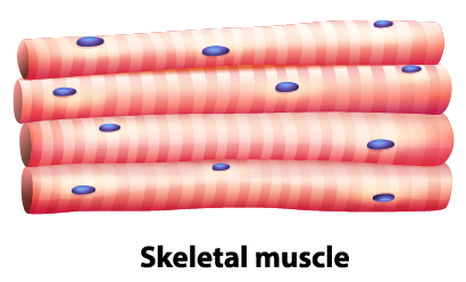
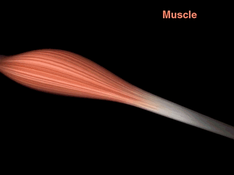
Skeletal Muscle
The Muscle Cell / Myocyte / Muscle Fiber
|
A myocyte (also known as a muscle cell or a muscle fiber) is the type of cell found in muscle tissue. Myocytes are long, tubular cells that develop from myoblasts to form muscles in a process known as myogenesis.
There are various specialized forms of myocytes: cardiac, skeletal, and smooth muscle cells, with various properties. |
|
Skeletal muscle is mostly under conscious / voluntary control of the central nervous system as part of the somatic division of the motor peripheral nervous system. Skeletal muscle tissue is the only type of muscle tissue that is under conscious (or voluntary) control via your somatic nervous system. Your brain sends signals down your spinal cord that connects with peripheral nerves to command the muscles to contract or to relax.
Skeletal muscle is the type of muscle that most people envision when the word 'muscle' is used. These are the noticeable muscles that can give shape to our skin and act to move the various parts of your body at will. Skeletal muscles are able to move parts of our body because they are attached to skeletal bones through structures of fibrous connective tissue called tendons.
Skeletal muscle is the type of muscle that most people envision when the word 'muscle' is used. These are the noticeable muscles that can give shape to our skin and act to move the various parts of your body at will. Skeletal muscles are able to move parts of our body because they are attached to skeletal bones through structures of fibrous connective tissue called tendons.
The Structure of Skeletal Muscle
The Structure of Skeletal Muscle
|

|
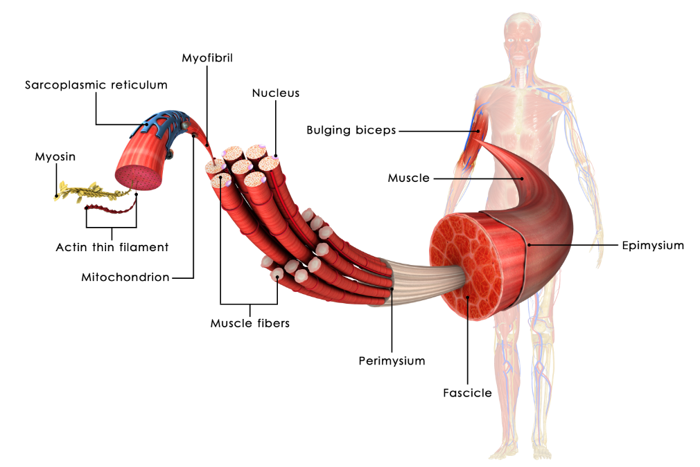
The Structure of a Muscle Fiber
• A myocyte (also known as a muscle cell or a muscle fiber) is the type of cell found in muscle tissue.
•Myocytes are long, tubular cells that develop from myoblasts to form muscles in a process known as myogenesis.
•Myocytes are long, tubular cells that develop from myoblasts to form muscles in a process known as myogenesis.
Skeletal muscle fibers are extremely long, cylindrical cells. A single muscle fiber can be up to a foot long in some muscles. This unique property is made possible, because the muscle fiber itself is actually formed from the fusion of literally hundreds of embryonic cells that get fused together during development.
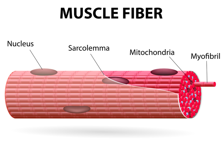
SKELETAL MUSCLE FIBERS ARE MULTI-NUCLEATED
Since skeletal muscle cells (muscle fibers) are made from the fusion of hundreds of embryonic cells, they contain many nuclei. In other words, they are multinucleated. You will notice that the nuclei lie on the outer portion of the muscle fiber when examining skeletal tissue under the microscope.
SKELETAL MUSCLE FIBERS ARE STRIATED
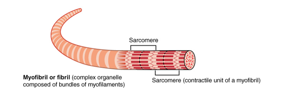
Skeletal muscle fibers have 'stripes' called striations. These striations are due to the presence of the myofibrils that make up the major portion of the muscle fiber.
The muscle fiber is a long cylinder that contains bundles of smaller, long cylinders, called myofibrils. Myofibrils contain the myofilaments necessary for muscle contraction. A myofibril is composed of repeating segments called sarcomeres. The sarcomere is the basic unit of contraction in skeletal muscle. The striations occur due to the presence of the sarcomeres of the myofibrils.
Striated muscle tissue consists of myocytes that appear to be striped (transversely) with alternating dark and light bands of color. These striations are visible when the muscle tissue is viewed histologically (on a slide). Each elongated, cylindrical unit is a skeletal muscle cell, called a muscle fiber or myocyte.
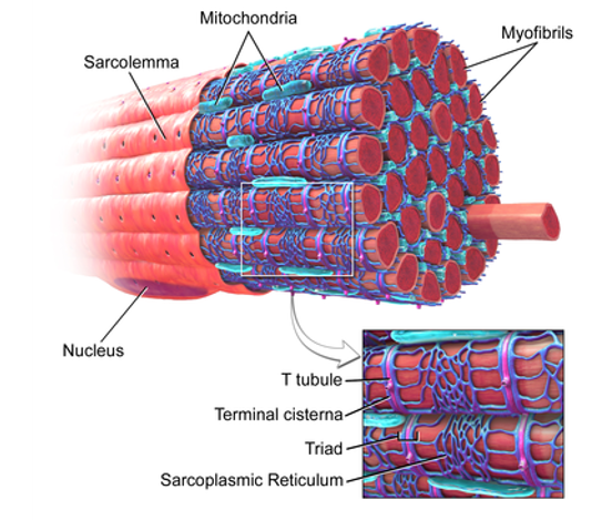
THE SARCOLEMMA
- The plasma membrane of skeletal muscle cells has a special name; the sarcolemma.
- The sarcolemma is unique, because it continues deep into the muscle cell to form the “T-tubules” or “transverse tubules”.
- The t-tubules are continuous with the sarcolemma.
- The fluid that fills the lumen (inner liquid) of the t-tubule is considered extracellular space.
- The intracellular fluid of a muscle fiber is referred to as the myoplasm or sarcoplasm. Within the sarcoplasm, there are numerous specialized structures which are unique to muscle fibers.
- The sarcoplasmic reticulum is a highly specialized structure that is tightly wrapped around individual myofibrils.
- The sarcoplasmic reticulum functions to store high concentration of Ca2+ which is releases upon sufficient depolarization triggered by the action of Release of Ca2+ from the sarcoplasmic reticulum is responsible for triggering muscular.
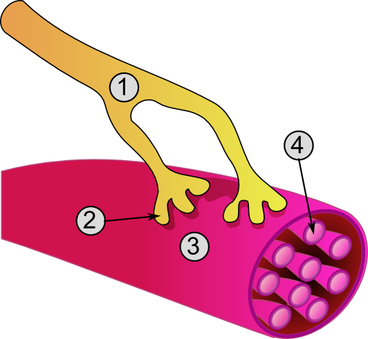
General structure of a muscle cell and neuromuscular junction:
|
Skeletal muscle is consciously (voluntarily) controlled by the (somatic) nervous system. The point at which the motor neuron synapses with (comes into close proximity to) the muscle fiber is called the neuromuscular junction. Structure of the
|
IMAGE Courtesy of CC BY-SA 3.0, https://commons.wikimedia.org/w/index.php?curid=282900
The Sliding Filament Model of Muscle Contraction
The striations of skeletal muscle fibers are due to the presence of repeated units called sarcomeres. The sarcomere is the functional unit of muscle contraction. The sarcomere contains filaments which slide past each other, contracting (shortening) the cell upon command from the nervous system.
The sarcomere is the functional unit of striated muscle. Let's look at the cross-bridge within the context of a single sarcomere to understand how contraction occurs.
Cross-Bridge Cycling
In order for the muscle to contract the actin and the myosin molecules of the thin and thick filaments have to interact with one another.
When the actin and myosin interact, they are said to be connected by cross-bridges. The cross-bridges refer to the myosin head groups that interact with a myosin-binding site on actin.
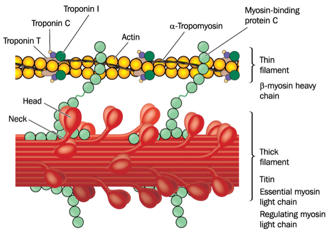
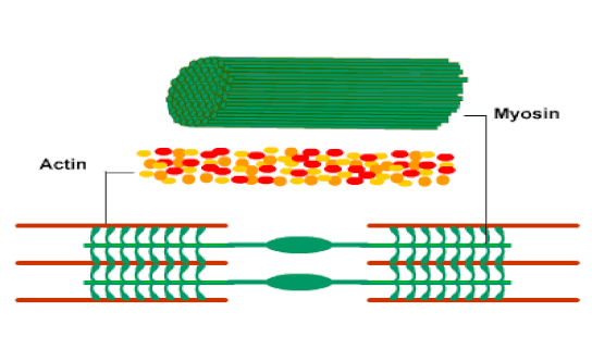
Those myofibrils are composed of contractile units called SARCOMERES.
PROTEINS of MUSCLE SARCOMERE
PROTEINS of MUSCLE SARCOMERE
- A. Contractile proteins
- 1. Myosin (gives rise to the thick filaments)
- 2. Actin (gives rise to the thin filaments)
- B. Regulatory proteins
- 1. Tropomyosin (in the absence of Ca2+, it covers the myosin-binding site of actin)
- 2.Troponin (Ca2+ sensor)
- Two regulatory proteins closely interact with actin. A regulatory protein called tropomyosin normally lies over the binding sites and prevents the interaction of the myosin head group with actin. When the myoplasmic Ca2+ concentration is low at its resting level, tropomyosin covers the myosin binding site of actin. The binding of Ca2+ to troponin moves the troponin-tropomyosin complex out of the way exposing the myosin binding site. This allows the myosin head to interact with actin. Actin makes up the thin filaments of the sarcomere. The actin units have myosin binding sites. When calcium is present, myosin is able to bind to the myosin binding site of on actin. It can do this because the myosin head has an actin binding site on it!
When calcium is not present, then muscle is relaxed. When the muscle is relaxed, tropomyosin covers the myosin binding sites on actin thin filaments which prevents cross-bridge formation. When the muscle receives enough stimulation from the nervous system to initiate muscle contraction, calcium is released from the sarcoplasmic reticulum into the sarcoplasm. Once calcium is in the sarcoplasm, calcium binds to tropomyosin and causes a change in conformation (shape) which pulls the troponin-tropomysin complex away from the myosin binding sites, allowing cross-bridge formation and muscular contraction to occur.
Events That lead to muscle fiber contraction
Muscle fiber contraction requires the following steps:
- Membrane Depolarization / Fiber Activation: The fiber must be activated or stimulated by a nerve (motor neuron)so that the membrane/sarcolemma becomes depolarized (the membrane potential becomes less negative due to an influx of positively charged sodium ions).
- Action Potential Generation: The change in membrane potential must be strong enough to generate an action potential in the sarcolemma.
- Action Potential Propagation: Once the action potential is initiated, it will be propagated along the sarcolemma.
- Calcium Release: Calcium is released from the sarcoplasmic reticulum.
The whole process really begins with the command from the central nervous system.
The decision to make a conscious movement is made in the brain.
The command to move is sent from the brain’s motor cortex and travels down the spinal cord, to the motor nerve (directly or indirectly).
The signal initiating movement of the muscle travels from the central nervous system to the motor nerve (motor neuron).
Excitation-Contraction Coupling
The signal reaches the terminal end (called the axon terminal) of the motor neuron causing the release of the neurotransmitter, acetylcholine.
The signal reaches the terminal end (called the axon terminal) causing calcium channels to open.
The influx of calcium in the axon terminal causes the release of the neurotransmitter, acetylcholine.
The signal reaches the terminal end (called the axon terminal) causing calcium channels to open.
The influx of calcium in the axon terminal causes the release of the neurotransmitter, acetylcholine.
Acetylcholine diffuses passively across the synaptic cleft and transiently binds to post- synaptic (nicotinic) receptors.
This binding causes the receptors to open, allowing an influx of sodium ions into the muscle fiber.
Events That lead to muscle fiber contraction
- Action potential arrives at axon terminal of the motor neuron at neuromuscular junction
- Acetylcholine (Ach) is released
- Ach binds to receptors on the sarcolemma
- Ion permeability of sarcolemma changes
- Local change in membrane voltage (depolarization) occurs
- Local depolarization (end plate potential) ignites AP in sarcolemma
- AP travels across the entire sarcolemma
- AP travels along T tubules
- SR releases Ca2+
- Ca2+ binds to troponin which exposes the myosin-binding sites on actin
- Myosin heads bind to actin; contraction begins
- Binding of myosin cross-bridges to the binding sites on G-actin molecules.
- Power stroke of the cross-bridges and movement of the thin filaments over the thick filaments.
- Continued cross-bridge cycling for as long as ATP is present and Ca2+ concentrations remain high in the myoplasm.
- Muscle shortening and/or tension development.
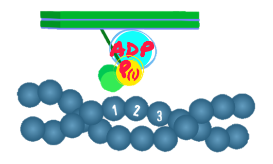
Cross-Bridge Cycle
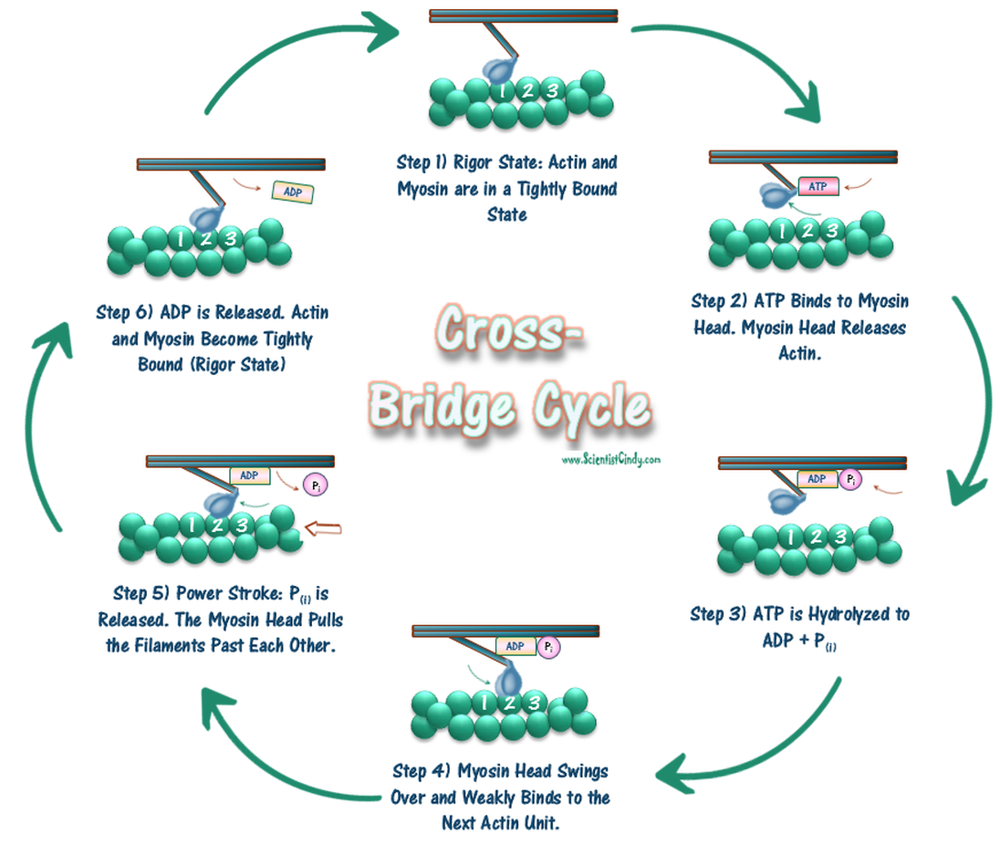
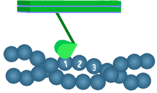
Step 1.
In this step, the myosin head interacts tightly with a G-actin of the thin filament.
The part of the myosin head group that interacts with actin is referred to as the actin- binding site, and the part of the G-actin molecule that interacts with myosin is referred to as the myosin-binding site. The myosin head makes a 45-degree angle with the thick filament. This is referred to as the rigor state because if there is no ATP present (such as after death), the thin and thick filaments maintain this tight interaction rendering the muscle very stiff (RIGORMORTIS).
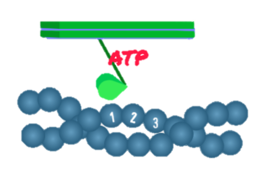
Step 2.
In this step, an ATP molecule binds to the nucleotide-binding site of the myosin head. Binding of ATP causes the release of the myosin head from the G-actin molecule.
In addition to the actin-binding site, the myosin head also has a nucleotide- binding site. This is a site where ATP and ADP interact with myosin.
Step 3.
Myosin is an ATPase (myosin ATPase) in that it has the ability to hydrolyze ATP to ADP and inorganic phosphate (Pi). In this step, the myosin head converts the bound ATP to ADP and Pi. Both ADP and Pi remain bound to the myosin head.
Myosin is an ATPase (myosin ATPase) in that it has the ability to hydrolyze ATP to ADP and inorganic phosphate (Pi). In this step, the myosin head converts the bound ATP to ADP and Pi. Both ADP and Pi remain bound to the myosin head.
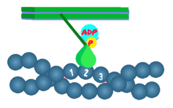
Step 4.
The energy released from the hydrolysis of ATP is used to change the conformation of the myosin head, so that now it makes a 90-degree angle with the thick filament. This change in conformation “energizes” the myosin head (i.e., it places it in a high-energy state). At this point, if sufficient Ca2+ is present in the cytoplasm (see Excitation-Contraction Coupling below), the myosin head attaches to a G-actin one or two positions away from the one bound in Step 1.
If there is not enough Ca2+ present in the cytoplasm, the myosin head remains in this energized 90-degree angle. A rise in cytoplasmic Ca2+ concentration is essential and evokes a series of events that facilitates the binding of myosin head to G-actin again. Please note that the relaxed state refers to the muscle cell and not to the conformation of the myosin molecule. At rest, most skeletal muscle fibers are in this “relaxed state”.
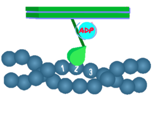
Step 5.
This is the power stroke step. Now Pi is released from the myosin head. As Pi is released, the energized 90-degree angle myosin head begins to assume its original 45- degree angle.
However, as it is bound to a G-actin of the thin filament, the change back to the 45-degree angle moves the actin thin filament toward the center of the sarcomere (M line)
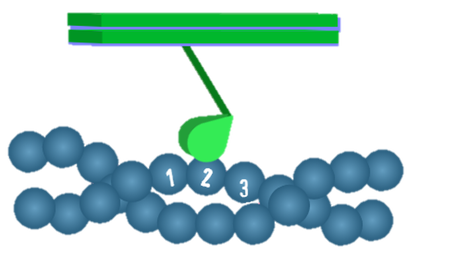
Step 6.
At this point, ADP is released from the myosin head, and the myosin head remains tightly bound to the G-actin. This brings us back to the beginning of the cycle at Step 1. If there is ATP around (and if the cytoplasmic Ca2+ concentration is high; see below), the cycle will repeat itself again and again. The repeated action of the cross- bridge cycle results in the sliding of the thin filaments over the thick filaments, which will lead to muscle shortening.
It is important to emphasize that the cross-bridge cycle can take place only if the cytoplasmic Ca2+ concentration is high. Thus, when skeletal muscles are at rest and the cytoplasmic Ca2+ concentration is low, the cross-bridge cycle does not take place. Instead, the myosin head groups remain in an “energized” state.
The cross-bridge cycle may be arbitrarily divided into six steps:
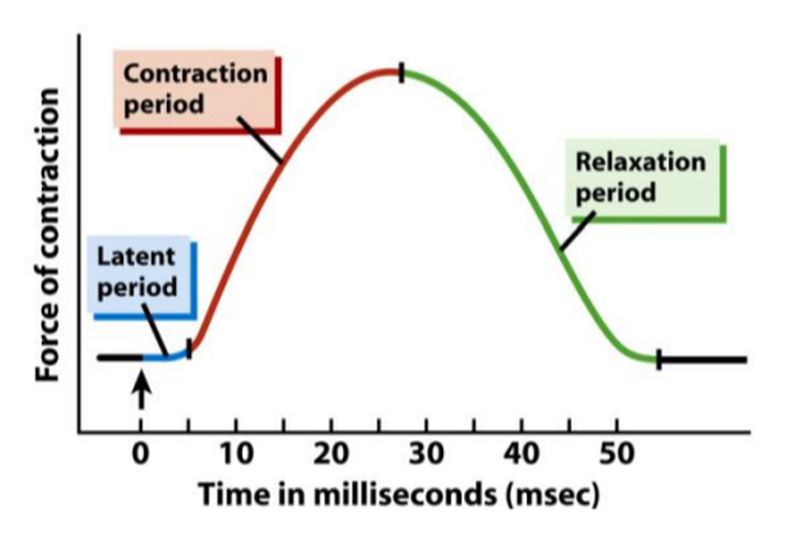
The Muscle Twitch
A muscle twitch is the minimum muscle response to a single action potential generated by a motor neuron. A single muscle twitch, which is a single contraction in response to a brief threshold stimulation. Threshold stimulation: the smallest amount of stimulation that result in sarcomere shortening. A threshold stimulation is the smallest amount of stimulation that will actually result in a contraction. If we administer a single threshold stimulus in a lab, we get a single muscle twitch in response. We can measure this with a Myogram. Excitatory input from the motor neuron must reach a minimum threshold (minimum level) to trigger the flood of calcium from the sarcoplasmic reticulum, in order for any muscular repose to occur.
A muscle twitch is the minimum muscle response to a single action potential generated by a motor neuron. A single muscle twitch, which is a single contraction in response to a brief threshold stimulation. Threshold stimulation: the smallest amount of stimulation that result in sarcomere shortening. A threshold stimulation is the smallest amount of stimulation that will actually result in a contraction. If we administer a single threshold stimulus in a lab, we get a single muscle twitch in response. We can measure this with a Myogram. Excitatory input from the motor neuron must reach a minimum threshold (minimum level) to trigger the flood of calcium from the sarcoplasmic reticulum, in order for any muscular repose to occur.
|
Every twitch has three distinct phases:
Latent period.
|
|
It is Important to be Able to Distinguish Between the Origin and the Insertion Point of a Muscle. This information will tell us the function of the muscle. Remember that structure equals function!
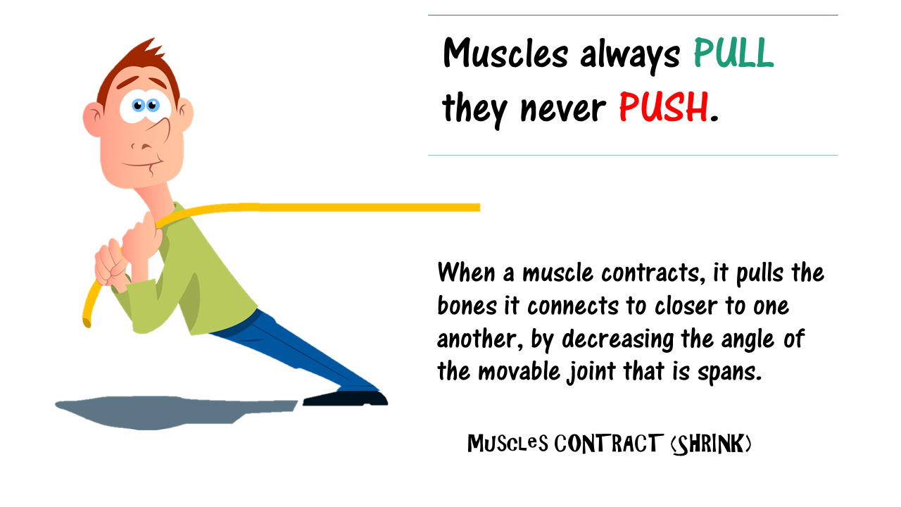
Skeletal muscles attach to at least 2 bones, and span one movable joint. The way that these muscles attach to the bones of your body, is through TENDONS. Muscles always PULL they never PUSH. When a muscle contracts, it pulls the bones it connects to closer to one another, by decreasing the angle of the movable joint that is spans.
Typically, when we contract a muscle, one of the bones the muscle attaches to moves a lot, while the other bone(s) the muscle attaches to remains relatively "fixed" in space.
Muscular contraction produces an action, or a movement of the appendage. We will use examples to describe how the origin and insertion affect the action of a skeletal muscle.
Skeletal muscles attach to at least 2 bones, and span one movable joint. The way that these muscles attach to the bones of your body, is through TENDONS. Muscles always PULL they never PUSH. When a muscle contracts, it pulls the bones it connects to closer to one another, by decreasing the angle of the movable joint that is spans.
Typically, when we contract a muscle, one of the bones the muscle attaches to moves a lot, while the other bone(s) the muscle attaches to remains relatively "fixed" in space.
- The origin is the attachment site that remains relatively "fixed in space" during muscle contraction
- The insertion is the attachment site that moves quite a bit during muscle contraction.
Muscular contraction produces an action, or a movement of the appendage. We will use examples to describe how the origin and insertion affect the action of a skeletal muscle.
Muscle contraction results in different types of movement. The particular movement is a direct result of the muscle attachment. Each of these actions can be described in one of two ways.
- You can describe a muscle's action in terms of the bone to which the muscle is attached to or the appendage that is moved. For example, the biceps brachii performs flexion of the forearm as the forearm is moved.
- You can describe a muscle's action in terms of the joint that is moved upon the contraction of that muscle. For example, that same muscle, the biceps brachii, performs flexion at the elbow, in which the elbow is the joint.
One way to describe muscle action is by the bone that is involved.
Muscle Functional Roles: The human body has over 500 muscles responsible for all types of movement. Each of these muscles has a name; for example, again, the biceps brachii and now the triceps brachii, responsible for both forearm flexion and forearm extension, respectively. When movement of a body part occurs, muscles work in groups rather than individually. Working together enhances a particular movement. During that particular movement, individual muscles will play different roles depending on their origin and insertion.
Muscle Functional Roles: The human body has over 500 muscles responsible for all types of movement. Each of these muscles has a name; for example, again, the biceps brachii and now the triceps brachii, responsible for both forearm flexion and forearm extension, respectively. When movement of a body part occurs, muscles work in groups rather than individually. Working together enhances a particular movement. During that particular movement, individual muscles will play different roles depending on their origin and insertion.
Groups of muscles are involved in most movements and names are used to describe the role of each muscle involved. Agonists, or prime movers, are responsible for the bulk of the action. Antagonist contractions are opposite that of the agonist and serve to control the action. Finally, synergist muscles enhance the action of the agonist.
Muscles can be described according to the Action of that Muscle, INCLUDING:
- agonists (or prime movers)
- antagonists
- synergists

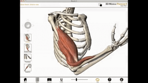
Let's take a look at forearm flexion and identify the roles of the different muscles involved. The biceps brachii is the agonist in forearm flexion. An agonist, or as I said before, a prime mover, is the muscle that is primarily responsible for the movement described: forearm flexion. The action makes sense when you consider the muscle's points of attachment.
The biceps brachii originates on the front of the scapula of the shoulder and inserts on the front of the radius in the forearm. Due to these attachments, contraction and muscle shortening of the biceps flexes the forearm.
The triceps is the antagonist, and its action opposes that of the agonist. The triceps brachii originates on the back of the scapula and humerus, and inserts on the back of the ulna in the forearm.
Due to these attachments, the triceps is stretched during forearm flexing. Stretching the muscle causes the triceps muscle to contract and, thus, slow flexion. It's important to note that the antagonist contraction is minor in comparison to the agonist contraction, and therefore it doesn't prevent the action of the agonist. Rather, antagonist contraction controls the movement by slowing it down and making it smooth.
The antagonist action helps control the muscle movement.
Agonists and antagonists are always functional opposites. Additionally, these muscles switch roles with opposite movements. Let's take a look at an example. The triceps brachii becomes the agonist - while the biceps brachii is the antagonist - when we extend our forearm.
A synergist is a muscle that enhances the action of the agonist. For example, the brachialis is a synergist of the biceps brachii during forearm flexion. The brachialis originates on the humerus, and it inserts on the front of the ulna. As these attachments of the brachialis are similar in nature to those of the biceps brachii, so is its action. Oftentimes, synergist muscles are needed to get a particular action started.
Skeletal muscles are attached to bones on each end by tendons. The origin is the fixed attachment, while the insertion moves with contraction. The action, or particular movement of a muscle, can be described relative to the joint or the body part moved.
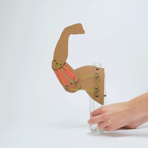
Muscles CONTRACT (SHRINK) - Muscles always PULL they never PUSH.
|
•When a muscle contracts, it pulls the bones it connects to closer to one another, by decreasing the angle of the movable joint that is spans.
Here’s Proof! •In this “muscle machine”, it is the “pulling action” of the biceps muscles that exerts the force that pulls the arm upwards. •AND…it is the “pulling action” of the triceps muscles that exerts the force that pulls the arm Downwards. |
|