Phage Plaque Assay
Phage Plaque Assay

The concentration (or titer) of viruses in the original sample can be calculated the same way we calculate the number or concentration of cells in an original cell culture.
When we are counting cells, we grow colonies from CFUs or colony-forming units. When we are dealing with viruses, we count the number of zones that show destrution of bacteria cells. The zones are called plaques and these plaques are formed from a single virus that we call a Plaque-forming unit of PFU.
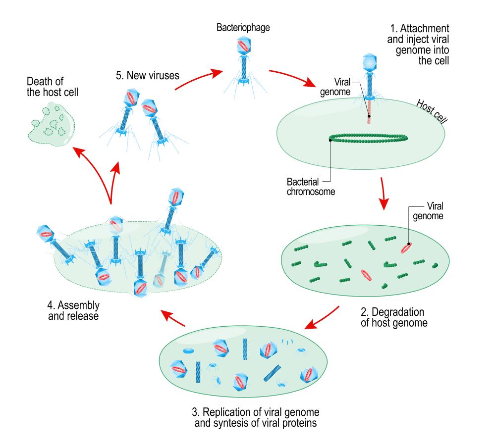
Viruses that we use for this study reproduce using the lytic cycle.
In a virus titer test, we are counting viruses indirectly, by counting the plaques the viruses form on the Petri dish.
- So, when we make a Petri dish for a viral titer test, we grow a monolayer of bacteria cells in e Petri dish first.
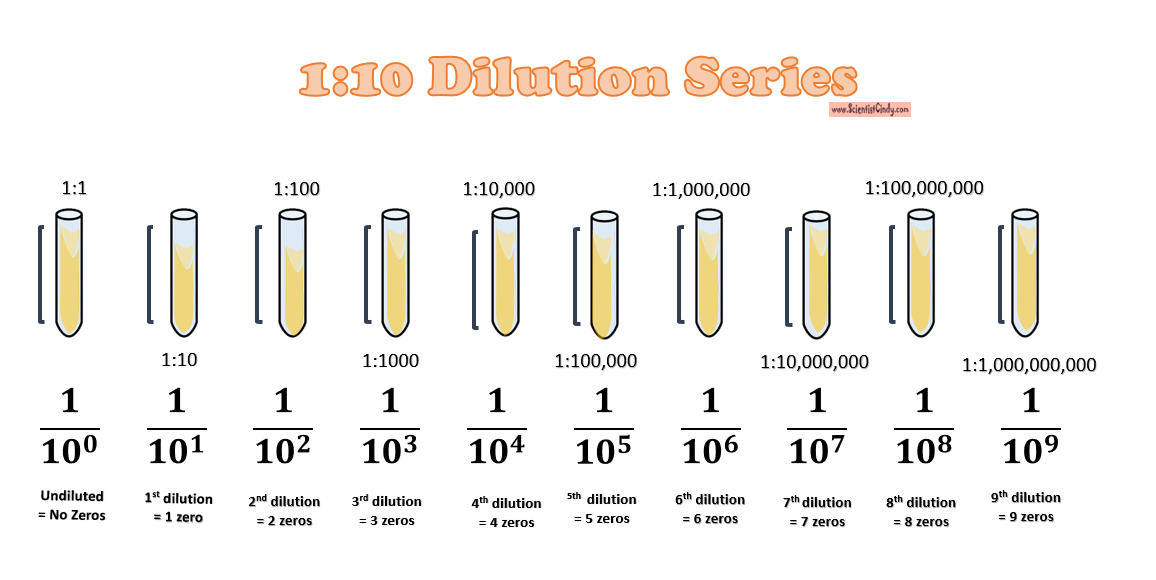
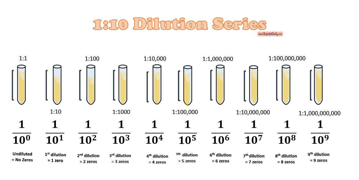
- Then we perform a 1 to 10 dilution series of our virus stock solution.
- We will create our pour plate using the most dilute sample of our virus.
- To create the pour plate, we pour the contents of our most dilute tube on top of the monolayer of bacteria cells on the dish.
- When the virus is pour on top of our bacteria cell monolayer, some of the bacteria cells come into contact with the virus and become infected.
- Infected cells act as a host for the virus. The virus uses the machinery of the host cell to reproduce itself. At the end of the lytic cycle, the host cell undergoes lysis (bursts open and dies) allowing the new viral progeny to go and infect nearby cell. As these new viruses infect new hosts, the lytic cycle repeats, forming more and more progeny to infect and kill more and more bacteria cells.
- Agar gel restricts the infection to only nearby cells, because the viruses cannot freely move in the semi-solid agar.
- We should make additional pour plate using a few other of the dilute tubes as well. Make sure to write the dilution used on the Petri dishes!
- The viruses will be incubated overnight at 37 degrees Celsius.
- This results in a noticeable circular zone of infected cells called a plaque.
- These plaque grow larger over time and can eventually be seen with the naked eye.
- We will then chose plates that grew between 10 and 100 PFUs.
- The results of a titer test with viruses vary slightly. So we will add the notation +/- 10% for all of our values.
- For example, if we grew 14 plaques on the plate made from the 10^ -6 dilution, the titer of the virus stock was 1.4 x 10^8 PFU/ml +/- 10%.

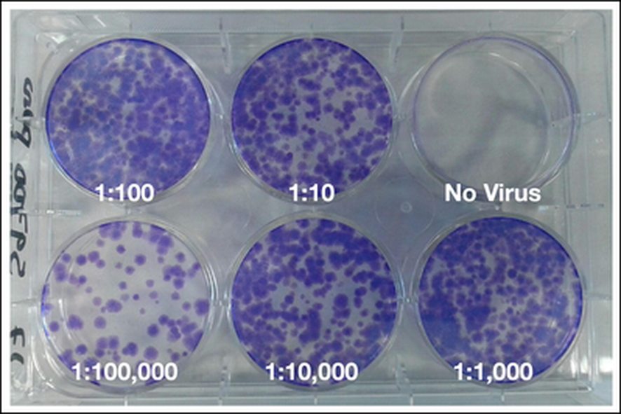
An example of plaques formed by a virus on a monolayer of cells is shown at left. In this image, the cells have been stained with crystal violet, and the plaques are readily visible where the cells have been destroyed by viral infection.


