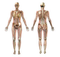
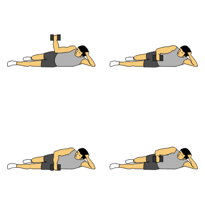
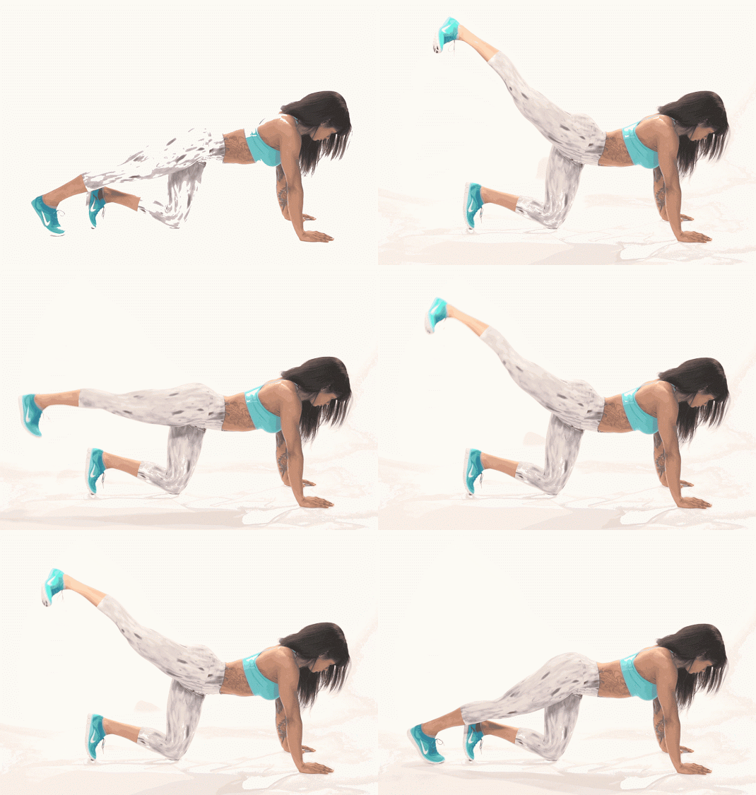
This Tutorial Will Focus on 4 Muscles Commonly Used to Administer Intramuscular Injections
Muscles (skeletal muscles) always connect to at least 2 bones and span 1 movable joint. This is because the function of skeletal muscle, is to allow for movement of the body. Without these rules, there would NO movement!
Origins and Insertions
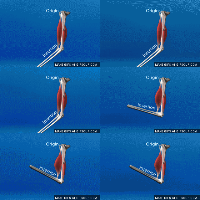
It is Important to be Able to Distinguish Between the Origin and the Insertion Point of a Muscle. This information reveals to us the functionality or actions of the muscle. Remember that structure equals function!
Skeletal muscles attach to at least 2 bones, and span one movable joint. The way that these muscles attach to the bones of your body, is through TENDONS.
Skeletal muscles attach to at least 2 bones, and span one movable joint. The way that these muscles attach to the bones of your body, is through TENDONS.
When we talk about "origins" and "insertions", we are talking about these attachments of the muscle to the bone (through the assistance of the tendons).
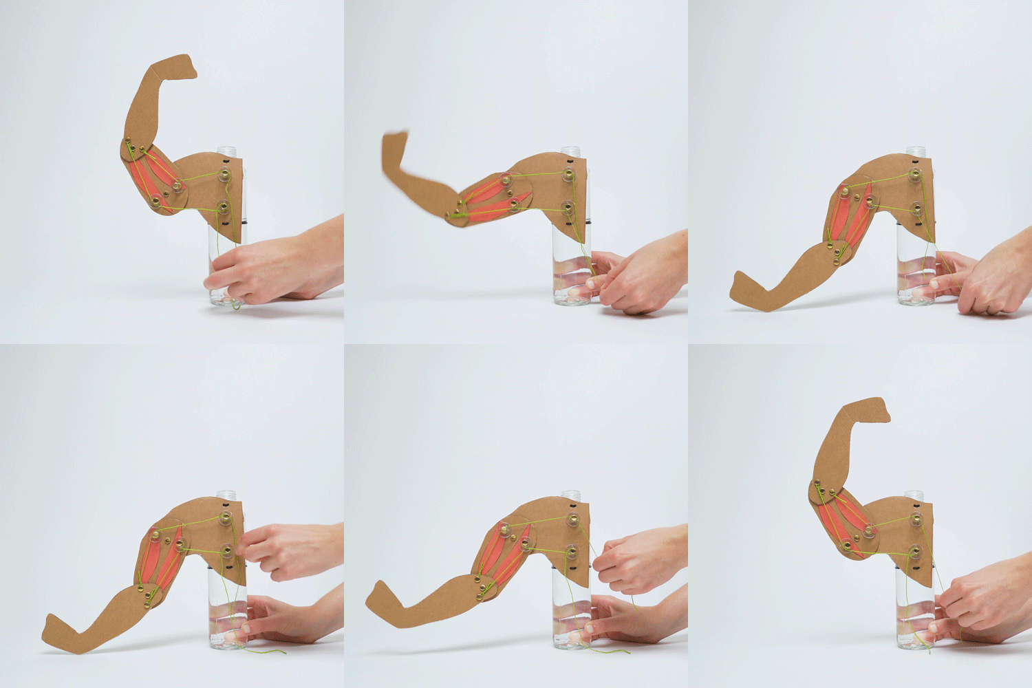
Muscles always PULL they never PUSH.
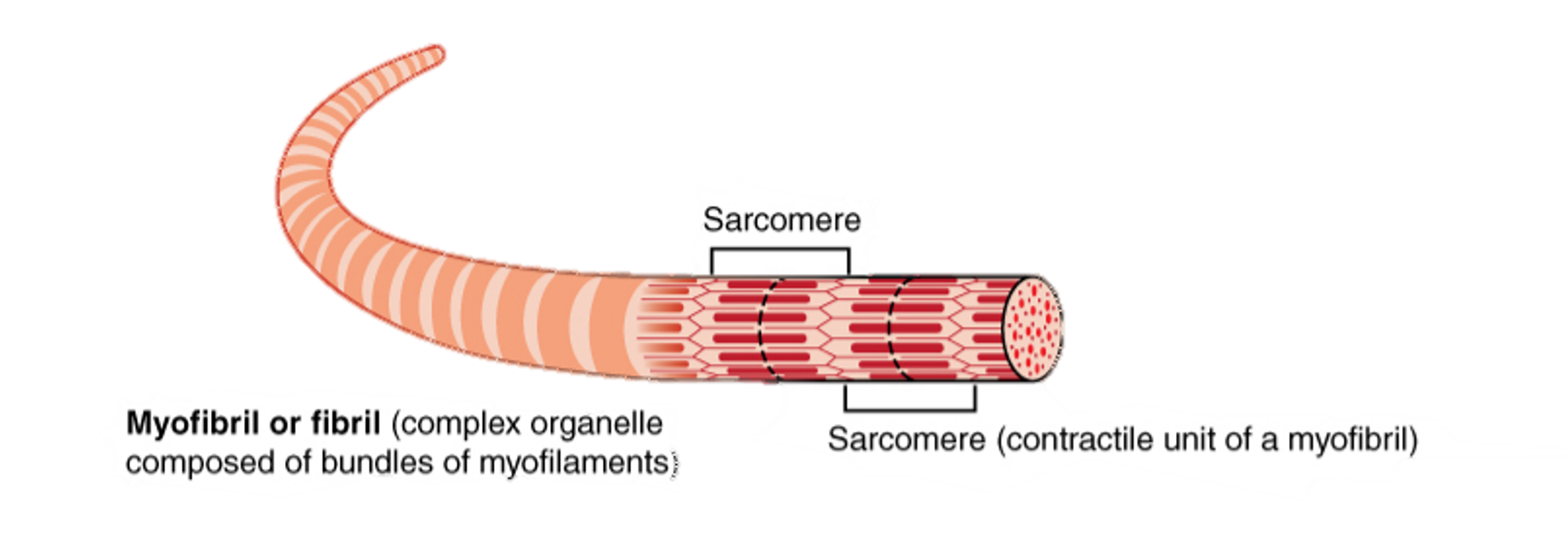
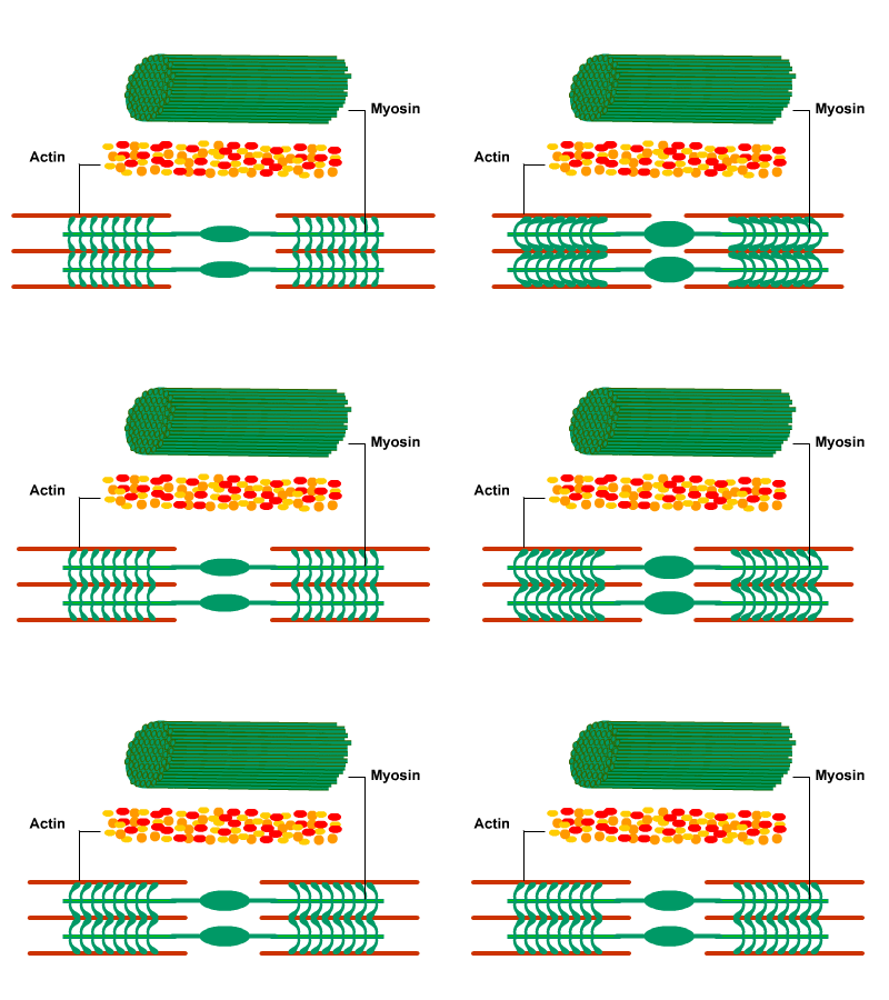
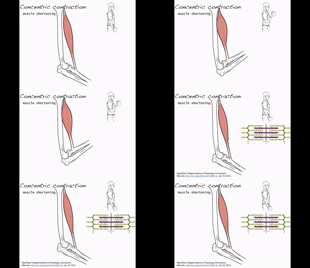
When muscles contract, the muscle is shortened. Muscles are composed of individual muscle fibers that are composed of repeating units called sarcomeres. When the muscle receives the command from the nervous system to contract, the actin and myosin filaments in the sarcomeres slide past one another, causing each of the activated sarcomere to become shorter. As these sarcomeres shorten, the entire muscle is shortened.
Since a muscle must connect to at least 2 bones and span 1 movable joint, let's see what happens when a muscle contracts.
Typically, when we contract a muscle, one of the bones the muscle attaches to moves a lot, while the other bone(s) the muscle attaches to remains relatively "fixed" in space.
- The origin is the attachment site that remains relatively "fixed in space" during muscle contraction
- The insertion is the attachment site that moves quite a bit during muscle contraction.
- The insertion is usually at the distal end, or the end that is further away from the body's center of mass, of the muscle.
- The origin is usually at the more proximal, or closer to the body's center of mass, relative to the insertion. For example, one could say the wrist is distal to the elbow. Conversely, you can say the elbow is proximal to the wrist.
Movements
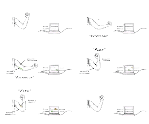
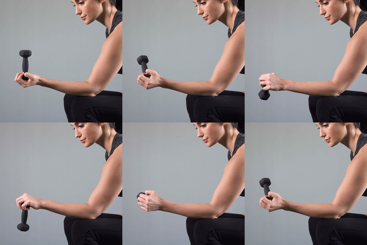
When a muscle contracts, it pulls the bones it connects to closer to one another, by decreasing the angle of the movable joint that is spans. Knowing the insertion and origin helps us identify the action of the muscle. The way in which these bones are able to move closer to each other is dictated by the type of joint that the muscle spans.
Muscular contraction produces an action, or a movement.
ROTATION
FLEXION AND EXTENSION
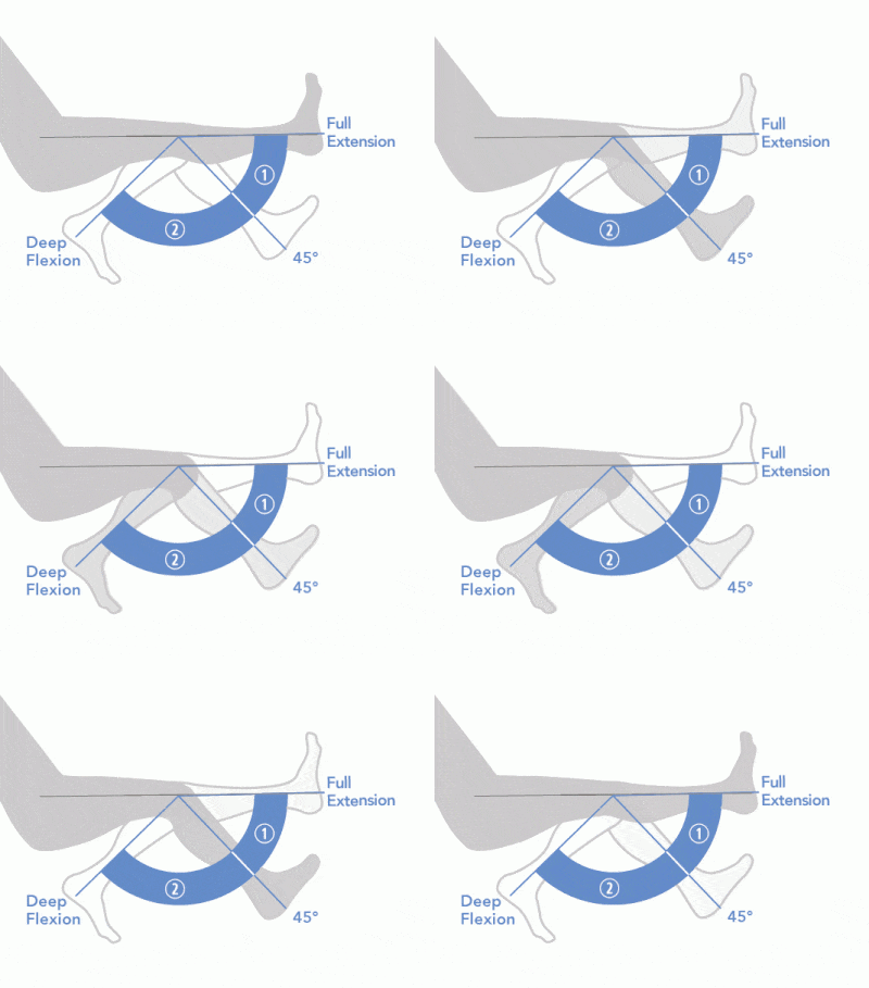
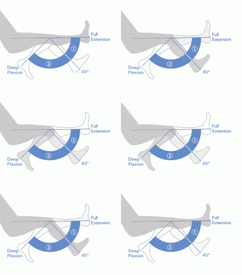
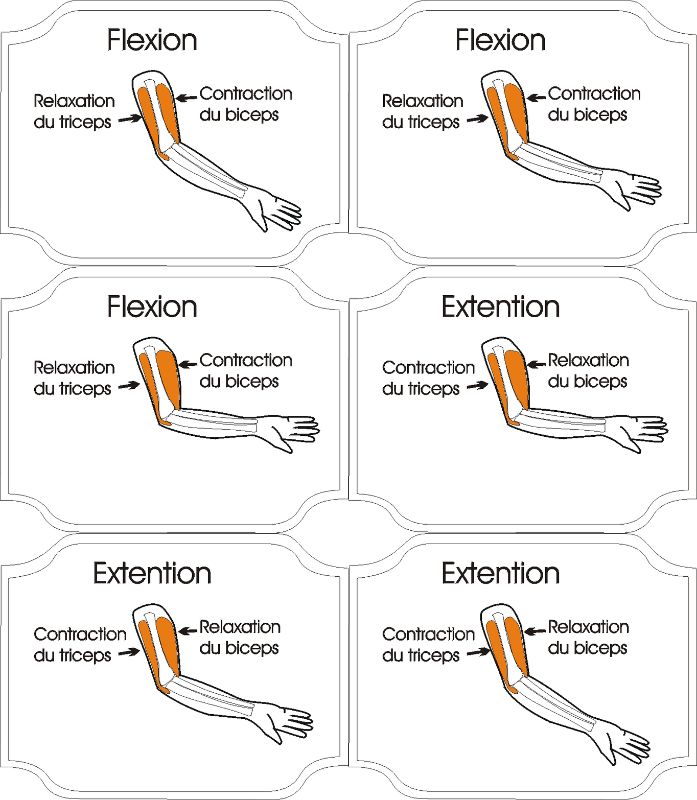
Flexion is movement that decreases the inner angle of the joint. A flexor is a muscle that causes flexion.
Extension is movement that increases the inner angle of the joint. An extensor is a muscle that causes extension.
Extension is movement that increases the inner angle of the joint. An extensor is a muscle that causes extension.
ABDUCTION AND ADDUCTION
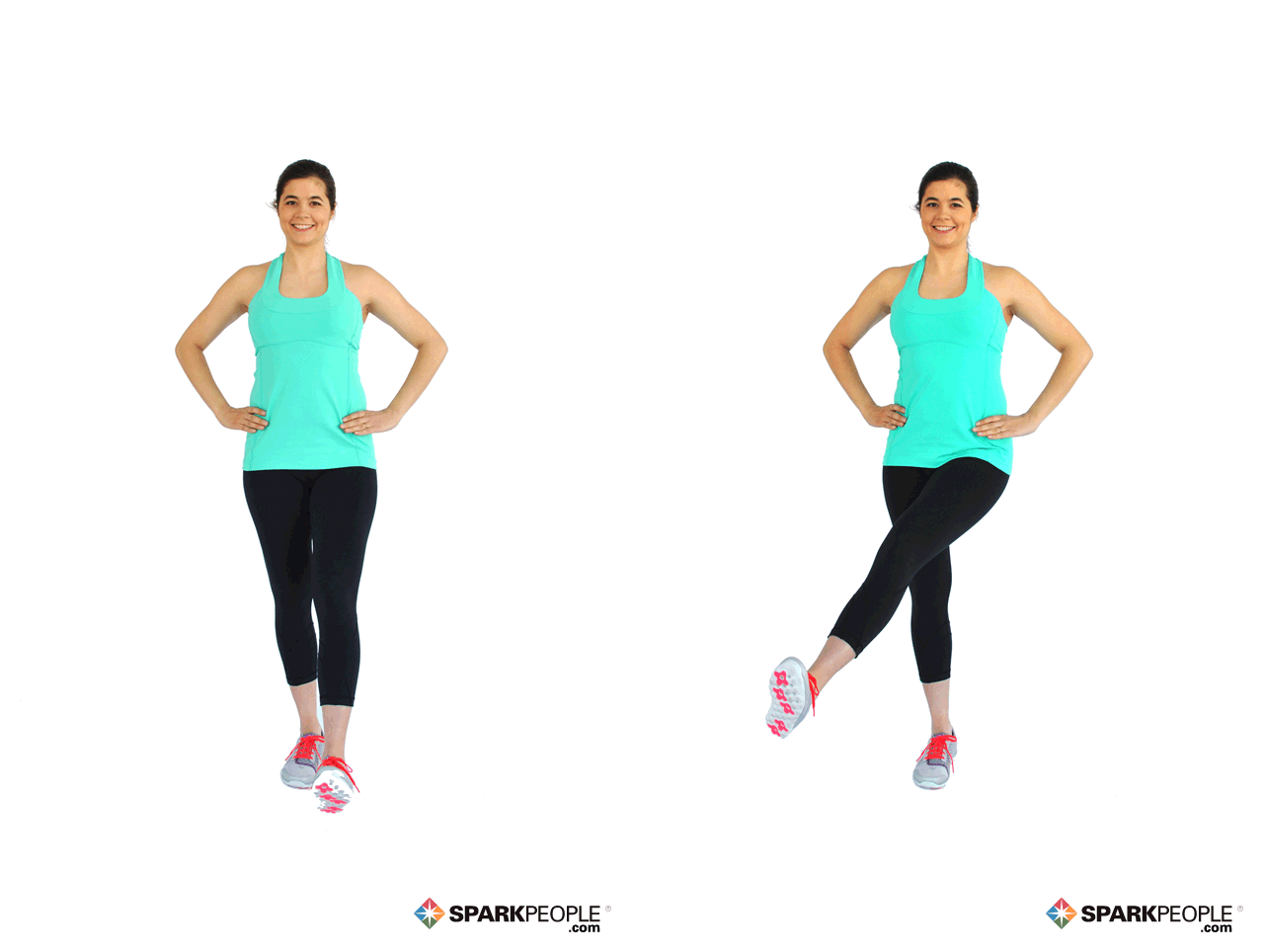
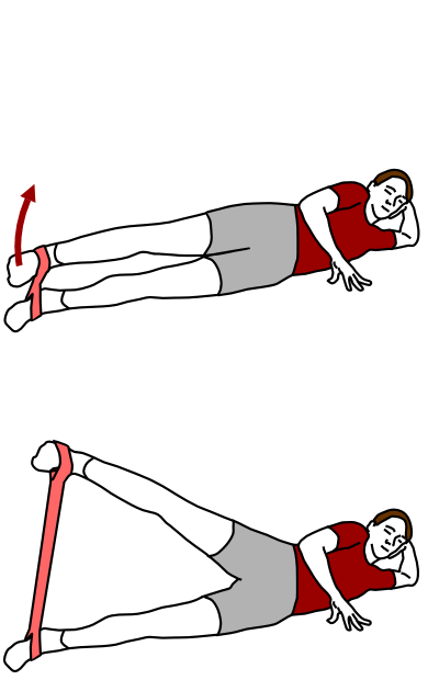
- Abduction is movement away from the mid-line of the body.
- Adduction is movement towards the mid-line of the body - also applies to movements inwards and across the body.
The adductor is a muscle that can act to cause an adduction movement.
You may be thinking, "how am I going to memorize all of these actions of the muscle?" The answer is simply YOU DON'T! You should not "memorize" the functions of the muscles. Instead you should know your anatomy and be able to infer the action of the muscle based on its origin and insertion, and by the position of the muscle relative to the joint it crosses.

Remember, a muscle attaches to at least 2 bones and spans at least 1 movable joint. In order to determine the function of the muscle, you need to see if that muscle passes in front or behind the joint it spans.
Remember that muscles by themselves, really only have 2 actions; they can either contract (shorten) or relax. This means that muscles will essentially either 'pull' on the bones it attaches to, or "not pull" on those bones.
Rules for determining muscle function/action:
Remember that muscles by themselves, really only have 2 actions; they can either contract (shorten) or relax. This means that muscles will essentially either 'pull' on the bones it attaches to, or "not pull" on those bones.
Rules for determining muscle function/action:
- When the muscle passes in front of (anterior to) the joint, it functions in FLEXION. Flexion decreases the angle of the joint.
- When the muscle passes in behind (posterior to) the joint, it functions in EXTENSION. Extension increases the angle of the joint.
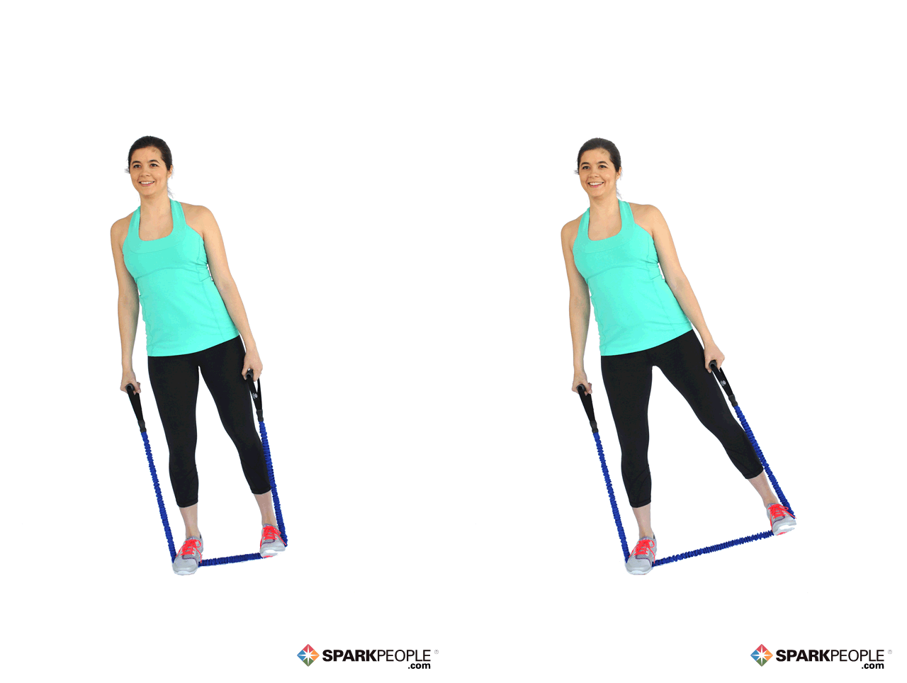
- When the muscle passes on the lateral side of the joint, it functions in ABDUCTION. Abduction acts to move a body part away from the midline.
- When the muscle passes on the medial side of the joint, it functions in ADDUCTION. Adduction acts to move a body part toward the midline.
Trapezius
Function:
- Elevation/Depression of the Scapulae
- upward rotation of the scapula
- Retraction of the Scapula
Innervation of the Accessory Nerve (Cranial Nerve XI)
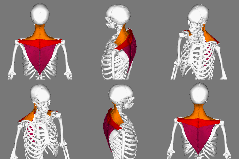
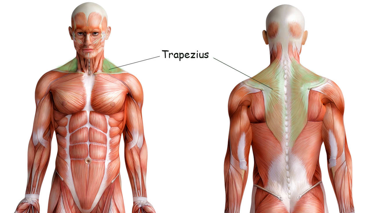
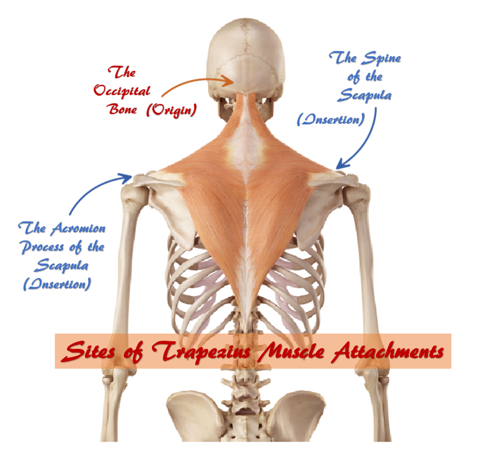
The trapezius muscles are the large major muscles of the upper back. The trapezius muscles function to move, rotate and stabilize the scapula (shoulder blade).
By Anatomography - en:Anatomography (setting page of this image)., CC BY-SA 2.1 jp, https://commons.wikimedia.org/w/index.php?curid=22182550
via GIPHY
By Anatomography - en:Anatomography (setting page of this image)., CC BY-SA 2.1 jp, https://commons.wikimedia.org/w/index.php?curid=22182550
via GIPHY
Trapezius Muscle Attachments
|
Origins -
|
Insertions -
|
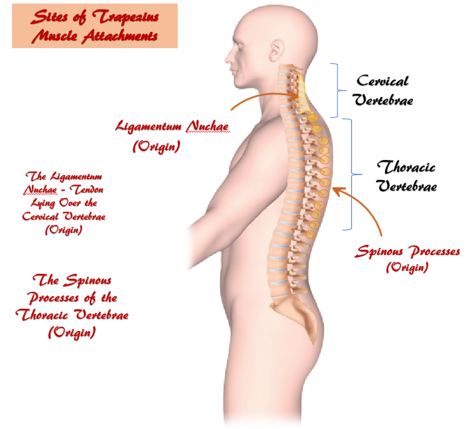
Origin: external occipital protuberance, spinous processes of vertebrae C7-T12, Nuchal ligament, Occipital Bone
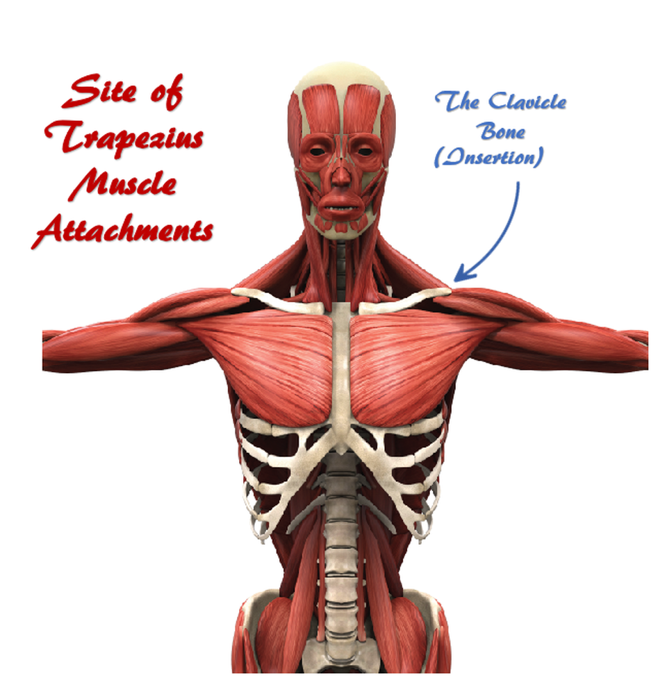
Insertion : nuchal ligament, medial superior nuchal line, posterior border of the lateral one-third of the clavicle, acromion process, and spine of scapula
Nerve: accessory nerve (motor)
Actions: The deltoid is the prime mover of arm abduction
Rotation, retraction, elevation, and depression of scapula
- Origin(s) of the muscle
- Insertion(s) of the muscle
- Actions of the muscle
- Nerve supply of the muscle
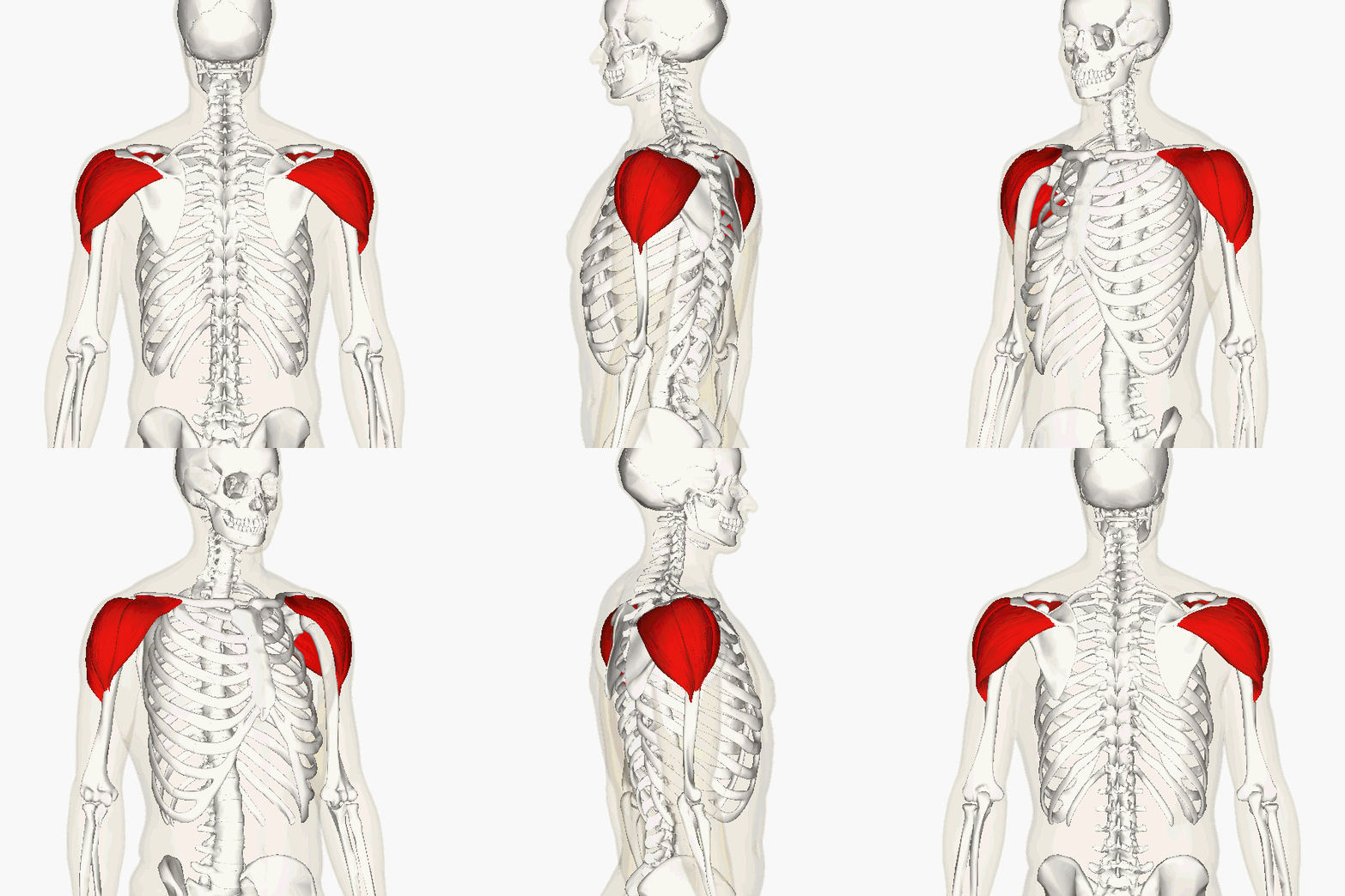
Deltoid
Origins -
- The Clavicle
- The Spine of the Scapula
- The Acromian Process of the Scapula
Insertions -
- The Deltoid Tuberosity of the Humerus
Primary Action -
- Arm Abduction (out to the sides)
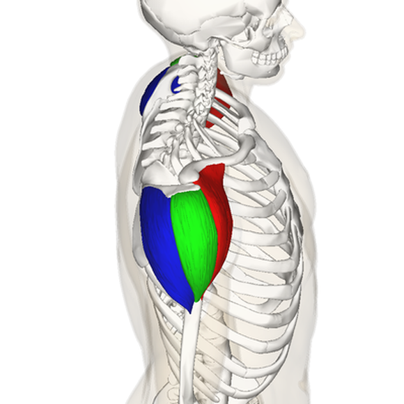
- The deltoid is the prime mover of arm abduction.
The Gluteus Medius
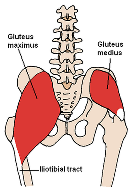
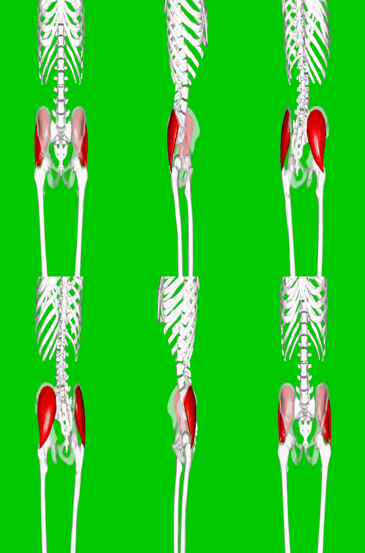
Here is an illustration comparing the locations of the gluteus maximus and the gluteus medius.
Gluteus Medius
A portion of the gluteus medius lies superior to gluteus maximus. The inferior portion of the gluteus medius lies underneath (deep to) the gluteus maximus. The gluteus medius is one of the three gluteal muscles which lie on the outer portion of the pelvis at the posterior aspect.
By Anatomography - en:Anatomography (setting page of this image), CC BY-SA 2.1 jp, https://commons.wikimedia.org/w/index.php?curid=27657573
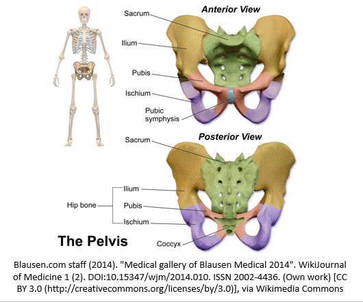
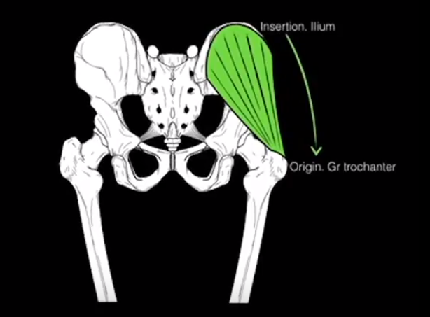
The gluteus medius originates at the ilium (iliac spine and the iliac crest) and inserts at the greater trochanter of the femur.
Innervated by the superior gluteal nerve, a branch of the sacral plexus (L4-S1)
Innervated by the superior gluteal nerve, a branch of the sacral plexus (L4-S1)
Watch This 2-minute Video on How to Give an
Intramuscular Injection in the Gluteus Medius
QUADRICEPS
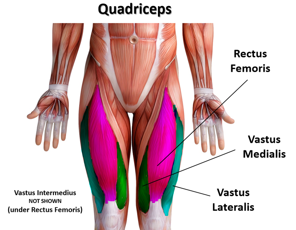
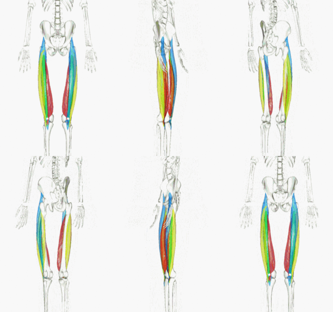
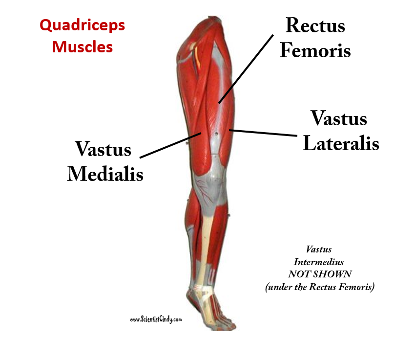
The 4 Heads of the Quadriceps
|
The 4 heads of the quadriceps muscles have 4 different origins, but the same insertion:
|
Rectus Femoris
Origins:
|
Vastus Medialis
Origin:
|
|
Vastus lateralis
Origins:
|
Vastus Intermedius
Origins:
|
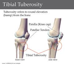
Insertion: Tibial tuberosity
Action: The quadriceps muscles are the prime movers of Knee Extension / Leg Extension
Action: The quadriceps muscles are the prime movers of Knee Extension / Leg Extension
|
The quadriceps femoris gets its name from the Latin words that mean "four-headed muscle of the femur". The quadraceps femoris refers to a muscle group that consists of 4 muscles of the anterior (front) thigh area (or the femoral area. The muscles are extensors of the knee.
Quick Overview of the Quadriceps
Rectus Femoris Function = Extends leg and flexes thigh (brings thigh up) The rectus femoris is the quadriceps muscle that lies at the middle of the front part of the thigh. It lies (wholly or partially) on top of the other three quadriceps muscles. It originates on the ilium. It is named from its straight-appearing orientation at the front of the thigh. |
Vastus Medialis Function = Extends leg; stabilize knee (patella) The vastus medialis is located on the front medial portion of the thigh. It is one of the quadraceps muscles. The vastus medialis originates medially along the entire length of the femur. It is connected to the other quadriceps muscle. Vastus Lateralis Function = Extends leg (brings leg straight) and stabilizes knee The vastus lateralis muscle is the largest head of the quadriceps group. It forms the lateral aspect of the thigh. Vastus Intermedius Function = Extends leg Vastus Intermedius Cannot be Viewed in the Muscular Leg Model Without Removing the Rectus Femoris. Vastus intermedius lies between vastus lateralis and vastus medialis on the front of the femur (i.e. on the top or front of the thigh), but underneath (deep) to the rectus femoris. Typically, it cannot be seen without dissection of the rectus femoris. |


