The Nervous System
Functional Divisions of the Nervous System
The nervous system can be divided on the basis of its functions.
There are two ways to consider how the nervous system is divided functionally.
- First, the basic functions of the nervous system are
- sensation
- integration
- response
- Secondly, control of the body can be somatic or autonomic—divisions that are largely defined by the structures that are involved in the response.
- There is also a region of the peripheral nervous system that is called the enteric nervous system that is responsible for a specific set of the functions within the realm of autonomic control related to gastrointestinal functions.
Basic Functions
Functions of the Nervous System
Sensory input
Sensory input
- Information gathered by sensory receptors about internal and external changes
- Processing and interpretation of sensory input done by brain or CNS
- Activation of effector organs (muscles and glands) produces a response
The nervous system is involved in receiving information about the environment around us (sensation) and generating
responses to that information (motor responses). The nervous system can be divided into regions that are responsible
for sensation (sensory functions) and for the response (motor functions). But there is a third function that needs to be
included. Sensory input needs to be integrated with other sensations, as well as with memories, emotional state, or learning
(cognition). Some regions of the nervous system are termed integration or association areas. The process of integration
combines sensory perceptions and higher cognitive functions such as memories, learning, and emotion to produce a
response.
- Sensation. The first major function of the nervous system is sensation—receiving information about the environment to gain input about what is happening outside the body (or, sometimes, within the body). The sensory functions of the nervous system register the presence of a change from homeostasis or a particular event in the environment, known as a stimulus.
- Response. The nervous system produces a response on the basis of the stimuli perceived by sensory structures. An obvious response would be the movement of muscles, such as withdrawing a hand from a hot stove, but there are broader uses of the term. The nervous system can cause the contraction of all three types of muscle tissue. For example, skeletal muscle contracts to move the skeleton, cardiac muscle is influenced as heart rate increases during exercise, and smooth muscle contracts as the digestive system moves food along the digestive tract. Responses also include the neural control of glands in the body as well, such as the production and secretion of sweat by the eccrine and merocrine sweat glands found in the skin to lower body temperature. Responses can be divided into those that are voluntary or conscious (contraction of skeletal muscle) and those that are involuntary (contraction of smooth muscles, regulation of cardiac muscle, activation of glands). Voluntary responses are governed by the somatic nervous system and involuntary responses are governed by the autonomic nervous system.
- Integration. Stimuli that are received by sensory structures are communicated to the nervous system where that information is processed. This is called integration. Stimuli are compared with, or integrated with, other stimuli, memories of previous stimuli, or the state of a person at a particular time. This leads to the specific response that will be generated. Seeing a baseball pitched to a batter will not automatically cause the batter to swing. The trajectory of the ball and its speed will need to be considered. Maybe the count is three balls and one strike, and the batter wants to let this pitch go by in the hope of getting a walk to first base. Or maybe the batter’s team is so far ahead, it would be fun to just swing away.
Controlling the Body
The nervous system can be divided into two parts mostly on the basis of a functional difference in responses.
The somatic nervous system (SNS) is responsible for conscious perception and voluntary motor responses. Voluntary motor response
means the contraction of skeletal muscle, but those contractions are not always voluntary in the sense that you have to want
to perform them. Some somatic motor responses are reflexes, and often happen without a conscious decision to perform
them.
The autonomic nervous system (ANS) is responsible for involuntary control of the body, usually for the sake of
homeostasis (regulation of the internal environment).
- Sensory input for autonomic functions can be from sensory structures tuned to external or internal environmental stimuli.
- The motor output extends to smooth and cardiac muscle as well as glandular tissue.
The enteric nervous system (ENS) is responsible for controlling the smooth muscle and glandular tissue in your digestive system. It is a large part of the PNS, and is not dependent on the CNS.
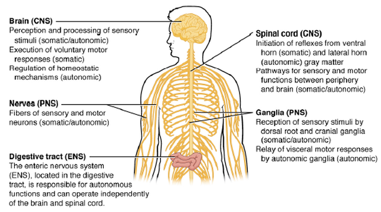
Somatic, Autonomic, and Enteric Structures of the Nervous System Somatic structures include the spinal nerves, both motor and sensory fibers, as well as the sensory ganglia (posterior root ganglia and cranial nerve ganglia). Autonomic structures are found in the nerves also, but include the sympathetic and parasympathetic ganglia. The enteric nervous system includes the nervous tissue within the organs of the digestive tract.
Image Courtesy of OpenStax Anatomy and Physiology https://openstax.org/details/books/anatomy-and-physiology
Image Courtesy of OpenStax Anatomy and Physiology https://openstax.org/details/books/anatomy-and-physiology
Divisions of the Nervous System
Two Main Divisions of the Nervous System
The nervous system is divided into the
The nervous system is divided into the
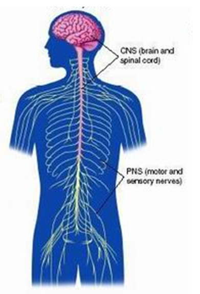
- Central nervous system (CNS) -
- The CNS includes the brain and spinal cord
- Peripheral nervous system (PNS) -
- The PNS includes everything outside of the CNS
The Peripheral Nervous System is further divided into the
- Sensory (Afferent) Division - input
- Motor (Efferent) Division - output
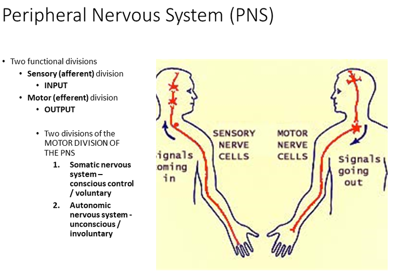
The Motor Division of the Peripheral Nervous System is subdivided into the
- Somatic nervous system – conscious control / voluntary
- Autonomic nervous system - unconscious / involuntary
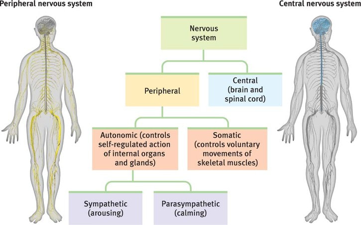
The Autonomic Nervous System is Subdivided into:
- Sympathetic Nervous System
- Parasympathetic Nervous System
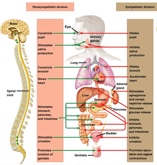
The sympathetic nervous system controls the body's fight or flight responses;
- releases adrenaline (epinephrine)
- pupils dilate
- increases heart rate
- increases rate of respiration
- releases additional glucose into the bloodstream
- inhibits digestion and inhibits urination
The Parasympathetic Nervous System governs the body's rest and digest responses
- pupils constrict
- slows heart rate
- decreases rate of respiration
- promotes digestion and urination
Nervous Tissue
By the end of this section, you will be able to:
• Describe the basic structure of a neuron
• Identify the different types of neurons on the basis of polarity
• List the glial cells of the CNS and describe their function
• List the glial cells of the PNS and describe their function
Nervous tissue is composed of two types of cells, neurons and glial cells. Neurons are the primary type of cell that most anyone associates with the nervous system. They are responsible for the computation and communication that the nervous system provides. They are electrically active and release chemical signals to target cells. Glial cells, or glia, are known to play a supporting role for nervous tissue. Ongoing research pursues an expanded role that glial cells might play in signaling, but neurons are still considered the basis of this function. Neurons are important, but without glial support they would not be able to perform their function.
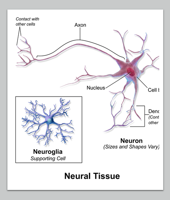
- Neurons (nerve cells)— the principle cell of the nervous system
- Neuroglia (supporting cells) - assist neurons in various ways.
- CNS neuroglia include oligodendrocytes for myelin.
- PNS neuroglia include Schwann cells for myelin.
Neurons
Neurons are the cells considered to be the basis of nervous tissue. They are responsible for the electrical signals that communicate information about sensations, and that produce movements in response to those stimuli, along with inducing thought processes within the brain. An important part of the function of neurons is in their structure, or shape. The three-dimensional shape of these cells makes the immense numbers of connections within the nervous system possible.
Neuron Classification by Shape
There are many neurons in the nervous system—a number in the trillions. And there are many different types of neurons. They can be classified by many different criteria. The first way to classify them is by the number of processes attached to the cell body. Using the standard model of neurons, one of these processes is the axon, and the rest are dendrites. Because information flows through the neuron from dendrites or cell bodies toward the axon, these names are based on the neuron's polarity
Unipolar cells have one process that includes both the axon and dendrite. Bipolar cells have two processes, the axon and a dendrite. Multipolar cells have more than two processes, the axon and two or more dendrites.
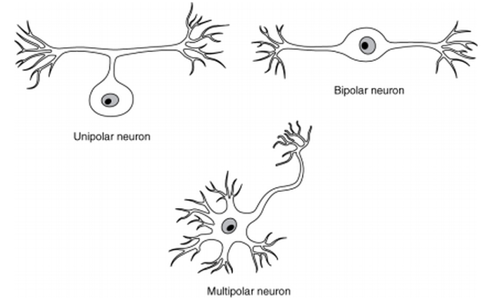
- Unipolar cells have only one process emerging from the cell. True unipolar cells are only found in invertebrate animals, so the unipolar cells in humans are more appropriately called “pseudo-unipolar” cells. Invertebrate unipolar cells do not have dendrites. Human unipolar cells have an axon that emerges from the cell body, but it splits so that the axon can extend along a very long distance. At one end of the axon are dendrites, and at the other end, the axon forms synaptic connections with a target.
- Unipolar cells are exclusively sensory neurons and have two unique characteristics.
- First, their dendrites are receiving sensory information, sometimes directly from the stimulus itself.
- Secondly, the cell bodies of unipolar neurons are always found in ganglia. The axon projects from the dendrite endings, past the cell body in a ganglion, and into the central nervous system.
- First, their dendrites are receiving sensory information, sometimes directly from the stimulus itself.
- Unipolar cells are exclusively sensory neurons and have two unique characteristics.
- Bipolar cells have two processes, which extend from each end of the cell body, opposite to each other. One is the axon and one the dendrite. Bipolar cells are not very common. They are found mainly in the olfactory epithelium (where smell stimuli are sensed), and as part of the retina.
- Multipolar neurons are all of the neurons that are not unipolar or bipolar. They have one axon and two or more dendrites (usually many more). With the exception of the unipolar sensory ganglion cells, and the two specific bipolar cells mentioned above, all other neurons are multipolar. Some cutting edge research suggests that certain neurons in the CNS do not conform to the standard model of “one, and only one” axon.
Neuron Classification by Function
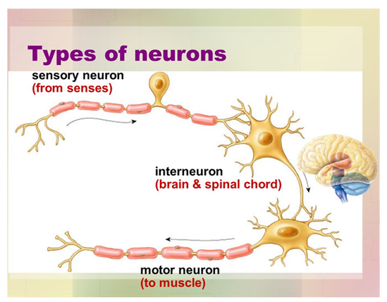
SENSORY (AFFERENT) NEURONS
- - Neurons carrying information TOWARD the CNS, are Sensory Neurons
- Transmit impulses from sensory receptors toward CNS
- Almost all are Unipolar
- Cell bodies in ganglia in PNS
- Most cell bodies in CNS (except some autonomic neurons
- Neurons carrying information AWAY from the CNS, are Motor Neurons
- Carry impulses from CNS to effectors which are glands or muscles that carry out the commands of the CNS
- If a neuron connects a sensory neuron to a motor neuron in the CNS, it is an Interneuron.
- Interneurons are sometimes called association neurons
- most are entirely within CNS
- Lie between motor and sensory neurons
- Shuttle signals through CNS pathways; most are entirely within CNS
Glial Cells
Glial cells, or neuroglia or simply glia, are the other type of cell found in nervous tissue. They are considered to be supporting cells, and many functions are directed at helping neurons complete their function for communication. The name glia comes from the Greek word that means “glue,” and was coined by the German pathologist Rudolph Virchow, who wrote in 1856: “This connective substance, which is in the brain, the spinal cord, and the special sense nerves, is a kind of glue (neuroglia) in which the nervous elements are planted.” Today, research into nervous tissue has shown that there are many deeper roles that these cells play. And research may find much more about them in the future.
Neuroglia support and maintain neurons in various ways.
- In the Central Nervous System (CNS) neuroglia called oligodendrocytes function to make the myelin sheath.
- In the Peripheral Nervous System (PNS) neuroglia called Schwann cells function to make the myelin sheath.

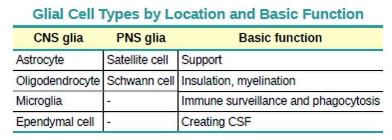
Glial Cells of the CNS
Glial Cells of the CNS The CNS has astrocytes, oligodendrocytes, microglia, and ependymal cells that support the neurons of the CNS in several ways.
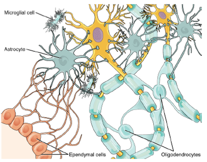
ASTROCYTES -
One cell providing support to neurons of the CNS is the astrocyte, so named because it appears to be star-shaped under the microscope (astro- = “star”). Generally, they are supporting cells for the neurons in the central nervous system.
Some ways in which they support neurons in the central nervous system are by maintaining the concentration of chemicals in the extracellular space, removing excess signaling molecules, reacting to tissue damage, and contributing to the blood-brain barrier (BBB).
- The blood-brain barrier is a physiological barrier that keeps many substances that circulate in the rest of the body from getting into the central nervous system, restricting what can cross from circulating blood into the CNS. Nutrient molecules, such as glucose or amino acids, can pass through the BBB, but other molecules cannot. This actually causes problems with drug delivery to the CNS. Pharmaceutical companies are challenged to design drugs that can cross the BBB as well as have an effect on the nervous system.
OLIGODENDROCYTES -
Also found in CNS tissue is the oligodendrocyte, sometimes called just “oligo,” which is the glial cell type that insulates axons in the CNS. There are a few processes that extend from the cell body. Each one reaches out and surrounds an axon to insulate it in myelin. One oligodendrocyte will provide the myelin for multiple axon segments, either for the same axon or for separate axons.
Also found in CNS tissue is the oligodendrocyte, sometimes called just “oligo,” which is the glial cell type that insulates axons in the CNS. There are a few processes that extend from the cell body. Each one reaches out and surrounds an axon to insulate it in myelin. One oligodendrocyte will provide the myelin for multiple axon segments, either for the same axon or for separate axons.
MICROGLIA -
Microglia are, as the name implies, smaller than most of the other glial cells. Ongoing research into these cells, although not entirely conclusive, suggests that they may originate as white blood cells, called macrophages, that become part of the CNS during early development.
Microglia are, as the name implies, smaller than most of the other glial cells. Ongoing research into these cells, although not entirely conclusive, suggests that they may originate as white blood cells, called macrophages, that become part of the CNS during early development.
EPENDYMAL CELLS -
The ependymal cell is a glial cell that filters blood to make cerebrospinal fluid (CSF), the fluid that circulates through the CNS. Ependymal cells line each ventricle at the choroid plexus which a specialized structure in the ventricles. Here, they filter and absorb
components of the blood to produce cerebrospinal fluid.
Glial Cells of the PNS
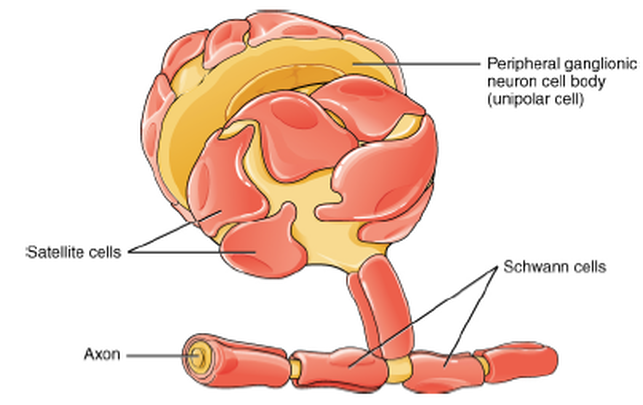
Satellite Cells -
One of the two types of glial cells found in the PNS is the satellite cell. Satellite cells are found in sensory and autonomic ganglia, where they surround the cell bodies of neurons. This accounts for the name, based on their appearance under the microscope. They provide support, performing similar functions in the periphery as astrocytes do in the CNS—except, of course, for establishing the BBB.
Schwann Cells -
The second type of glial cell is the Schwann cell, which insulate axons with myelin in the periphery. Schwann cells are different than oligodendrocytes, in that a Schwann cell wraps around a portion of only one axon segment and no others. The nucleus and cytoplasm of the Schwann cell are on the edge of the myelin sheath.
Neurons
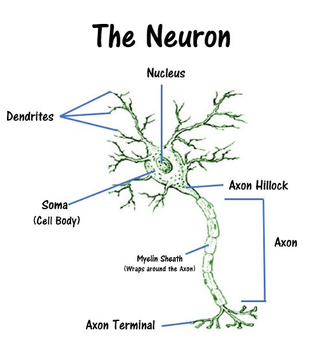
Neurons are the cells considered to be the basis of nervous tissue. They are responsible for the electrical signals that communicate information about sensations, and that produce movements in response to those stimuli, along with inducing thought processes within the brain. An important part of the function of neurons is in their structure, or shape. The three-dimensional shape of these cells makes the immense numbers of connections within the nervous system possible. Parts of a Neuron As you learned in the first section, the main part of a neuron is the cell body, which is also known as the soma (soma = “body”).
- The cell body contains the nucleus and most of the major organelles. But what makes neurons special is that they have many extensions of their cell membranes, which are generally referred to as processes.
- Neurons are usually described as having one, and only one, axon—a fiber that emerges from the cell body and projects to target cells. That single axon can branch repeatedly to communicate with many target cells. It is the axon that propagates the nerve impulse, which is communicated to one or more cells.
- The other processes of the neuron are dendrites, which receive information from other neurons at specialized areas of contact called synapses. The dendrites are usually highly branched processes, providing locations for other neurons to communicate with the cell body. Information flows through a neuron from the dendrites, across the cell body, and down the axon. This gives the neuron a polarity—meaning that information flows in this one direction. Figure 12.8 shows the relationship of these parts to one another.
Neurons
contain four distinct regions:
contain four distinct regions:
- the cell body
- the dendrites
- the axon
- the axon terminals
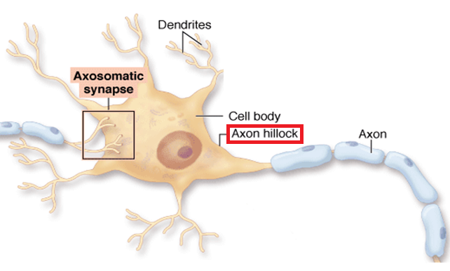
Neuronal Structures
The cell body contains the nucleus and most of the organelles. Most neurons have multiple dendrites, which extend out-ward from the cell body. Dendrites are specialized to receive chemical signals (neurotransmitters) from the axon terminal(s) of other neurons. Electric disturbances generated in the dendrites will diffuse to the initial segment of the axon, called the axon hillock (trigger zone). If the axon hillock is depolarized to threshold, the axon hillock will initiate an action potential. Almost every neuron has a single axon. Axons are specialized for the conduction of electrical-chemical signals called action potentials. Axons carry the action potential away from the cell body toward the axon terminal.
The Cell Body
- The Cell Body Is the Control Center
- The main part of a neuron is the cell body, which is also known as the soma (soma = “body”). The cell body contains the nucleus and most of the major organelles. But what makes neurons special is that they have many extensions of their cell membranes, which are generally referred to as processes.
- The cell body (cell soma) of a neuron resembles a typical cell, with a nucleus and all organelles needed to direct cellular activity.
- The cell body with its nucleus is essential to the well-being of the cell because it contains DNA that is the template for protein synthesis.
Dendrites
The other processes of the neuron are dendrites, which receive information from other neurons at specialized areas of contact called synapses. The dendrites are usually highly branched processes, providing locations for other neurons to communicate with the cell body. Information flows through a neuron from the dendrites, across the cell body, and down the axon. This gives the neuron a polarity—meaning that information flows in this one direction.
- Dendrites Receive Incoming Signals
The other processes of the neuron are dendrites, which receive information from other neurons at specialized areas of contact called synapses. The dendrites are usually highly branched processes, providing locations for other neurons to communicate with the cell body. Information flows through a neuron from the dendrites, across the cell body, and down the axon. This gives the neuron a polarity—meaning that information flows in this one direction.
- Dendrites are thin, branched processes that receive incoming information from neighboring cells.
- The primary function of dendrites in the peripheral nervous system is to receive incoming information and transfer it to an integrating region within the neuron.
- Within the CNS, dendrite function is more complex. Dendritic spines can function as independent compartments, sending signals back and forth with other neurons in the brain.
The Axon
- Axons Carry Outgoing Signals
- Most peripheral neurons have a single axon that originates from a specialized region of the cell body called the axon hillock.
- The primary function of an axon is to transmit outgoing electrical signals from the integrating center of the neuron to target cells at the end of the axon.
- At the distal end of the axon, the electrical signal usually causes secretion of a chemical messenger molecule (neurotransmitter).
The Synapse
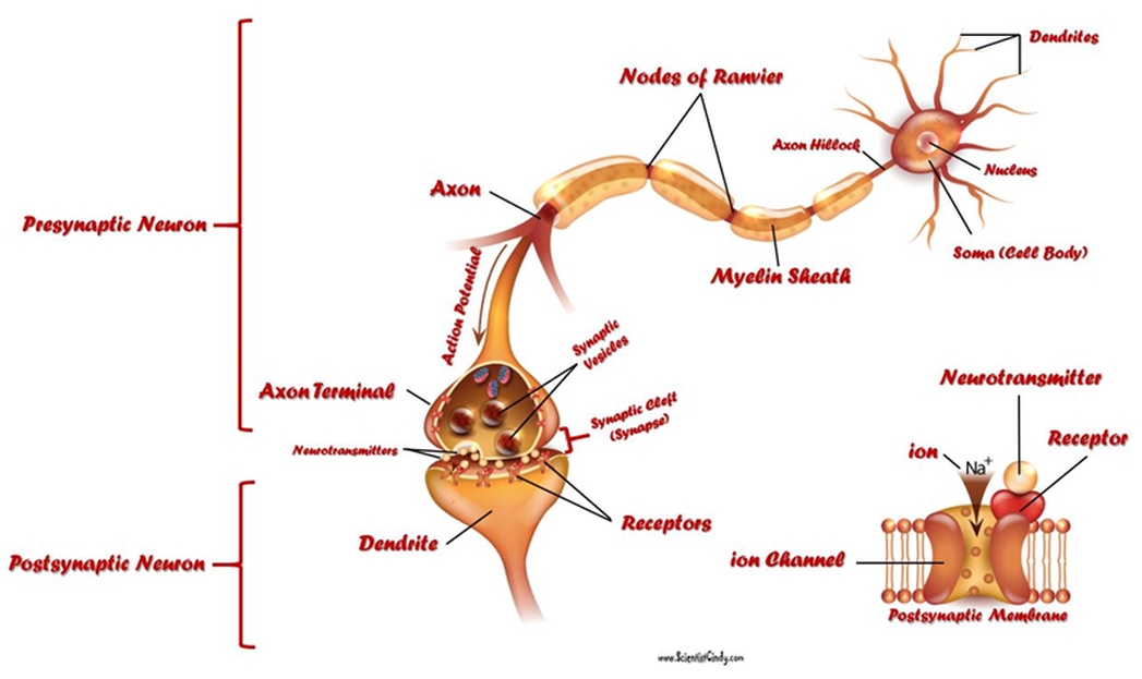
- Synapses are tiny gaps that exist between neurons. Neurotransmitters are released from the presynaptic terminal and passively diffuse across the synapse where they can then transiently bind to post-synaptic receptors located on the dendrites of the post-synaptic neuron.
The Axon Hillock
- Where the axon emerges from the cell body, there is a special region referred to as the axon hillock. This is a tapering of the cell body toward the axon fiber. Within the axon hillock, the cytoplasm changes to a solution of limited components called axoplasm. Because the axon hillock represents the beginning of the axon, it is also referred to as the initial segment.
Axon Terminals
- At the end of the axon is the axon terminal, where there are usually several branches extending toward the target cell, each of which ends in an enlargement called a synaptic end bulb. These bulbs are what make the connection with the target cell at the synapse.
- Axon terminals release neurotransmitters in response the an action potential.
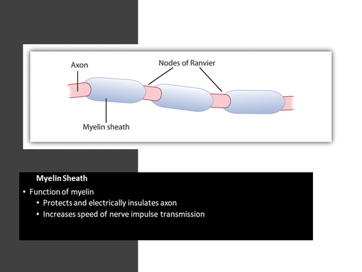
Myelin Sheath
Many axons are myelinated
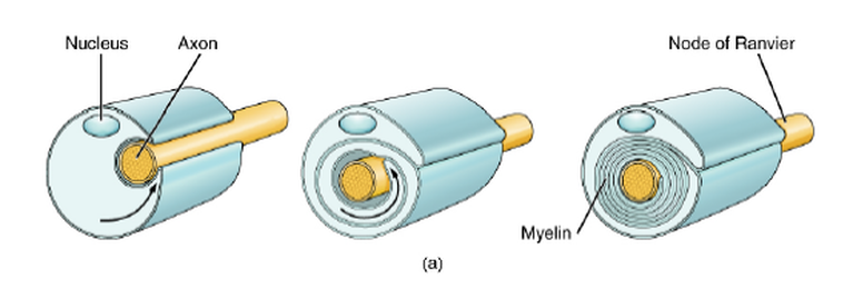
Many axons are wrapped by an insulating substance called myelin, which is actually made from glial cells. Myelin acts as insulation much like the plastic or rubber that is used to insulate electrical wires. A key difference between myelin and the insulation on a wire is that there are gaps in the myelin covering of an axon. Myelin The insulation for axons in the nervous system is provided by glial cells, oligodendrocytes in the CNS, and Schwann cells
in the PNS. Whereas the manner in which either cell is associated with the axon segment, or segments, that it insulates is different, the means of myelinating an axon segment is mostly the same in the two situations. Myelin is a lipid-rich sheath that surrounds the axon and by doing so creates a myelin sheath that facilitates the transmission of electrical signals along
the axon. The lipids are essentially the phospholipids of the glial cell membrane. Myelin, however, is more than just the membrane of the glial cell. It also includes important proteins that are integral to that membrane. Some of the proteins help to hold the layers of the glial cell membrane closely together.
Many axons are myelinated
Many axons are wrapped by an insulating substance called myelin, which is actually made from glial cells. Myelin acts as insulation much like the plastic or rubber that is used to insulate electrical wires. A key difference between myelin and the insulation on a wire is that there are gaps in the myelin covering of an axon. Myelin The insulation for axons in the nervous system is provided by glial cells, oligodendrocytes in the CNS, and Schwann cells
in the PNS. Whereas the manner in which either cell is associated with the axon segment, or segments, that it insulates is different, the means of myelinating an axon segment is mostly the same in the two situations. Myelin is a lipid-rich sheath that surrounds the axon and by doing so creates a myelin sheath that facilitates the transmission of electrical signals along
the axon. The lipids are essentially the phospholipids of the glial cell membrane. Myelin, however, is more than just the membrane of the glial cell. It also includes important proteins that are integral to that membrane. Some of the proteins help to hold the layers of the glial cell membrane closely together.
- Function of myelin
- Protects and electrically insulates axon
- Increases speed of nerve impulse transmission
- Formation of myelin
- The myelin sheath is formed in the PNS by Schwann cells
- The myelin sheath is formed in the CNS by oligodendrocytes.
- Structure of the myelin
- The myelin sheath contains gaps called nodes of Ranvier.
Nodes of Ranvier
Each gap is called a node of Ranvier and is important to the way that electrical signals travel down the axon. The length of the axon between each gap, which is wrapped in myelin, is referred to as an axon segment.
Each gap is called a node of Ranvier and is important to the way that electrical signals travel down the axon. The length of the axon between each gap, which is wrapped in myelin, is referred to as an axon segment.
- periodic gaps in the insulating sheath (myelin) on the axon of certain neurons that serves to facilitate the rapid conduction of nerve impulses.
The Function of Nervous Tissue
By the end of this section, you will be able to:
• Distinguish the major functions of the nervous system: sensation, integration, and response
• List the sequence of events in a simple sensory receptor–motor response pathway
Having looked at the components of nervous tissue, and the basic anatomy of the nervous system, next comes an
understanding of how nervous tissue is capable of communicating within the nervous system.
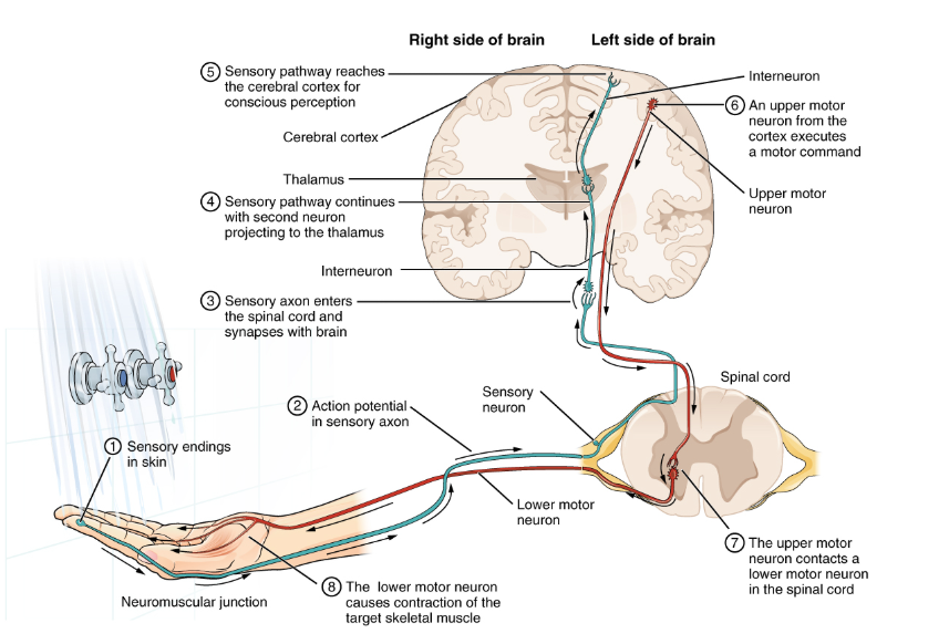
Testing the Water; An Example of Nervous System Function
(1) The sensory neuron has endings in the skin that sense a stimulus such as water
temperature. The strength of the signal that starts here is dependent on the strength of the stimulus.
(2) The graded potential from the sensory endings, if strong enough, will initiate an action potential at the initial segment of the axon (which is immediately adjacent to the sensory endings in the skin).
(3) The axon of the peripheral sensory neuron enters the spinal cord and contacts another neuron in the gray matter. The contact is a synapse where another graded potential is caused by the release of a chemical signal from the axon terminals.
4) An action potential is initiated at the initial segment of this neuron and travels up the sensory pathway to a region of the brain called the thalamus. Another synapse passes the information along to the next neuron.
(5) The sensory pathway ends when the signal reaches the cerebral cortex.
(6) After integration with neurons in other parts of the cerebral cortex, a motor command is sent from the precentral gyrus of the frontal cortex.
(7) The upper motor neuron sends an action potential down to the spinal cord. The target of the upper motor neuron is the dendrites of the lower motor neuron in the gray matter of the spinal cord.
(8) The axon of the lower motor neuron emerges from the spinal cord in a nerve and connects to a muscle through a neuromuscular junction to cause contraction of the target muscle.
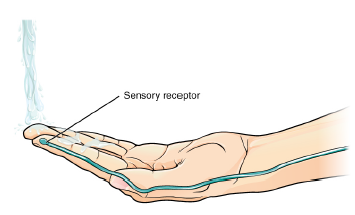
Found in the skin of your fingers or toes is a type of sensory receptor that is sensitive to temperature, called a thermoreceptor. When you place your hand under the shower the cell membrane of the thermoreceptors changes its electrical state (voltage). The amount of change is dependent on the strength of the stimulus (how hot the water is). This is called a graded potential. If the stimulus is strong, the voltage of the cell membrane will change enough to generate an electrical signal that will travel down the axon. The voltage at which such a signal is generated is called the threshold, and the resulting electrical signal is called an action potential. In this example, the action potential travels—a process known as propagation—along the axon from the axon hillock to the axon terminals and into the synaptic end bulbs. When this signal reaches the end bulbs, it causes the release of a signaling molecule called a neurotransmitter.
The Sensory Input Receptors in the skin sense the temperature of the water.
When the molecular signal binds to the receptor, the cell membrane of the target neuron changes its electrical state and a new graded potential begins. If that graded potential is strong enough to reach threshold, the second neuron generates an action potential at its axon hillock. The target of this neuron is another neuron in the thalamus of the brain, the part of the CNS that acts as a relay for sensory information. At another synapse, neurotransmitter is released and binds to its receptor.
The thalamus then sends the sensory information to the cerebral cortex, the outermost layer of gray matter in the brain, where conscious perception of that water temperature begins.
Within the cerebral cortex, information is processed among many neurons, integrating the stimulus of the water temperature with other sensory stimuli, with your emotional state (you just aren't ready to wake up; the bed is calling to you), memories (perhaps of the lab notes you have to study before a quiz). Finally, a plan is developed about what to do, whether that is to turn the temperature up, turn the whole shower off and go back to bed, or step into the shower. To do any of these things, the cerebral cortex has to send a command out to your body to move muscles.
The Motor Response On the basis of the sensory input and the integration in the CNS, a motor response is formulated and executed
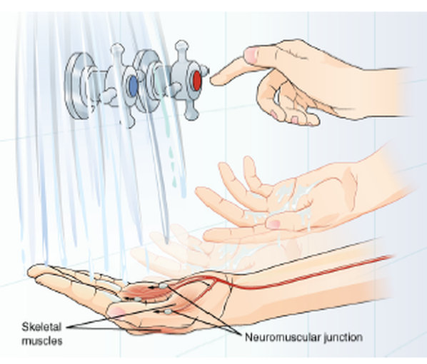
A region of the cortex is specialized for sending signals down to the spinal cord for movement. The upper motor neuron is in this region, called the precentral gyrus of the frontal cortex, which has an axon that extends all the way down the spinal cord. At the level of the spinal cord at which this axon makes a synapse, a graded potential occurs in the cell membrane of a lower motor neuron. This second motor neuron is responsible for causing muscle fibers to contract. The axon terminates on muscle fibers at the neuromuscular junction. Acetylcholine is released at this specialized synapse, which causes the muscle action potential to begin, following a large potential known as an end plate potential. When the lower motor neuron excites the muscle fiber, it contracts. All of this occurs in a fraction of a second, but this story is the basis of how the nervous system functions.
Ion Channels
There are many types of ion channels. The 2 principle types of ion channels that cause changes in the membrane potential are...
- Ligand-gated ion channels
- Voltage-gated ion channels
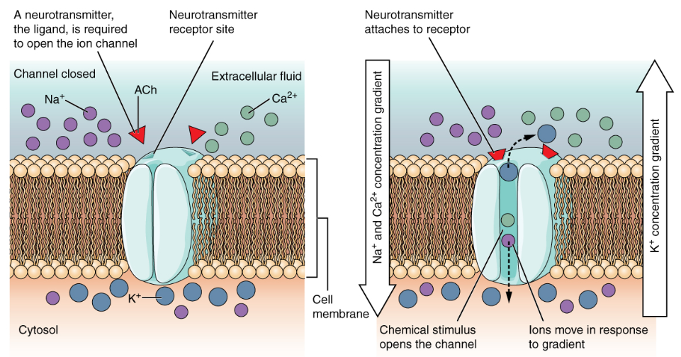
Ligand-gated ion channels
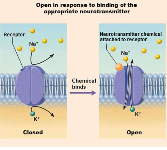
A ligand-gated channel opens because a signaling molecule, a ligand, binds to the extracellular region of the channel.
In the dendrites, at the post-synaptic membrane, specialized ligand-gated sodium ion channels, also known as receptors, open in response to the binding of a neurotransmitter. When a neurotransmitter binds to these receptors, the "gate" opens and sodium flows into the neuron down its electrochemical gradient.
The influx of sodium ions into the post-synaptic neuron leads to a localized depolarization event, also known as a graded membrane potential.
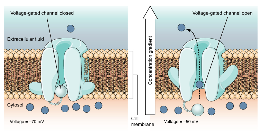
Voltage-gated ion channels
A voltage-gated channel is a channel that responds to changes in the electrical properties of the membrane in which it is embedded. Normally, the inner portion of the membrane is at a negative voltage. When that voltage becomes less negative, the channel begins to allow ions to cross the membrane. Voltage-gated channels open when the transmembrane voltage changes around them.
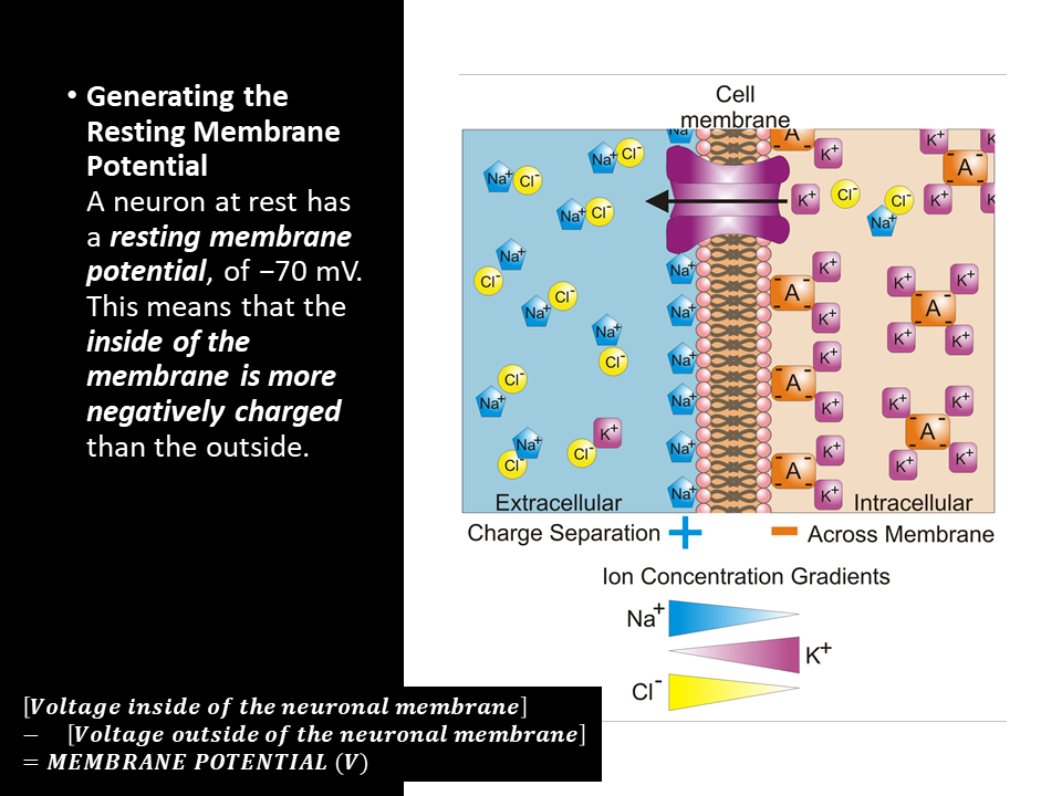
The Membrane Potential
The electrical state of the cell membrane can have several variations. These are all variations in the membrane potential. A potential is a distribution of charge across the cell membrane, measured in millivolts (mV). The standard is to compare the inside of the cell relative to the outside, so the membrane potential is a value representing the charge on the intracellular side of the membrane based on the outside being zero, relatively speaking.
- Depends on the electrochemical gradients AND the membrane permeability
- Ions are charged atoms. Ions carry an electrical charge.
- A sodium ion (Na+) carries a +1 charge
- A potassium ion (K+) carries a +1 charge
- A calcium ion (Ca2+) carries a +2 charge
The Resting Membrane Potential
Resting membrane potential describes the steady state of the cell, which is a dynamic process that is balanced by ion leakage and ion pumping.Without any outside influence, it will not change.A neuron at rest has a resting membrane potential, of −70 mV. This means that the inside of the membrane is more negatively charged than the outside.
Why is there a resting membrane potential??
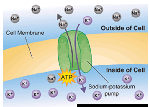
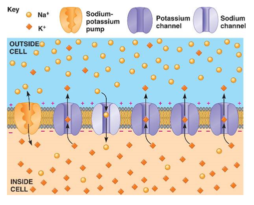
The resting membrane potential is produced by the action of the sodium-potassium pump, which forces 3 Na+ OUT OF the neuron and at the same time it pushes only 2 K+ INTO the neuron!
The action of the sodium-potassium pump requires energy (in the form of ATP) since it pushes these ions against their concentration gradients.
Sodium-Potassium Pump
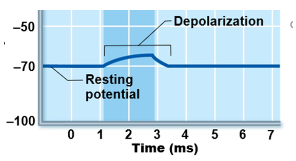
Graded Potentials
Graded Potentials Graded potentials are temporary changes in the membrane voltage, the
characteristics of which depend on the size of the stimulus. Some types of stimuli cause depolarization of the membrane, whereas others cause hyperpolarization. It depends on the specific ion channels that are activated in the cell membrane.
characteristics of which depend on the size of the stimulus. Some types of stimuli cause depolarization of the membrane, whereas others cause hyperpolarization. It depends on the specific ion channels that are activated in the cell membrane.
Ligand-Gated Ion Channels
Graded potentials are changes in membrane potential that vary in size, as opposed to being all-or-none like we observe in action potentials. They arise from the summation of the individual actions of ligand-gated ion channels (receptors). Graded potentials decrease over time and they dissipate as they passively diffuse away from post-synaptic site. Graded potentials occur at the postsynaptic dendrite as a result of presynaptic neuron firing and release of neurotransmitter. The magnitude of a graded potential is determined by the strength of the stimulus, or how long neurotransmitters remain available for binding in the synaptic cleft.
Graded Potentials are short-lived, localized changes in membrane potential
- Magnitude varies with stimulus strength
- Stronger stimulus leads to more voltage changes; farther current flows
- Can lead to a localized depolarization or hyperpolarization
- Triggered by stimulus that opens gated ion channels
- Current flows but dissipates quickly and decays
- Graded potentials are signals only over short distances
Action Potentials (AP)
By the end of this section, you will be able to:
• Describe the components of the membrane that establish the resting membrane potential
• Describe the changes that occur to the membrane that result in the action potential
The functions of the nervous system—sensation, integration, and response—depend on the functions of the neurons underlying these pathways. To understand how neurons are able to communicate, it is necessary to describe the role of an excitable membrane in generating these signals. The basis of this communication is the action potential, which demonstrates how changes in the membrane can constitute a signal. Looking at the way these signals work in more variable circumstances involves a look at graded potentials, which will be covered in the next section.
Action Potentials (AP) are an 'all or none' signal sent from the axon hillock down the axon to the axon terminal.
- Principle way neurons send signals
- Principal means neuronal communication
- Occurs only in muscle cells and axons of neurons
- Do not decay over distance as graded potentials do
Threshold Stimulus, Initiation of the Action Potential at the Axon Hillock
To get an electrical signal started, the membrane potential has to change.
This starts with a channel opening for Na+ in the membrane. Because the concentration of Na+ is higher outside the cell than inside the cell by a factor of 10, ions will rush into the cell that are driven largely by the concentration gradient. Because sodium is a positively charged ion, it will change the relative voltage immediately inside the cell relative to immediately outside. The resting potential is the state of the membrane at a voltage of -70 mV, so the sodium cation entering the cell will cause it to become less negative. This is known as depolarization, meaning the membrane potential moves toward zero.
The concentration gradient for Na+ is so strong that it will continue to enter the cell even after the membrane potential has become zero, so that the voltage immediately around the pore begins to become positive. The electrical gradient also plays a role, as negative proteins below the membrane attract the sodium ion. The membrane potential will reach +30 mV by the time sodium has entered the cell.
As the membrane potential reaches +30 mV, other voltage-gated channels are opening in the membrane. These channels are specific for the potassium ion. A concentration gradient acts on K+, as well. As K+ starts to leave the cell, taking a positive charge with it, the membrane potential begins to move back toward its resting voltage. This is called repolarization,
meaning that the membrane voltage moves back toward the -70 mV value of the resting membrane potential.
Repolarization returns the membrane potential to the -70 mV value that indicates the resting potential, but it actually overshoots that value. Potassium ions reach equilibrium when the membrane voltage is below -70 mV, so a period of hyperpolarization occurs while the K+ channels are open. Those K+ channels are slightly delayed in closing, accounting for
this short overshoot.
The Action Potential is Initiated at the Axon Hillock.
Action potentials occur only when the membrane at the axon hillock is sufficiently depolarized to reach threshold which is about -55 mV. Voltage-gated sodium channels located at the axon hillock open at -55 mV. There and initiate an action potential.
Action potentials occur only when the membrane at the axon hillock is sufficiently depolarized to reach threshold which is about -55 mV. Voltage-gated sodium channels located at the axon hillock open at -55 mV. There and initiate an action potential.
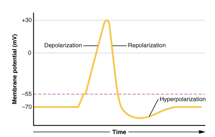
Phases of the Action Potential
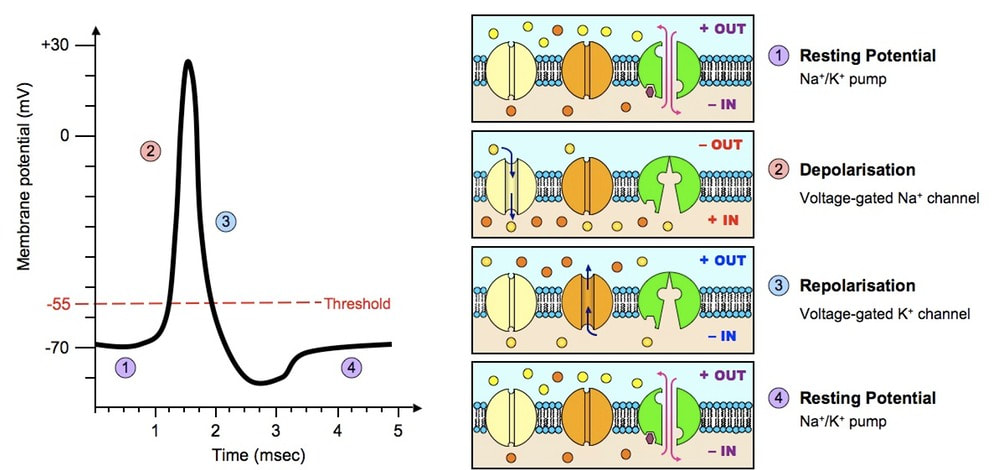
- RESTING PHASE - The membrane potential starts out at −70 mV (resting membrane potential). The resting membrane potential is maintained by the sodium-potassium pump which pumps 3 sodium ions out of the cell, while pumping in 2 positively charged potassium ions.
- DEPOLARIZATION PHASE - When threshold (-55 mV) is reached at the axon hillock the action potential is initiated which results in a massive sodium ion influx through activated voltage-gated sodium ion channels. This causes the membrane potential rapidly rises to a peak potential of +40 mV.
- REPOLARIZATION PHASE - The voltage-gated Na+ channels become INACTIVATED at the peak of the action potential which rapidly repolarizes the membrane back toward resting membrane potential (-70 mV). Repolarization is caused by K+ diffusing out of the cell through voltage-gated potassium channels that do not inactivate after the voltage-gated Na+ channels are INACTIVATED.
- Hyperpolarization is when the membrane potential becomes more negative, beyond the resting membrane potential.
REPOLARIZATION and HYPERPOLARIZATION
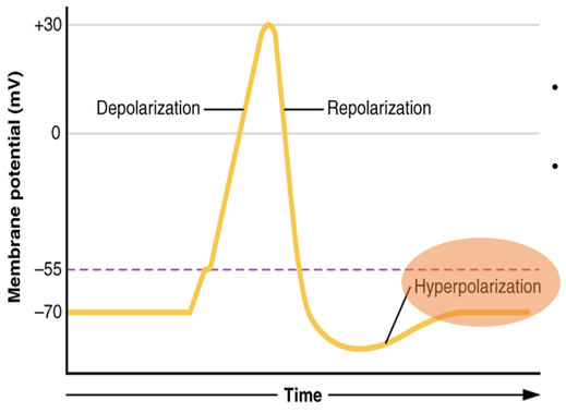
The REPOLARIZATION and HYPERPOLARIZATION phases of the action potential are caused by voltage-gated K+ that do not inactivate.
- K+ has an electrochemical gradient that causes it to leave the neuron when potassium channels are open.
- This loss of positive charge, hyperpolarizes the cell. The potassium channels eventually close, which brings the membrane potential back to its resting level.
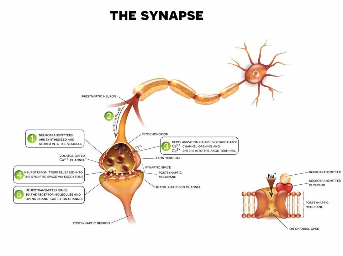
Neurotransmission
The abbreviated version of the steps of neurotransmission are as follows:
- An action potential reaches the axon terminal.
- Synaptic vesicles that are filled with neurotransmitters fuse to the presynaptic membrane which allows the neurotransmitters to move out into the synaptic cleft.
- These neurotransmitters then passively diffuse across the synaptic cleft and then bind to receptors that are located on the postsynaptic membrane (located on the dendrite of the postsynaptic neuron).
- When the neurotransmitter binds to the receptor, ion channels open leading to an influx of positive charge.
- If there is a sufficient amount of stimulation (positive charge) sensed at the axon hillock, an action potential (in the form of positive charge) is generated.
- The action potential travels down the axon toward the axon terminal.
- Repeat!
Action potentials are “all or none.”
Either the membrane reaches the threshold and everything occurs as described above, or the membrane does not reach the threshold and nothing else happens. All action potentials peak at the same voltage (+30 mV), so one action potential is not bigger than another. Stronger stimuli will initiate multiple action potentials more quickly, but the individual signals are not bigger. Thus, for example, you will not feel a greater sensation of pain, or have a stronger muscle contraction, because of the size of the action potential because they are not different sizes.
The depolarization and repolarization of an action potential are dependent on two types of channels (the voltage-gated Na+ channel and the voltage-gated K+ channel).
The voltage-gated Na+ channels
The voltage-gated Na+ channel actually has two gates. One is the activation gate, which opens when the membrane potential crosses -55 mV. The other gate is the inactivation gate, which closes after a specific period of time—on the order of a fraction of a millisecond. When a cell is at rest, the activation gate is closed and the inactivation gate is open. However, when the threshold is reached, the activation gate opens, allowing Na+ to rush into the cell. Timed with the peak of depolarization, the inactivation gate closes. During repolarization, no more sodium can enter the cell. When the membrane potential passes -55 mV again, the activation gate closes. After that, the inactivation gate re-opens, making the channel ready to start the whole process over again.
The voltage-gated K+ channel
The voltage-gated K+ channel has only one gate, which is sensitive to a membrane voltage of -50 mV. However, it does not open as quickly as the voltage-gated Na+ channel does. It might take a fraction of a millisecond for the channel to open once that voltage has been reached. The timing of this coincides exactly with when the Na+ flow peaks, so voltage-gated K+ channels open just as the voltage-gated Na+ channels are being inactivated. As the membrane potential repolarizes and the voltage passes -50 mV again, the channel closes—again, with a little delay. Potassium continues to leave the cell for a short while and the membrane potential becomes more negative, resulting in the hyperpolarizing overshoot. Then the channel closes again and the membrane can return to the resting potential because of the ongoing activity of the non-gated channels and the Na+/K+ pump.
INTERACTIVE LINKS
- 1. In 2003, the Nobel Prize in Physiology or Medicine wasawarded to Paul C. Lauterbur and Sir Peter Mansfield fordiscoveries related to magnetic resonance imaging (MRI). This is a tool to see the structures of the body (not just the nervous system) that depends on magnetic fields associated with certain atomic nuclei. The utility of this technique in the nervous system is that fat tissue and water appear as different shades between black and white. Because white matter is fatty (from myelin) and gray matter is not, they can be easily distinguished in MRI images. Visit the Nobel Prize website (http://openstaxcollege.org/l/nobel_2) to play an interactive game that demonstrates the use of this technology and compares it with other types of imaging technologies. Also, the results from an MRI session are compared with images obtained from x-ray or computed tomography. How do the imaging techniques shown in this game indicate the separation of white and gray matter compared with the freshly dissected tissue shown earlier?
- Visit this site (http://openstaxcollege.org/l/troublewstairs) to read about a woman that notices that herdaughter is having trouble walking up the stairs. This leadsto the discovery of a hereditary condition that affects thebrain and spinal cord. The lectromyography and MRI testsindicated deficiencies in the spinal cord and cerebellum,both of which are responsible for controlling coordinated movements. To what functional division of the nervous system would these structures belong?