An Anatomical Walk Through the Human Skull by ScientistCindy
Virtual Flashcards - Click on the Slide Show to Control! You May Also Download the PDF Version. Beware, there are 149 slides. ENJOY!
| skull_anatomy_flash_cards_2017_09_23.pdf | |
| File Size: | 6365 kb |
| File Type: | |
The Axial Skeleton
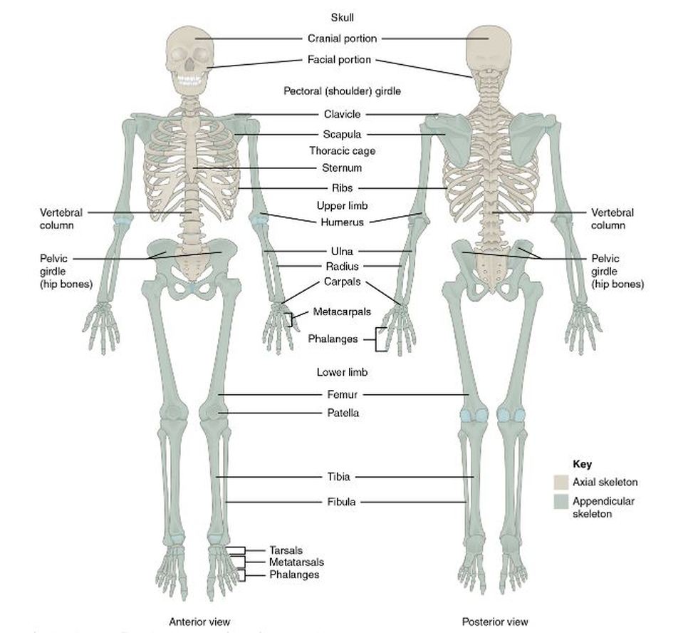
The human skeleton is divided into 2 categories
- the axial skeleton and
- the appendicular skeleton
The axial skeleton forms the long axis of your body. The axial skeleton is divided into 3 categories; 1) the skull, 2) the vertebral column, and 3) the thoracic cage. The function of the axial skeleton is to support the head, neck, and trunk, and to protect the brain, spinal cord, and the thoracic organs.
The Skull
The skull is extremely complex and has unique features that are not found elsewhere in the skeleton. Most skull bones are flat bones that are joined together as a suture. A suture is an immobile joint.
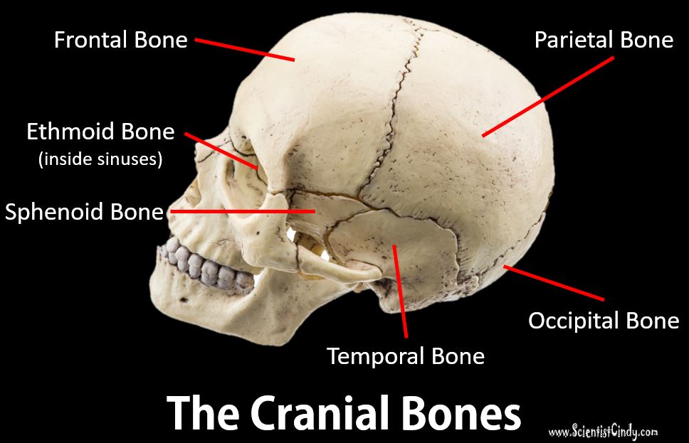
The skull bones fall into 1 of 2 categories;
- The cranial bones
- The facial bones.
The Cranial Bones
Your cranial bones have the important job of protecting your very valuable brain! In addition to this, the cranial bones also have "attachment sites" for muscles that allow for the head and neck to move.
Structures (Bone Markings) of the Cranial Bones:
CRANIAL SUTURES
|
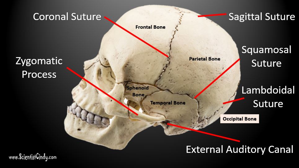
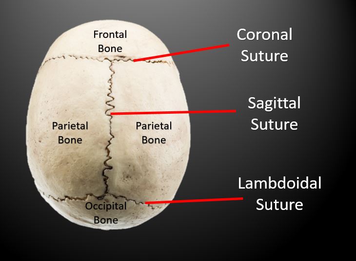
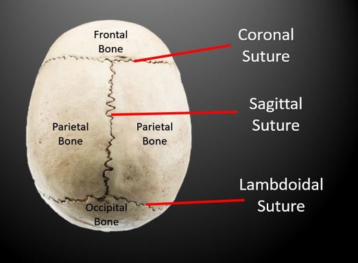
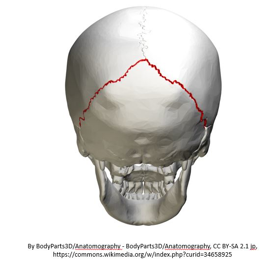
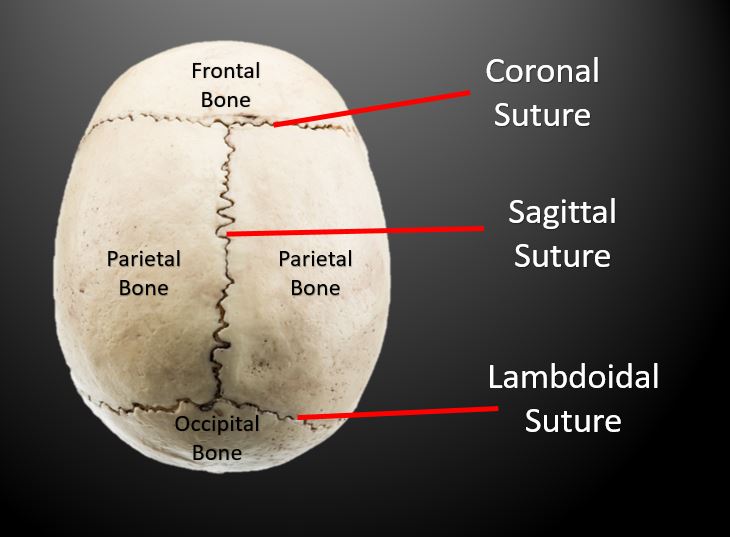
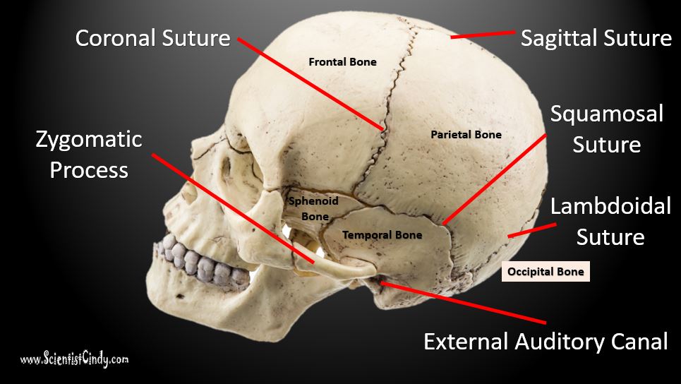
The longest sutures—the coronal, sagittal, squamous, and lambdoid sutures—connect the cranial bones. Most other skull sutures connect facial bones and are named according to the specific bones they connect. The largest cranial bones are the parietal bones. The parietal bones join with the other cranial bones, making the largest sutures of the cranium.
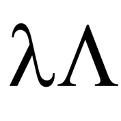
The sagittal suture joins the two parietal bones together. The coronal suture joins the parietal bone with the frontal bone. The coronal suture is on the coronal anatomic plane, hence the name! The squamosal (or squamous) suture joins the parietal bone with the each of the temporal bones on either side of the head, laterally and inferior to the parietal bone. The lambdoid suture joins the parietal bones to the occipital bone, which lies posterior. The shape of this suture is named after the Greek letter "lambda" because (supposedly) it resembles the shape. |
|
THE FRONTAL BONE HAS 1 STRUCTURE YOU SHOULD KNOW
1) THE FRONTAL SINUSES
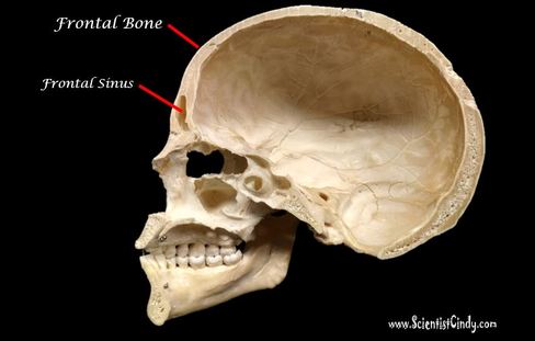
This is considered a MEDIAL VIEW or SAGITTAL VIEW of the skull. The frontal bone contains a cavity called the frontal sinus.
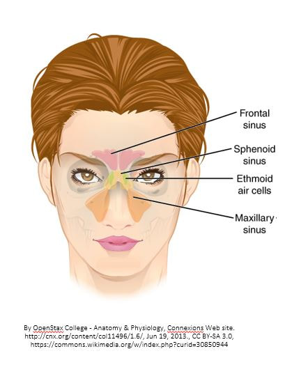
The frontal sinus is located behind the brow ridges. These sinuses (like all sinuses) air-filled, mucous- lined spaces within the bones of the face and skull.
The frontal sinus is located behind the brow ridges. These sinuses (like all sinuses) air-filled, mucous- lined spaces within the bones of the face and skull.
THE PARIETAL BONE HAS 2 STRUCTURES YOU SHOULD KNOW
1) THE CORONAL SUTURE
2) THE SAGITTAL SUTURE
THE TEMPORAL BONE HAS 5 STRUCTURES YOU SHOULD KNOW
1) THE SQUAMOSAL SUTURE
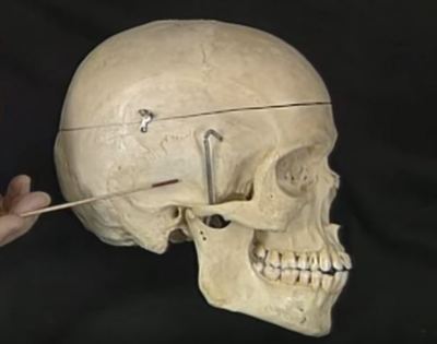
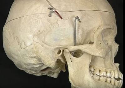
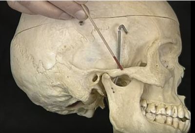
2) THE ZYGOMATIC PROCESS OF THE TEMPORAL BONE
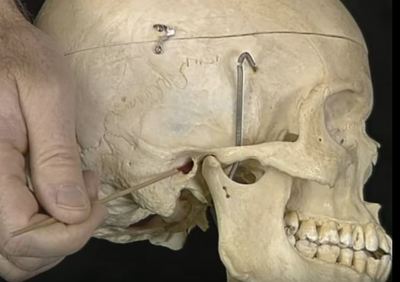
3) THE EXTERNAL AUDITORY MEATUS
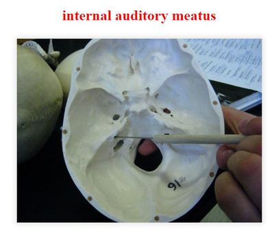
4) THE INTERNAL AUDITORY MEATUS
5) THE MANDIBULAR FOSSA
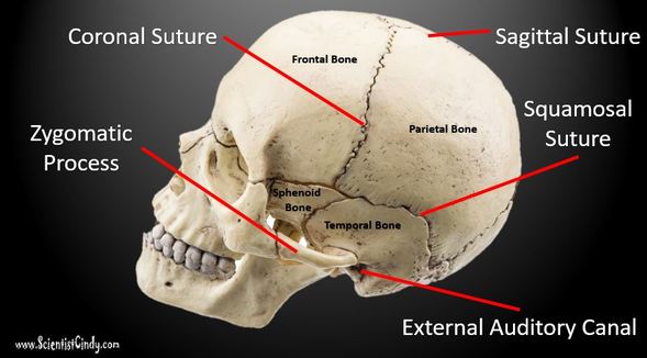
- SQUAMOSAL SUTURE - The temporal bone is associated with several features. The temporal bone is connected to the parietal bone (which lies superior to the temporal bone) by the squamosal suture. A suture is an immobile joint.
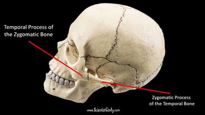
- ZYGOMATIC PROCESS - The zygomatic process is a "bar-like" bone of your cheek bone. It connects the temporal bone to the zygomatic bone of the face.
- EXTERNAL AUDITORY MEATUS - this is the outer ear canal!
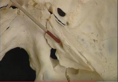
- INTERNAL AUDITORY MEATUS - this is the inner ear canal!.
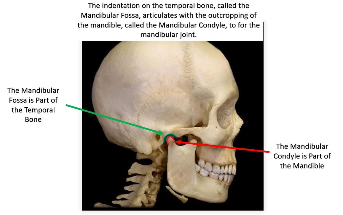
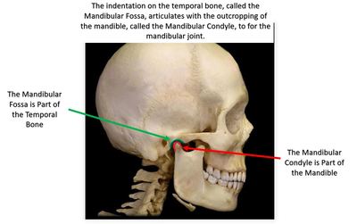
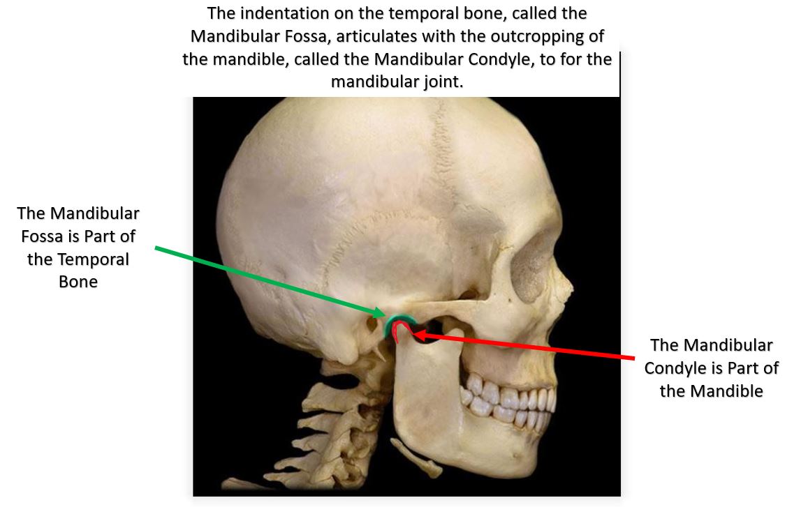
- MANDIBULAR FOSSA - The mandibular fossa is a depression located on the temporal bone that connects (articulates) with the mandible (jaw bone).
Gallery of Temporal Bone Structures
THE SPHENOID BONE HAS 2 STRUCTURES YOU NEED TO KNOW;
1) THE SELLA TURCICA
2) THE OPTIC FORAMEN
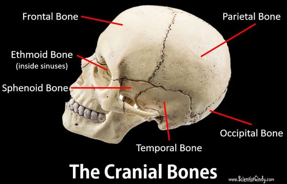
BUT... Before we talk about the structures of the sphenoid bone, let's look at the sphenoid bone as a whole.
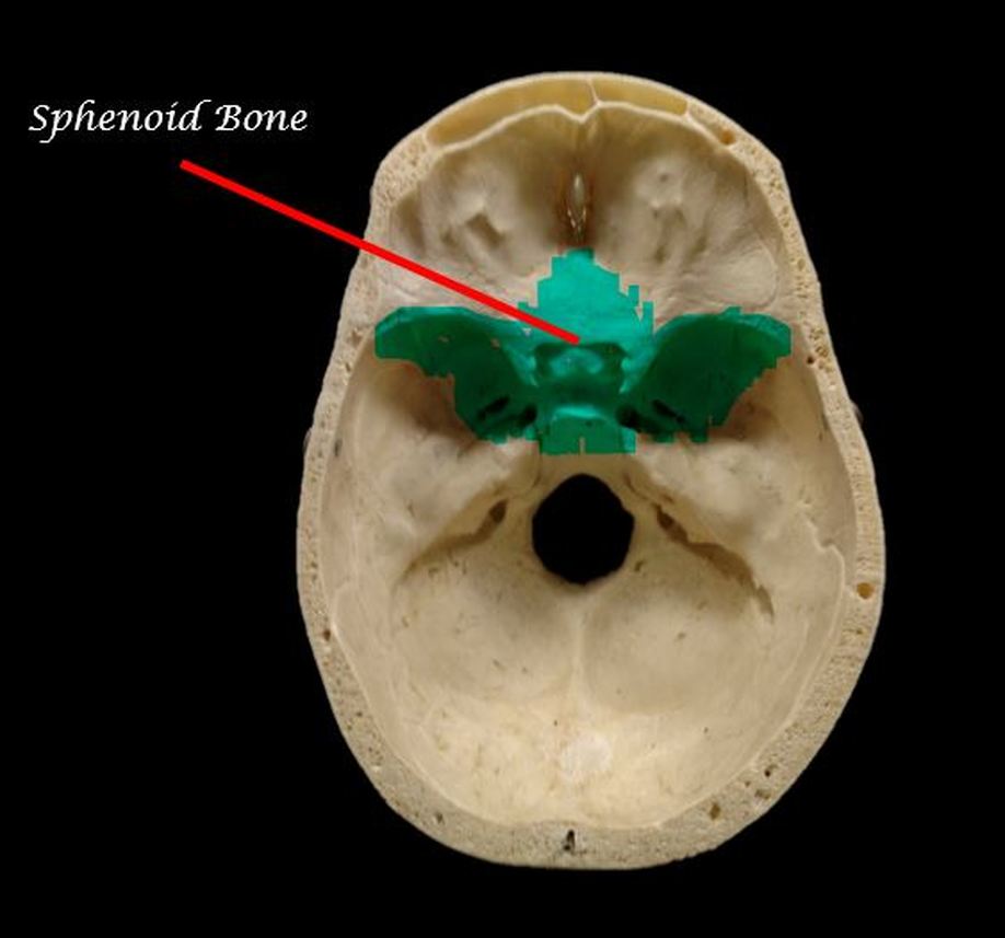
The sphenoid bone is visible from the outside of the skull, but it's most interesting structures are only visible while looking inside the cranial cavity.
The sphenoid bone is visible from the outside of the skull, but it's most interesting structures are only visible while looking inside the cranial cavity.
|
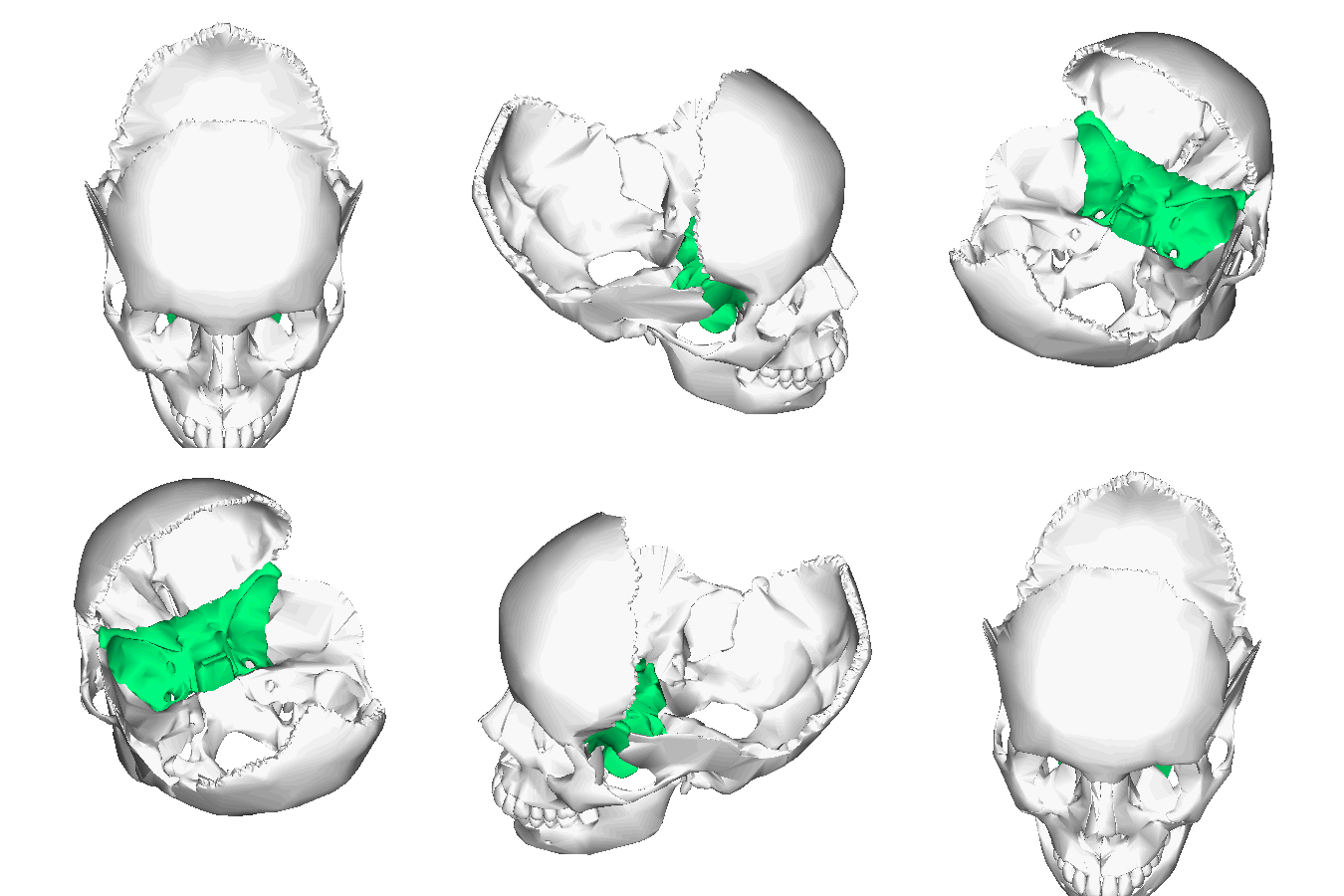
The sphenoid bone has its best view from inside the cranial cavity. At this view it looks like a butterfly! The cranial base of the skull is supported by sphenoid bone, which is looks like a butterfly with out-stretched wings.
|
Animation of the sphenoid bone was provided courtesy of Anatomography - en: Anatomography (setting page of this image), CC BY-SA 2.1 jp, https://commons.wikimedia.org/w/index.php?curid=23256846
|
1) THE SELLA TURCICA
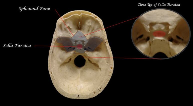
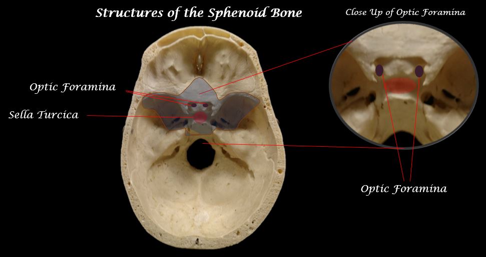
- SELLA TURCICA - Sella turcica is a tiny chamber within the sphenoid bone that surrounds the pituitary gland.. JUST FYI - The pituitary gland is small, but mighty! It is an endocrine gland about the size of a pea which is protected by the small bony cavity of the sella turcica. The pituitary functions to regulate levels of stress, growth, reproductive cycles, blood pressure, sex hormones, temperature regulation, pain relief and metabolism.
Interior of Skull - View of the Sella Turcica Structure of the Sphenoid Bone
2) THE OPTIC FORAMEN (OPTIC CANAL)
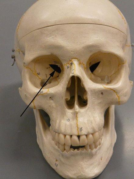
- OPTIC FORAMEN (foramina is the plural form) - The optic foramen (or the optic canal) is a passageway inside the sphenoid bone that allows the optic nerve as well as arteries and veins. to travel between the optic cavity, through the sphenoid bone, and into the cranial cavity.
THE ETHMOID BONE HAS 5 STRUCTURES YOU SHOULD KNOW.
1) THE CRISTA GALLI
2) THE CRIBRIFORM PLATE
3) THE PERPENDICULAR PLATE
4) THE NASAL CONCHA (CONCHAE)
5) THE ETHMOID SINUSES
|
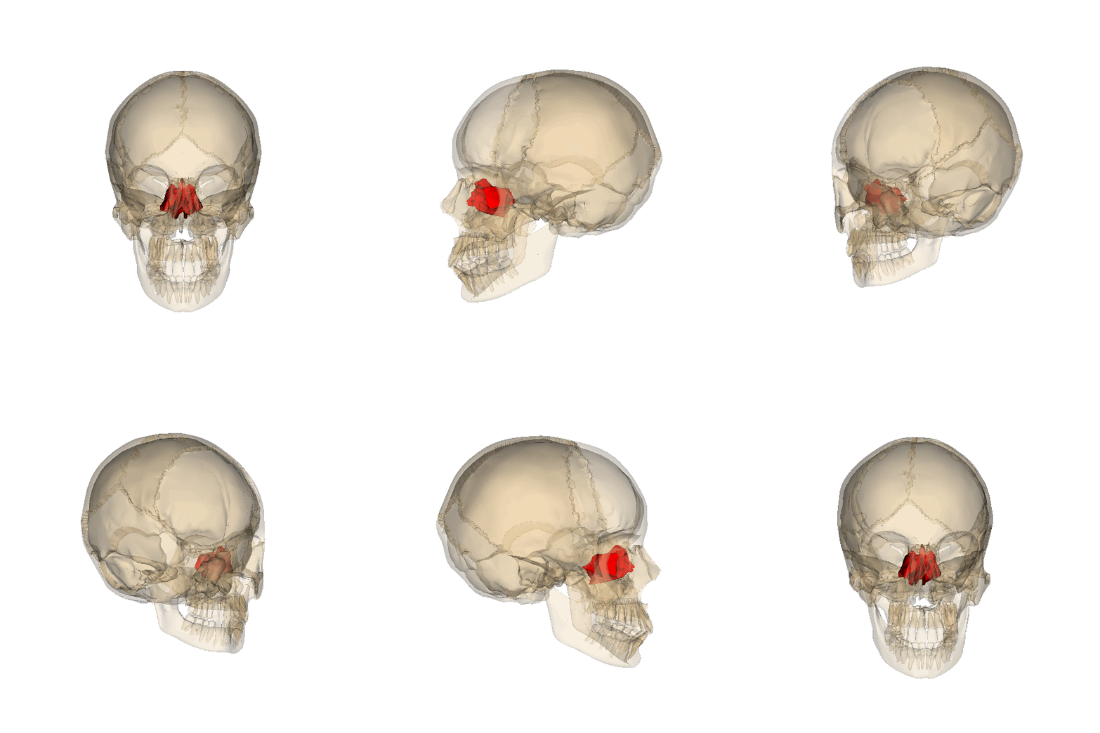
The ethmoid bone from Greek word for "sieve". This bone separates the nasal cavity from the brain. The ethmoid bone traverses the anterior part of the skull and much of its structure is obscured. We can view some of the structures of the ethmoid bone in the inferior, anterior, interior and medial views of the skull.
|
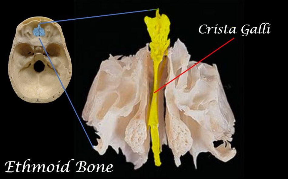
1) THE CRISTA GALLI
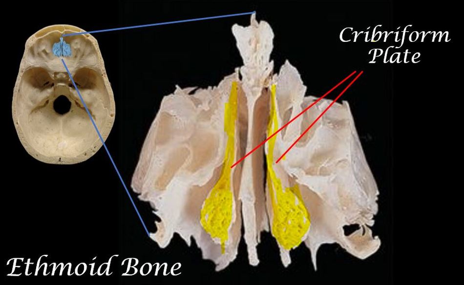
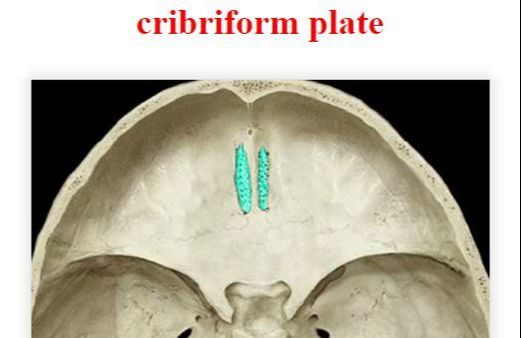
2) THE CRIBRIFORM PLATE
|
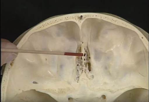
The cribriform plate is a sieve-like (spaghetti strainer) structure that is part of the ethmoid bone. The cribiform plate functions to supports the olfactory bulb (sense of smell). Projecting upward from the midline of this plate is a thick, smooth, triangular process, the crista galli, so called from its resemblance to a rooster's comb. |
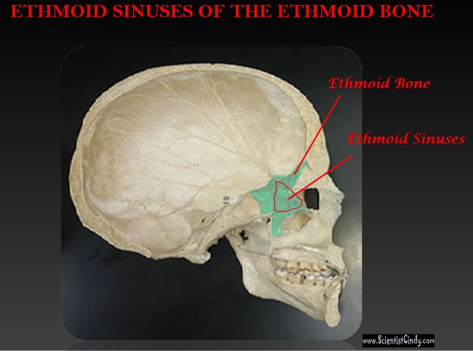
4) THE ETHMOID SINUSES
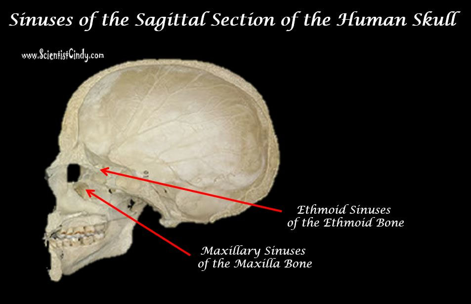
The ethmoid sinuses are only able to be viewed medially with the sagittal section of the skull, such as shown here.
Your ethmoid sinuses are located near the bridge of your nose. Sinuses help to filter, clean, and humidify inspired air. They also keep your head from becoming too heavy. Ultimately, mucus that goes through the sinuses will drain to the nose.
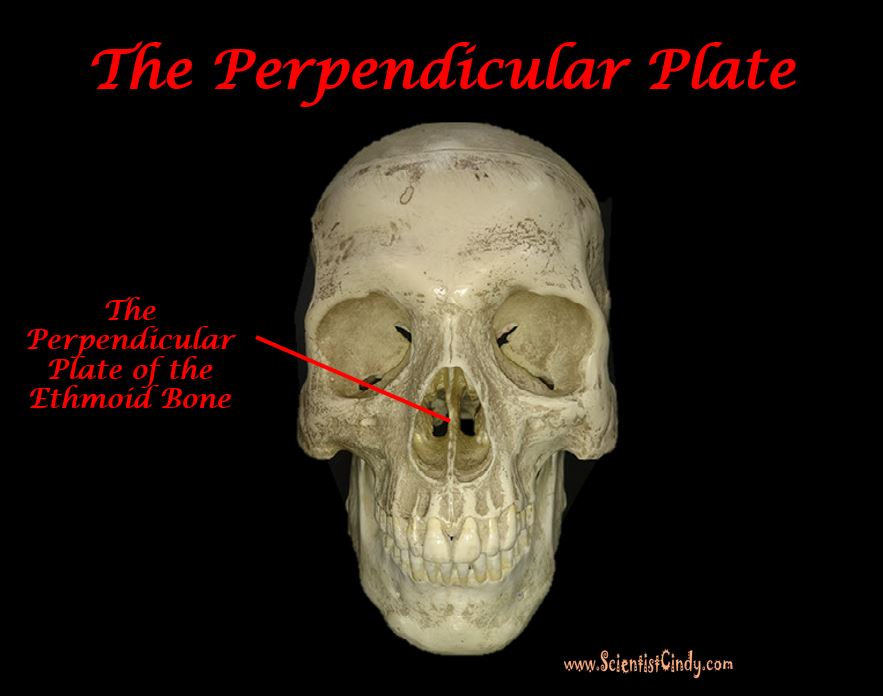
3) THE PERPENDICULAR PLATE
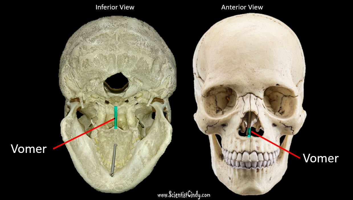
The perpendicular plate of the ethmoid bone runs vertically as a thin, flat, polygonal-shaped bone. The function of the perpendicular plate forms the upper portion of the nasal septum. The vomer forms the inferior part of the nasal septum, and the perpendicular plate forms the superior portion of the septum .
The top portion of the plate contains several grooves and canals which act to hold the olfactory nerves (nerves for the sense of smell).
The top portion of the plate contains several grooves and canals which act to hold the olfactory nerves (nerves for the sense of smell).
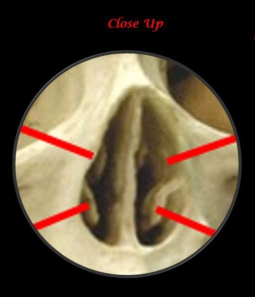
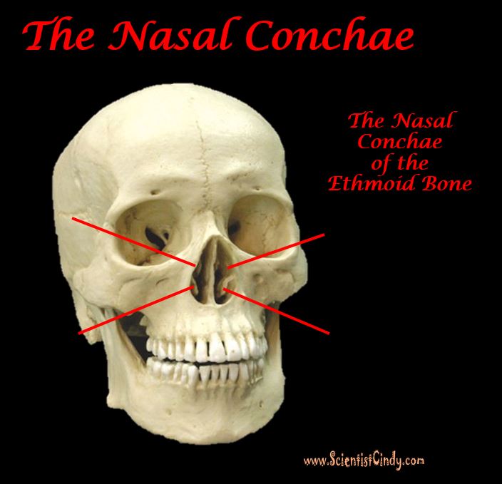
5) THE NASAL CONCHA
The Nasal Concha (Turbinate) is a a labyrinth of several thin body elements that appear "scroll-like". The nasal concha forms the upper chambers of your nasal cavities, thereby increasing the surface area of the nasal cavities. This increase in surface area allows for the rapid warming and humidification of air as it passes from the nasal cavities to the lungs.
THE OCCIPITAL BONE HAS 3 STRUCTURES THAT YOU NEED TO KNOW!
1) THE LAMBOIDAL SUTURE
2) FORAMEN MAGNUM
3) THE OCCIPITAL CONDYLE
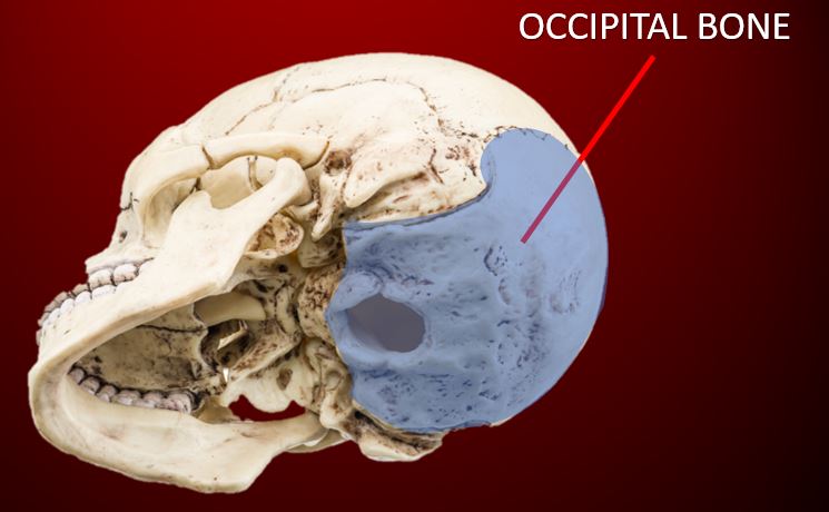
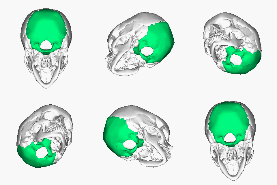
BUT... before we begin talking about the structures of the occipital bone, take a moment to remember the structure of the occipital bone. The occipital lobe makes up the inferior and posterior regions of the cranial cavity.
Occipital Bone - Superior View
By Anatomography - en:Anatomography (setting page of this image), CC BY-SA 2.1 jp,
ttps://commons.wikimedia.org/w/index.php?curid=23254580
ttps://commons.wikimedia.org/w/index.php?curid=23254580
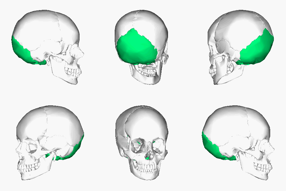
1) LAMBDOIDAL SUTURE
The lambdoidal suture is the immobile joint (suture) that connects the occipital bone (which is located at the posterior portion of the skull) to the parietal bones that lie above it (superior and anterior).
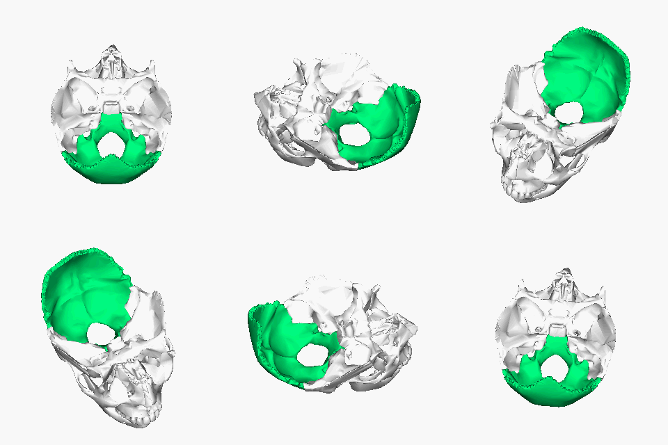
2) FORAMEN MAGNUM
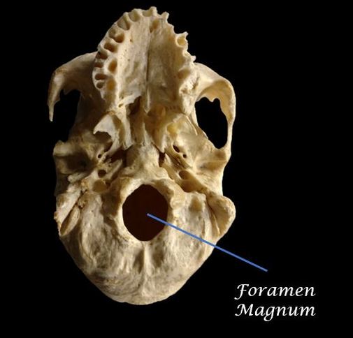
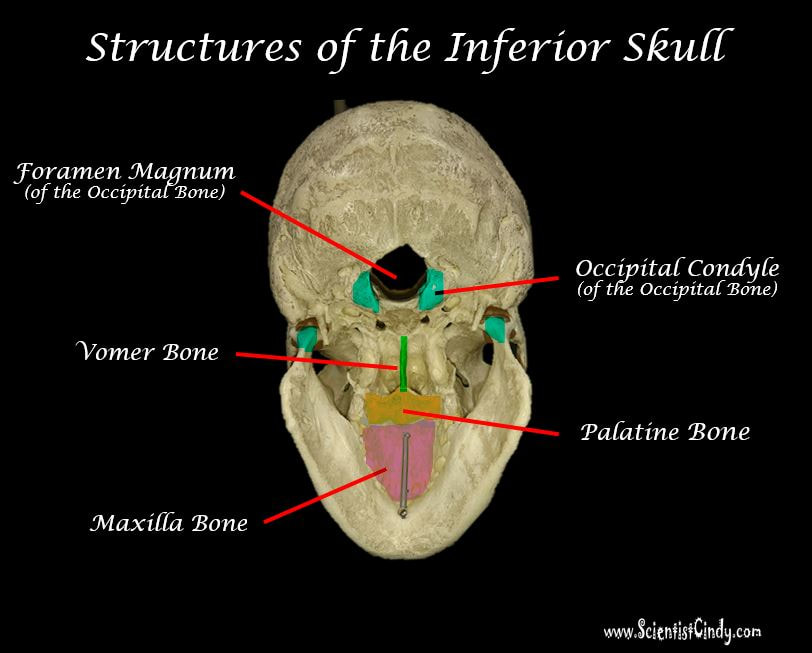
The foramen magnum literally translates as "the Great Hole" from its Latin roots. The foramen magnum lives up to its name as the large oval hole (foramen) at the base of the occipital bone.
You may recall that the dorsal body cavity contains the brain and the spinal cord, and that there is no physical separation between the cranial cavity of the dorsal cavity and the spinal cavity of the dorsal cavity. The spinal cord exits the cranial cavity through the foramen magnum, to travel down the length of the spinal or vertebral cavity.
You may recall that the dorsal body cavity contains the brain and the spinal cord, and that there is no physical separation between the cranial cavity of the dorsal cavity and the spinal cavity of the dorsal cavity. The spinal cord exits the cranial cavity through the foramen magnum, to travel down the length of the spinal or vertebral cavity.
3) THE OCCIPITAL CONDYLES
|
|
The occipital condyles are found at either side of the foramen magnum. The term "condyle" refers to a round protrusion that forms part of a joint. They act as a surface of articulation for the atlas which is the C1 vertebra. The condyles are kidney-shaped. You may also notice that the anterior aspect of the occipital condyles are closer to one another than the posterior aspect. |
Specialized Structures of the Fetal Skull

Fontanelle Definition = a membrane-covered space at the junction of a main suture line.
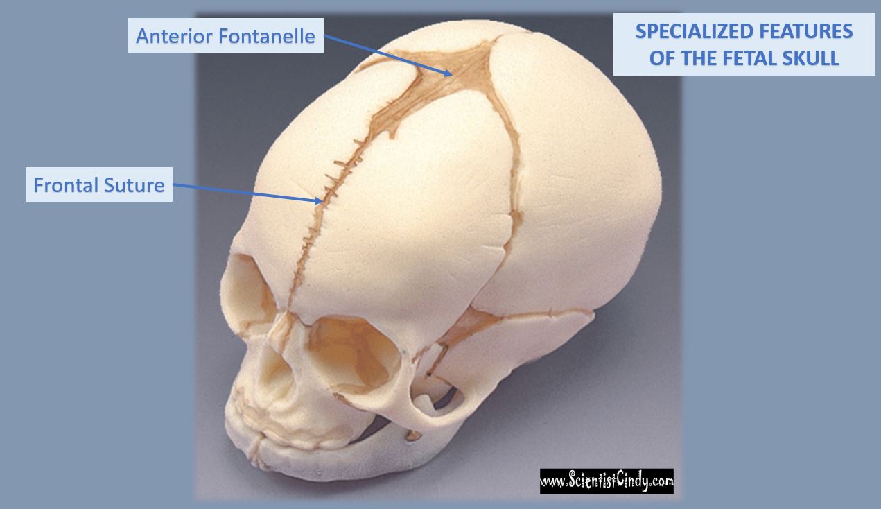
The fetal skull contains cranial bones that have not yet joined together. The spaces that lie between the large cranial bones (the frontal bone, the parietal bone, the temporal bone and the occipital bone) are the membrane-bound fontanelle. This is why new borns have a "soft spot" on their head.
The fetal skull contains cranial bones that have not yet joined together. The spaces that lie between the large cranial bones (the frontal bone, the parietal bone, the temporal bone and the occipital bone) are the membrane-bound fontanelle. This is why new borns have a "soft spot" on their head.
- The anterior fontanelle is the largest of the fontanelles. It forms a "diamond-shape" structure and can take between 12 and 18 months to form the coronal suture that joins the frontal bone to the parietal bones. The anterior fontanelle closes at about 12-18 months and is larger than the other.
- The posterior and sphenoidal fontanelle appear more "triangular-shaped" and close within a couple of months. The posterior fontanelle will close to form the lambdoidal suture that joins the occipital bone with the parietal bones.
- The sphenoidal fontanelles (one lying laterally at each side of the cranium) will close to form the squamosal suture. The squamosal suture is bordered anteriorly by the frontal bone, superiorly by the parietal bone, posteriorly by the squamous portion of the temporal bone, and inferiorly by the greater wing of the sphenoid bone that joins the temporal lobes with the sphenoid.
THE FRONTAL SUTURE - Besides the fontanelles, the fetal skull contains a frontal suture that runs down the midline of the frontal bone. In the adult skull, we see the frontal bone as one bone, completely absent of sutures. The frontal suture completely fuses between 3-months and 9-months of age.
The Facial Bones
|
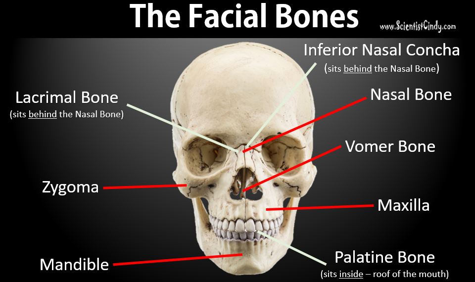
The facial bones include the following bones:
|
The facial bones perform the following functions:
|
The Mandible Has 1 Structure You Need to Know;
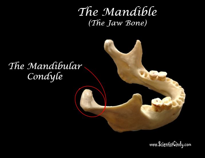
1) The Mandibular Condyle
In addition to the facial bones, you should be able to locate and identify the mandibular condyle.
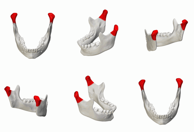
The mandibular condyle articulates with the mandibular fossa, to create the joint that allows your jaw bone to move.
The Maxilla Bone
THE MAXILLA HAS 1 STRUCTURE YOU NEED TO KNOW
1) THE MAXILLARY SINUSES
1) THE MAXILLARY SINUSES
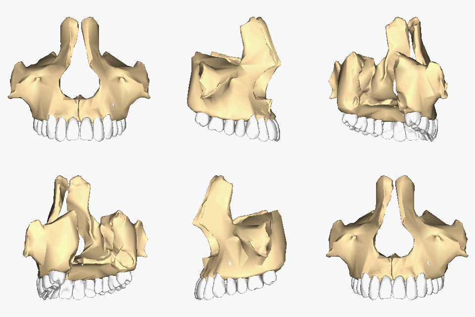
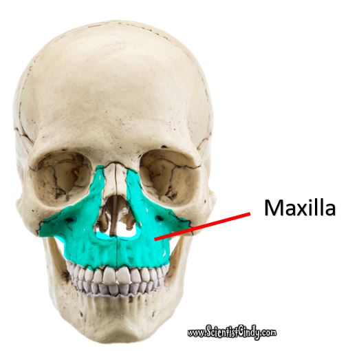
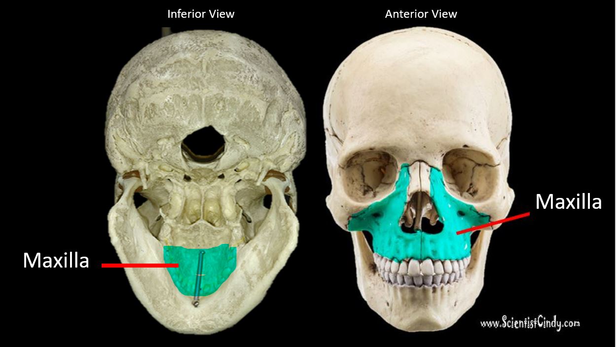
The maxilla is able to be viewed in the skull anteriorly, inferiorly and medially in the sagittal section of the human skull.
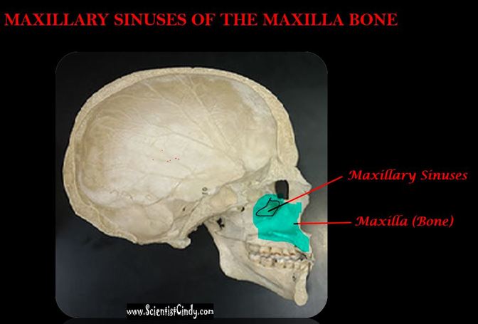
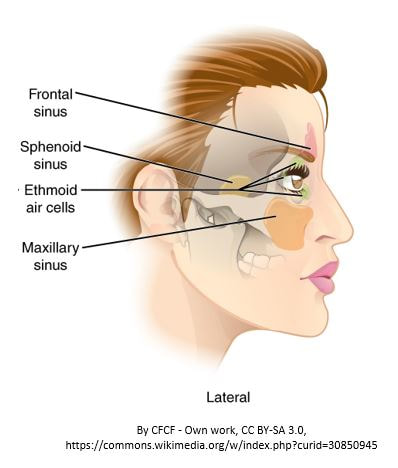
There is only one structure that you should know that is associated with the maxilla. This structure is the maxillary sinuses. The maxillary sinuses lie laterally from the midline and will only be in sagittal sections that are a little bit away from midsagittal.
The maxillary sinuses are observed as a hollow chamber existing within the maxilla (upper jaw bone).
The maxillary sinuses are observed as a hollow chamber existing within the maxilla (upper jaw bone).
The Palatine Bone
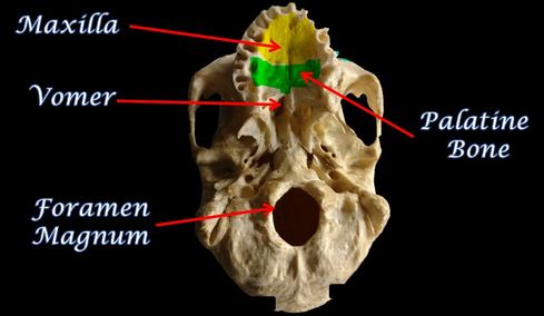
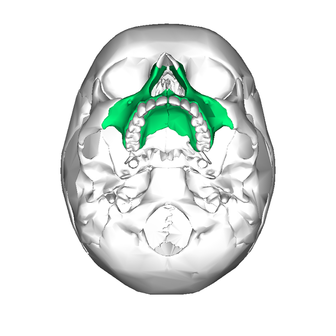
The roof of your mouth is formed by the "hard palate". The hard palate is made up of the maxilla (anterior) and the palatine bones (posterior).
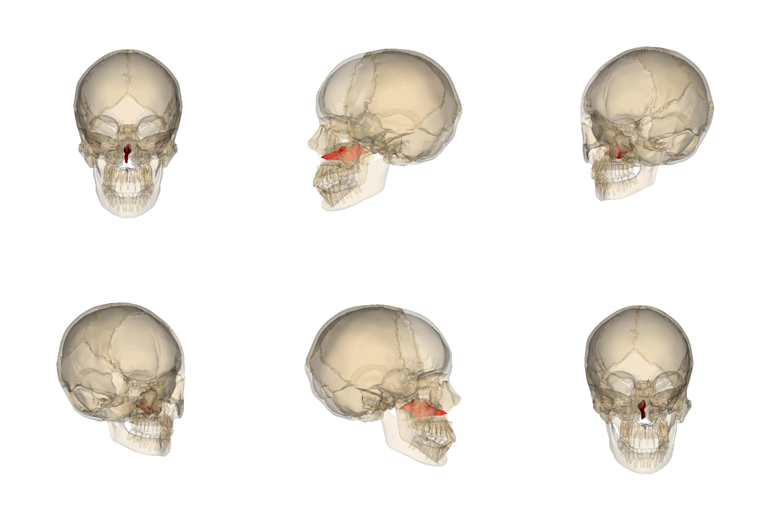
The palatine bones are two irregular bones of the facial skeleton. The palatine bones are located at the posterior portion of the nasal cavity, between the maxilla and part of the sphenoid bone. The human palatine articulates with six bones: the sphenoid, ethmoid, maxilla, inferior nasal concha, vomer and the other palatine bony.
The palatine bones are two irregular bones of the facial skeleton. The palatine bones are located at the posterior portion of the nasal cavity, between the maxilla and part of the sphenoid bone. The human palatine articulates with six bones: the sphenoid, ethmoid, maxilla, inferior nasal concha, vomer and the other palatine bony.
The Zygomatic Bone Has 1 Structure to Know;
The Temporal Process of the Zygomatic Bone
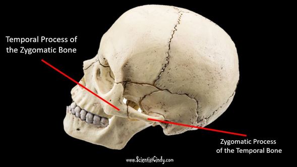
The temporal process of the zygomatic bone is the projection from the zygomatic bone that travels anteriorly to join with the zygomatic process of the temporal bone to form the zygomatic arch. The zygomatic arch is known as the cheek bones! The term, "zygomatic" translates as "bar", which is pretty accurate to their appearance.
The Nasal Bone
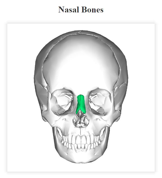
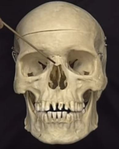
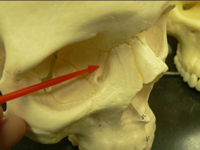
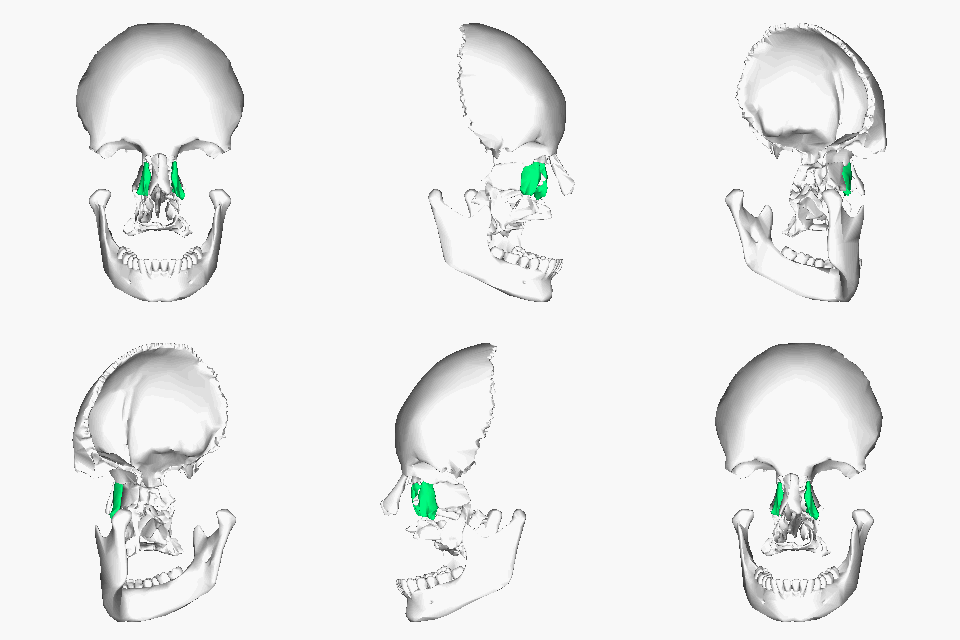
The nasal bones are two small oblong bones that lie side by side to form the "bridge" of your nose.
Vomer Bone
|
|


