|
|
|
Membrane Transport
When we discuss membrane transport, we are referring to the specific properties of the cell membrane and how different particles can move across that membrane. Thus, understanding membrane transport, requires an understanding of the cell membrane itself. So, this is where we will begin; at the cell membrane! |
The Cell Membrane (Plasma Membrane)
- The Main Function of the Cell Membrane is PROTECTION -
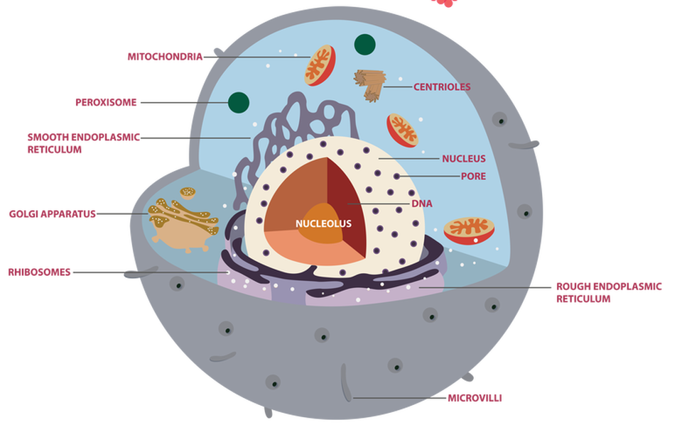
The cell membrane is also called the plasma membrane. You can think of the membrane as the "skin" of the cell. Anything outside of the cell is considered "extracellular" and the contents inside the cell are considered "intracellular". The cell membrane protects the cell by creating a barrier between what is inside the cell and what is outside the cell.
In addition to this, the cell membrane does something our skin can’t do... It regulates what comes into the cell and what goes out of the cell. For this reason, we consider the cell membrane to be “SELECTIVELY PERMEABLE” which means that it allows some substances to enter or exit the cell, but not others. This is a very important function.
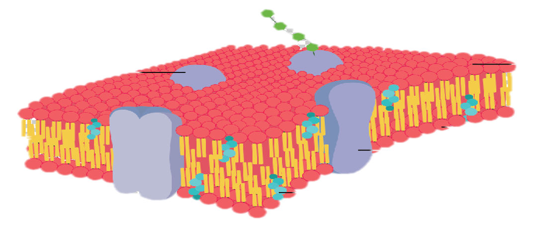
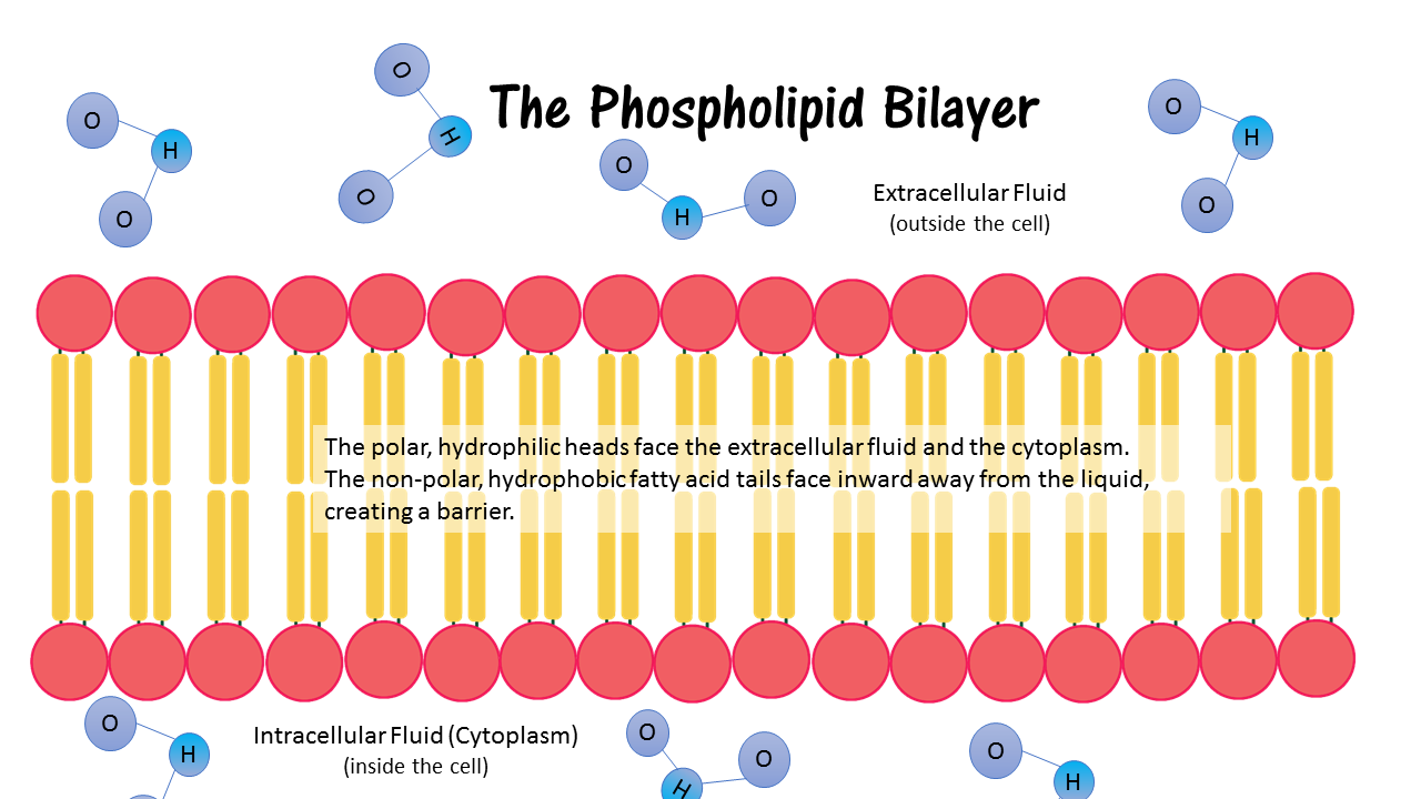
The Cell Membrane is selectively permeable due to its structure. The cell membrane is made up of a phospholipid bilayer.
The Cell Membrane is selectively permeable due to its structure. The cell membrane is made up of a phospholipid bilayer.
Phospholipid
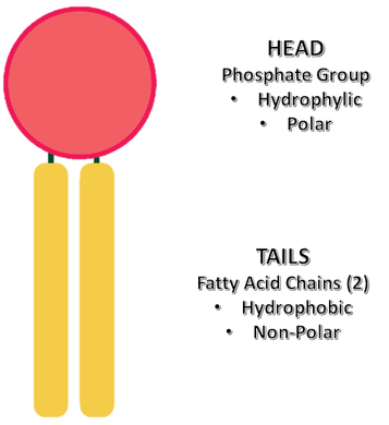
The phospholipid bilayer of the cell membrane has a unique structure. It is made up of an inner layer and an outer layer of phospholipids that are oriented with their 'tails' facing each other.
Phospholipids are considered amphiphilic, because they contain a polar, hydrophillic head that consists of a phosphate group and two nonpolar, hydrophobic fatty acid chains as 'tails'.
When the phospholipids form the cell membrane, the polar, hydrophillic (water-loving) heads are oriented towards the liquid outside the cells (extracellular fluid) and the liquid inside the cell (extracellular fluid). The tails of the phospholipids are oriented towards each other, away from the liquid, since they are made up of hydrophobic (water-fearing) fatty acid chains. This formation creates a barrier between the extracellular matrix and the intracellular fluid (cytology).
Click Below to View a Video on the Mighty Membrane
Types of Membrane Transport
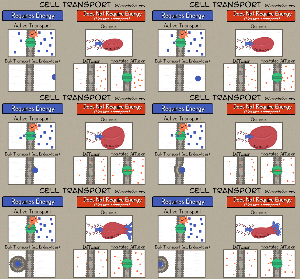
ANIMATED GIF Courtesy of AmoebaSisters see more www.amoebasisters.com/gifs.html
There are a lot of ways that substances are permitted to pass through the cell membrane. There are 2 main categories of membrane transport; active and passive.
ACTIVE TRANSPORT mechanisms require energy.
PASSIVE TRANSPORT mechanisms do not require energy.
There are a lot of ways that substances are permitted to pass through the cell membrane. There are 2 main categories of membrane transport; active and passive.
ACTIVE TRANSPORT mechanisms require energy.
PASSIVE TRANSPORT mechanisms do not require energy.
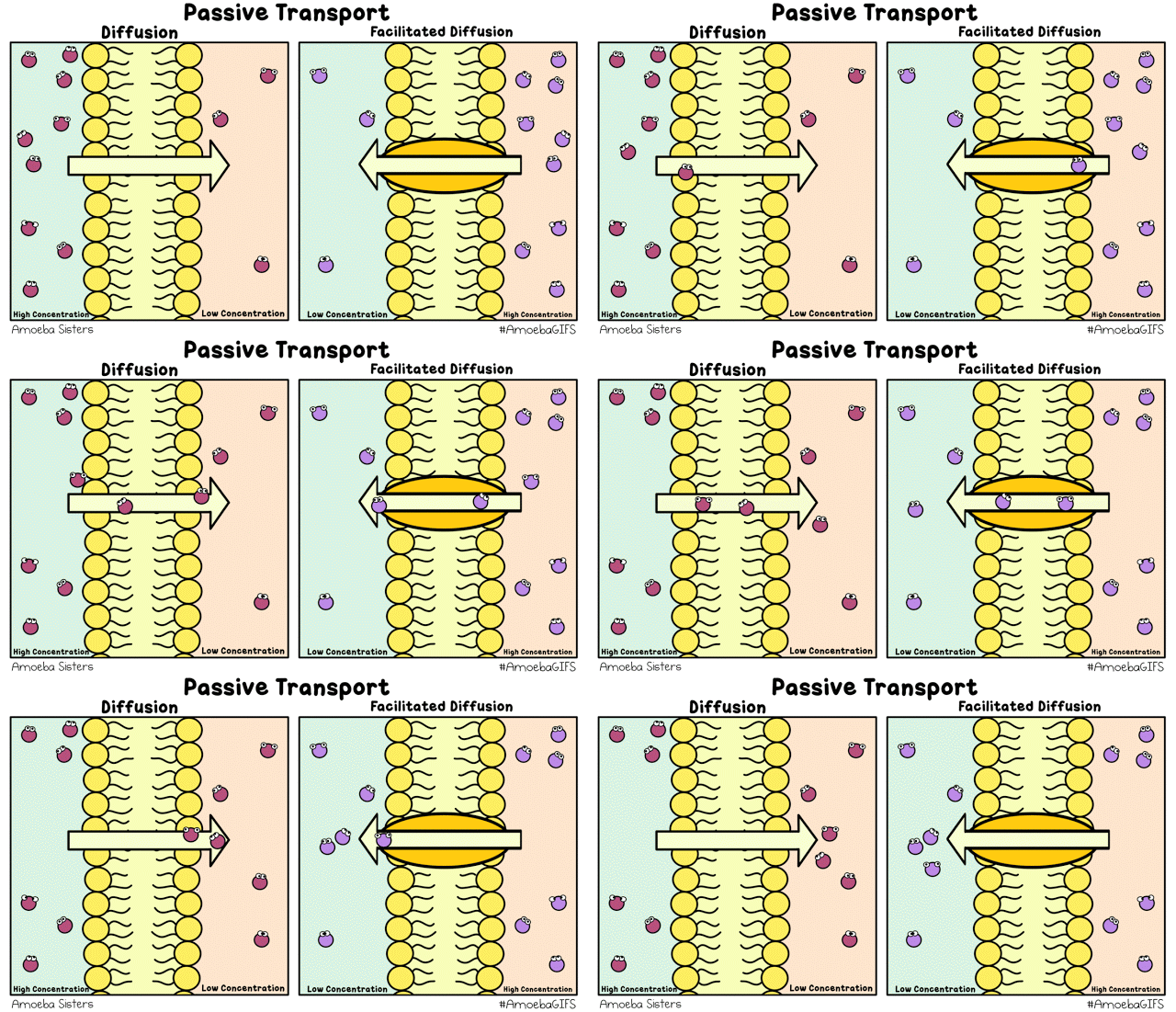
Passive Membrane Transport Mechanisms
The 3 types of passive membrane transport diffusion, facilitated diffusion and osmosis. In all of these transport mechanisms, substances move from an area of higher concentration to an area of lower concentration. When we have a difference in the concentration of a substance, we call this a concentration gradient. In passive transport mechanisms, molecules are traveling "down" their concentration gradient, from an area of high concentration to an area of low concentration.
Diffusion
Diffusion is a natural process that does not require energy. Small, charged molecules freely diffuse across the cell membrane.
Facilitated Diffusion
For molecules that do not freely diffuse across the membrane, specialized membrane proteins can allow for diffusion of these molecules across the membrane.This mechanism of transport is called facilitated diffusion.
Osmosis
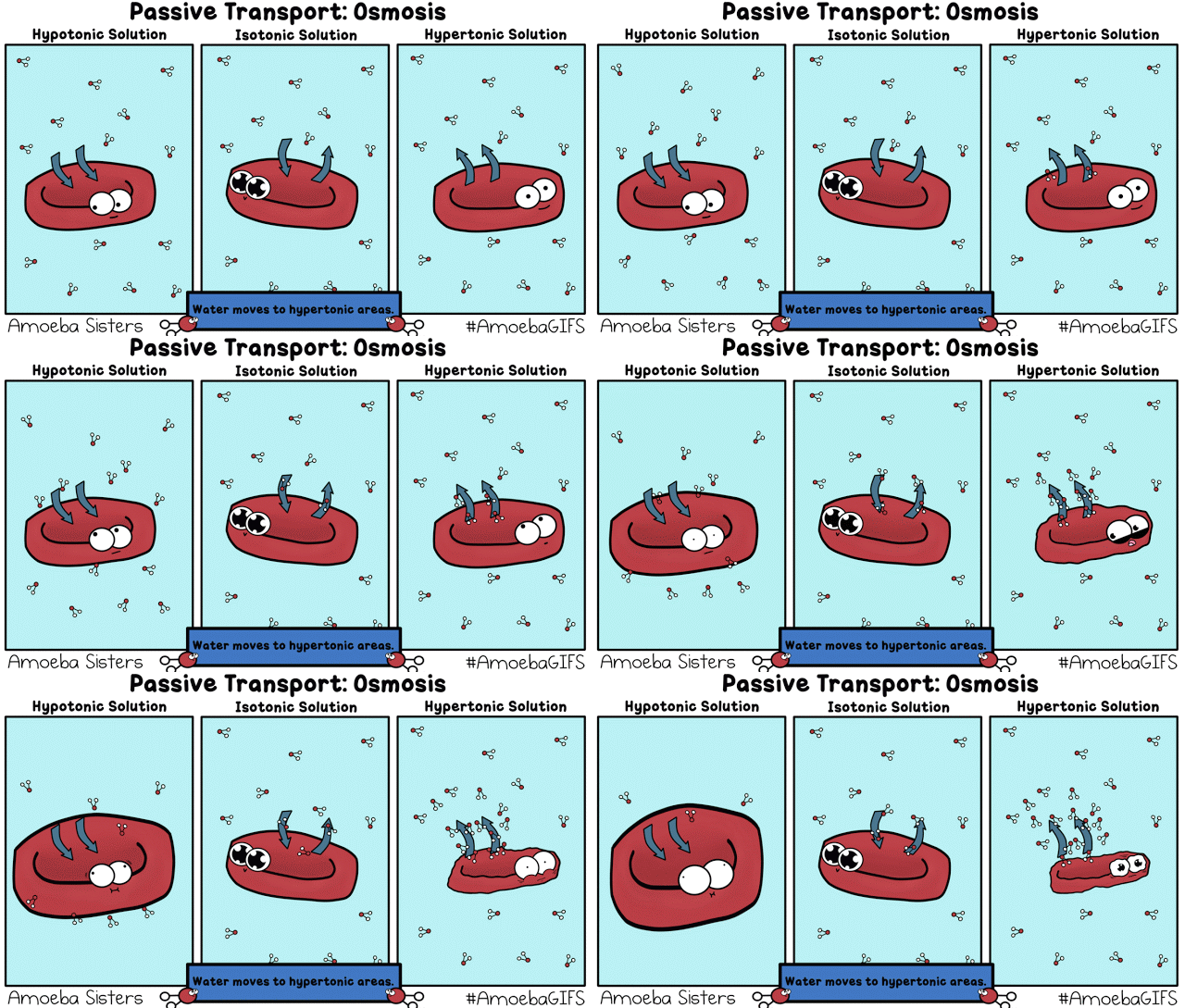
Diffusion of water is so important to biological processes and to osmoregularity, that it is given its own name; osmosis!
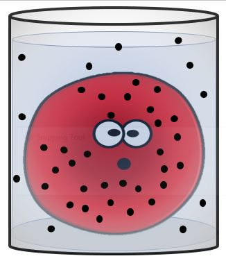
A hypotonic solution, is a solution that has a lower concentration of solutes (dissolved particles) than the liquid inside the cell. Since the cell membrane is only semi-permeable, the solutes cannot move across the membrane, but the water can! So, when a cell is placed into a hypotonic solution, water will rush into the cell causing it to bloat and swell. If the solution is very hypotonic, the cell can burst open! OUCH!
An isotonic solution is a solution that has the same concentration of solutes as the liquid inside of the cell. In this situation, since the concentration is the same inside the cell as it is outside the cell, water is happy! There will be no net movement of water flowing into or out of the cell. In this situation, the cell maintains its shape and is healthy and happy!
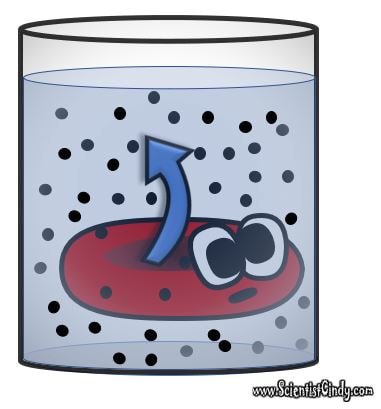
A hypertonic solution is a solution that contains a higher concentration of dissolved substance than the liquid inside of the cell. When a cell is placed in a hypertonic solution, water will move out of the cell, causing the cell to shrink and shrivel up!
|
|
|
Active Membrane Transport Mechanisms
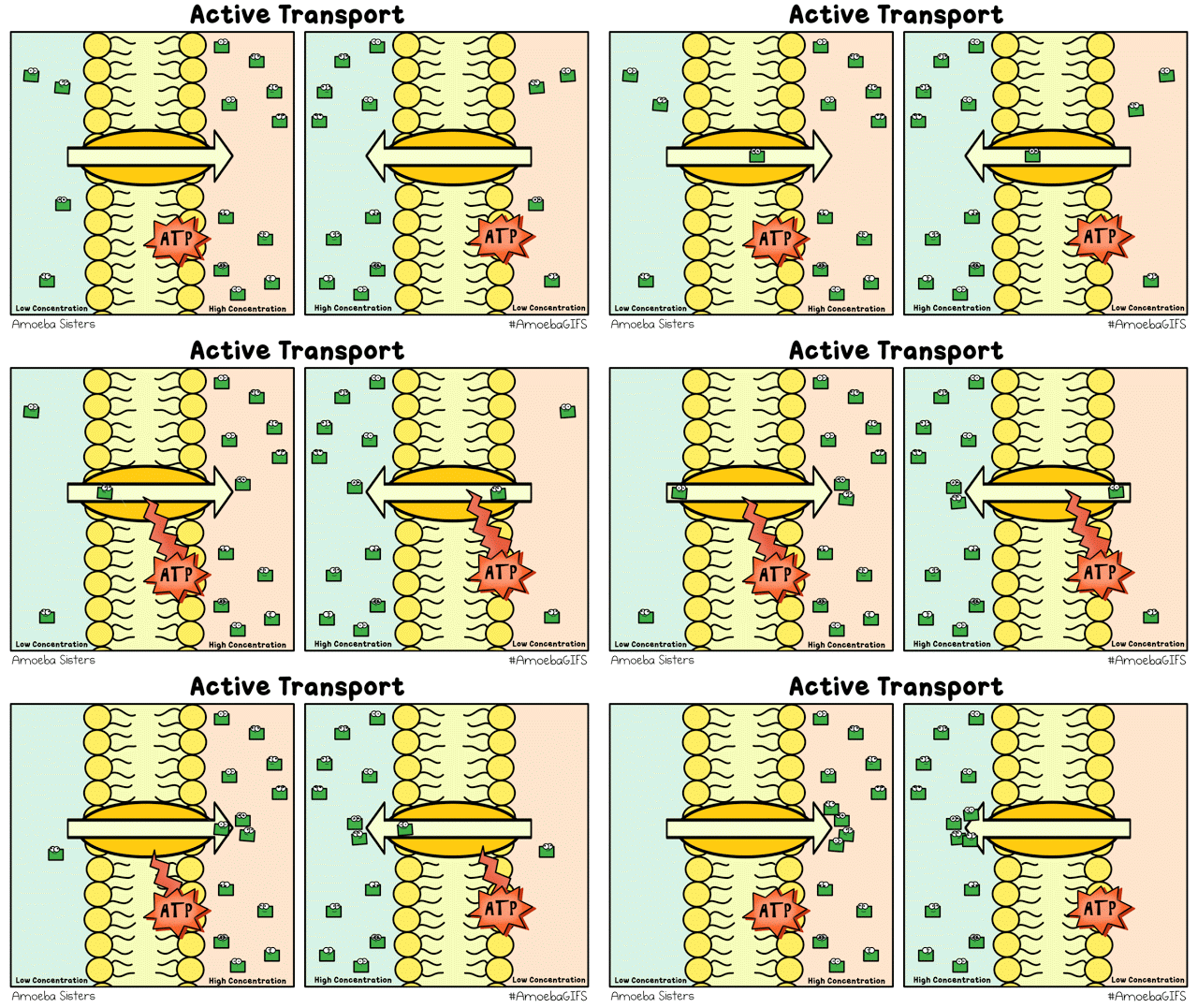
Active transport mechanisms require energy and act to move the molecule(s) against their concentration gradient, from an area of lower concentration to an area of higher concentration. Active transport requires an energy source and a transmembrane protein that allows passage of the molecule(s) against its concentration gradient.
Another type of active membrane transport is vesicular transport. This type of transport is explained in more detail below!
Vesicular Transport
exocytosis = secretion and endocytosis = uptake
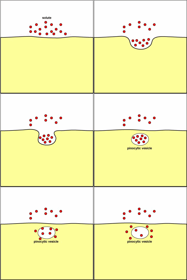
EndocytosisEndocytosis is the process by which contents from the extracellular matrix are taken up into the cell through a mechanism that involves the cell membrane essentially "pinching off" part of itself to form a vesicle that surrounds the particles being transported into the cell. |
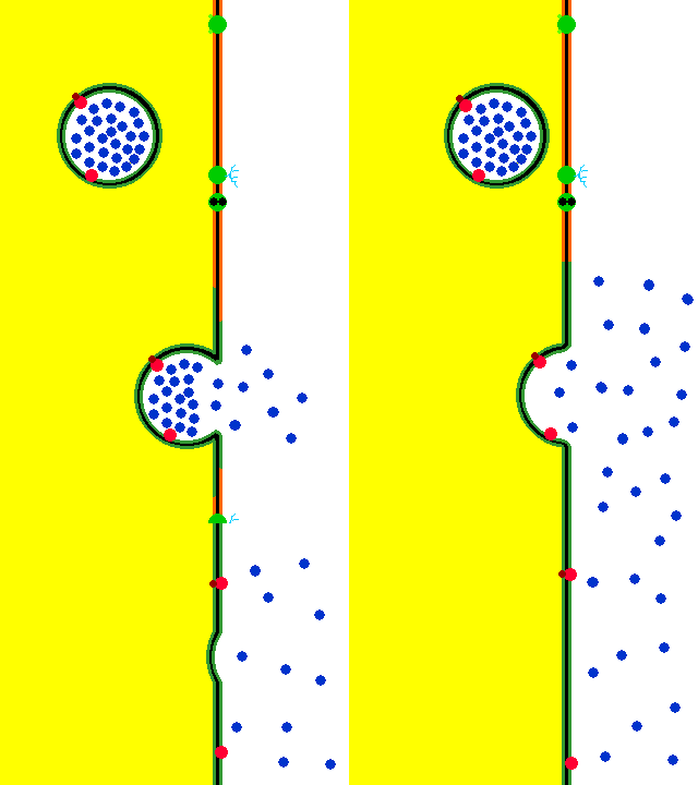
ExocytosisExocytosis is when a vesicle from inside the cell fuses with the membrane and the contents are released into the extracellular fluid.
Vesicular transport is a vital part of the cell's function. Vesicles perform a variety of functions, including transport, metabolism, and temporary storage .
|
There are 3 different types of endocytosis.
- Phagocytosis – “cell eating” - In phagocytosis, one cell will engulf the target and then proceed to digest or destroy the contents of the vesicle. Phagocytosis is demonstrated by white blood cells that are appropriately named, "phagocytes".
- Pinocytosis – “cell drinking” - Cells tend to sample small amounts of the extracellular fluid via pinocytosis. Pinocytosis is when the endocytosis results in a vesicle having only extracellular fluid as its contents.
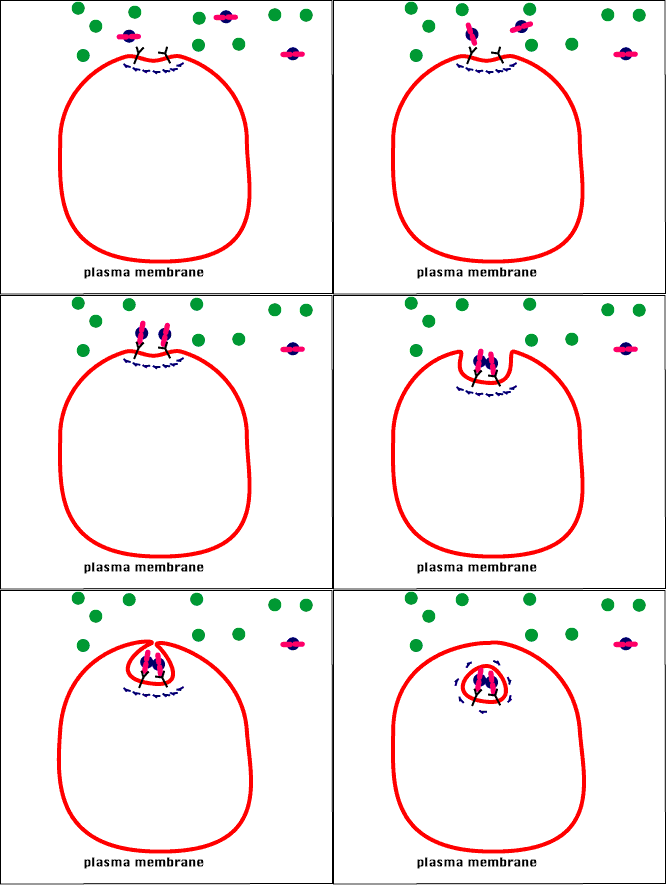
- Receptor-mediated endocytosis - This occurs when the plasma proteins bind only to certain molecules via a "lock and key" mechanism. The binding triggers the section of the membrane to "invaginate" and form a vesicle around the molecule that transports it into the cell from the extracellular fluid.
Receptor-Mediated EndocytosisGIF Courtesy of Source s10.lite.msu.edu
|
Endocytosis and Exocytosis |
Vesicle
The Main Function of the Vesicle is Transport
By SuperManu - Own work, CC BY-SA 3.0, https://commons.wikimedia.org/w/index.php?curid=2918850

The vesicle consists of a small amount of fluid (and sometimes particles) surrounded by a phospholipid bilayer. This bilayer is made up of the same phospholipids that are found in the cell membrane of the plasma membrane. this is due to the fact that vesicles are actually made from the cell membrane itself!
.
Osmosis, diffusion and filtration in the human body
Example 1) The Cardiovascular System: Blood Vessels
Capillary Exchange and Capillary Dynamics are important concepts for understanding how gases and nutrients are moved from the blood to the cells of the body. Capillary dynamics is governed by two opposing forces; hydrostatic pressure and osmotic pressure.
|
What is capillary exchange?
|
|
|
What is capillary dynamics?
Capillary Dynamics is concerned with how the permeability of the capillary wall changes under different conditions. Capillary dynamics is governed by the following forces:
|
Diffusion
Diffusion is the process by which a substance moves from an areas of high concentration (or pressure) to a lower concentration (or pressure). Small uncharged molecules such as glucose, oxygen and carbon dioxide are allowed to freely diffuse across the thin capillary walls according to the driving force caused by a concentration gradient (a difference in concentration between one area to another area).
Transcytosis
Impermeable substances can be transported across the capillary walls through a type of facilitate diffusion (a type of diffusion that requires energy) called transcytosis.
Trancytosis occurs by the processes of ligand-gated endocytosis, which allows a substance that cannot cross the cell membrane to enter the cells that make up the one cell-layer thick capillary walls, and then be carried from one side of the cell to the other in a vesicle and be release out the other side of the cell through exocytosis.
Trancytosis occurs by the processes of ligand-gated endocytosis, which allows a substance that cannot cross the cell membrane to enter the cells that make up the one cell-layer thick capillary walls, and then be carried from one side of the cell to the other in a vesicle and be release out the other side of the cell through exocytosis.
Bulk Flow
Bulk flow is the process by which small, lipid-insoluble materials can cross the the capillary wall. The movement of materials across the wall is dependent on pressure and is bi-directional depending on the net filtration pressure derived from the four Starling forces that modulate capillary dynamics.
Capillary Dynamics
The four Starling forces modulate capillary dynamics.
Capillary Dynamics: Osmotic pressure exerted by solids in the interstitial fluid drives water from the interstitial fluid into the blood vessels (capillaries). The pumping of the heart forces blood to flow into blood vessels. This force creates the blood pressure in the vessels that is needed to push blood around the body. This blood pressure is a type of hydrostatic pressure. This pressure is usually higher in the blood vessels than it is in the interstitial fluid, so the driving force due to hydrostatic pressue moves blood plasma from the blood vessels into the interstitial fluid that bathes the cells and tissues of the body.
In conditions where plasma proteins are reduced (e.g. from being lost in the urine or from malnutrition), or blood pressure is significantly increased, a change in net filtration pressure and an increase in fluid movement across the capillary result in excess fluid build-up in the tissues (edema).
Transcytosis
Transcytosis is a process by which molecules are transported into the capillaries.
LEARNING OBJECTIVESDescribe the process of transcytosis in capillary exchange
KEY TAKEAWAYSKey Points
Substances are transported through the endothelial cells themselves within vesicles. This mechanism is mainly used by large molecules, typically lipid-insoluble preventing the use of other transport mechanisms. The substance to be transported is endocytosed by the endothelial cell into a lipid vesicle which moves through the cell and is then exocytosed to the other side. Vesicles are capable of merging, allowing for their contents to mix, and can be transported directly to specific organs or tissues.
PathologyDue to the function of transcytosis, it can be a convenient mechanism by which pathogens can invade a tissue. Transcytosis has been shown to be critical to the entry of Cronobacter sakazakiiacross the intestinal epithelium and the blood-brain barrier.
Listeria monocytogenes has been shown to enter the intestinal lumen via transcytosis across goblet cells. Shiga toxin secreted by entero-hemorrhagic E. coli has been shown to be transcytosed into the intestinal lumen. These examples illustrate that transcytosis is vital to the process of pathogenesis for a variety of infectious agents.
Transcytosis in PharmaceuticalsPharmaceutical companies are currently exploring the use of transcytosis as a mechanism for transporting therapeutic drugs across the human blood-brain barrier. Exploiting the body’s own transport mechanism can help to overcome the high selectivity of this barrier, which blocks the uptake of most therapeutic antibodies into the brain and central nervous system.
Bulk Flow: Filtration and ReabsorptionCapillary fluid movement occurs as a result of diffusion (colloid osmotic pressure), transcytosis, and filtration.
LEARNING OBJECTIVESExplain the process of filtration and reabsorption in capillaries
KEY TAKEAWAYSKey Points
Bulk Flow ProcessBulk flow is used by small, lipid-insoluble solutes in water to cross the the capillary wall and is dependent on the physical characteristics of the capillary. Continuous capillaries have a tight structure reducing bulk flow. Fenestrated capillaries permit a larger amount of flow and discontinuous capillaries allow the largest amount of flow.
The movement of materials across the capillary wall is dependent on pressure and is bidirectional depending on the net filtration pressure derived from the four Starling forces.
When moving from the bloodstream into the interstitium, bulk flow is termed filtration, which is favored by blood hydrostatic pressure and interstitial fluid oncotic pressure. When moving from the interstitium into the bloodstream, the process is termed reabsorption and is favored by blood oncotic pressure and interstitial fluid hydrostatic pressure.
Modern evidence shows that in most cases, venular blood pressure exceeds the opposing pressure, thus maintaining a positive outward force. This indicates that capillaries are normally in a state of filtration along their entire length.
The Kidneys and Bulk FlowThe kidney is a major site for bulk flow transport. Blood that enters the kidneys is filtered by nephrons, the functional unit of the kidney. Each nephron begins in a renal corpuscle composed of a glomerulus containing numerous capillaries enclosed in a Bowman’s capsule. Proteins and other large molecules are filtered out of the oxygenated blood in the glomerulus and pass into Bowman’s capsule and the tubular fluid contained within. Blood continues to flow around the nephron until it reaches another capillary-rich region the peritubular capillaries, where the previously filtered molecules are reabsorbed from the tubule of the nephron.
Tubular reabsorption is the process by which solutes and water are removed from the tubular fluid and transported into the blood. Reabsorption is a two-step process beginning with the active or passive extraction of substances from the tubule fluid into the renal interstitium, and then the transport of these substances from the interstitium into the bloodstream
Tubular Secretion: Diagram showing the basic physiologic mechanisms of the kidney and the three steps involved in urine formation.
Capillary Dynamics
The four Starling forces modulate capillary dynamics.
- Oncotic or colloid osmotic pressure is a form of osmotic pressure exerted by proteins in the blood plasma or interstitial fluid.
- Hydrostatic pressure is the force generated by the pressure of fluid within or outside of capillary on the capillary wall.
Capillary Dynamics: Osmotic pressure exerted by solids in the interstitial fluid drives water from the interstitial fluid into the blood vessels (capillaries). The pumping of the heart forces blood to flow into blood vessels. This force creates the blood pressure in the vessels that is needed to push blood around the body. This blood pressure is a type of hydrostatic pressure. This pressure is usually higher in the blood vessels than it is in the interstitial fluid, so the driving force due to hydrostatic pressue moves blood plasma from the blood vessels into the interstitial fluid that bathes the cells and tissues of the body.
In conditions where plasma proteins are reduced (e.g. from being lost in the urine or from malnutrition), or blood pressure is significantly increased, a change in net filtration pressure and an increase in fluid movement across the capillary result in excess fluid build-up in the tissues (edema).
Transcytosis
Transcytosis is a process by which molecules are transported into the capillaries.
LEARNING OBJECTIVESDescribe the process of transcytosis in capillary exchange
KEY TAKEAWAYSKey Points
- Transcytosis is the process by which various macromolecules are transported across the endothelium of the capillaries.
- Due to this function, transcytosis can be a convenient mechanism for pathogens to invade a tissue.
- transcytosis: The process whereby macromolecules are transported across the interior of a cell via vesicles.
Substances are transported through the endothelial cells themselves within vesicles. This mechanism is mainly used by large molecules, typically lipid-insoluble preventing the use of other transport mechanisms. The substance to be transported is endocytosed by the endothelial cell into a lipid vesicle which moves through the cell and is then exocytosed to the other side. Vesicles are capable of merging, allowing for their contents to mix, and can be transported directly to specific organs or tissues.
PathologyDue to the function of transcytosis, it can be a convenient mechanism by which pathogens can invade a tissue. Transcytosis has been shown to be critical to the entry of Cronobacter sakazakiiacross the intestinal epithelium and the blood-brain barrier.
Listeria monocytogenes has been shown to enter the intestinal lumen via transcytosis across goblet cells. Shiga toxin secreted by entero-hemorrhagic E. coli has been shown to be transcytosed into the intestinal lumen. These examples illustrate that transcytosis is vital to the process of pathogenesis for a variety of infectious agents.
Transcytosis in PharmaceuticalsPharmaceutical companies are currently exploring the use of transcytosis as a mechanism for transporting therapeutic drugs across the human blood-brain barrier. Exploiting the body’s own transport mechanism can help to overcome the high selectivity of this barrier, which blocks the uptake of most therapeutic antibodies into the brain and central nervous system.
Bulk Flow: Filtration and ReabsorptionCapillary fluid movement occurs as a result of diffusion (colloid osmotic pressure), transcytosis, and filtration.
LEARNING OBJECTIVESExplain the process of filtration and reabsorption in capillaries
KEY TAKEAWAYSKey Points
- Bulk flow is a process used by small lipid-insoluble proteins to cross the capillary wall.
- Capillary structure plays a large role in the rate of bulk flow, with continuous capillaries limiting flow and discontinuous capillaries facilitating the greatest amount of flow.
- When moving from the blood to the interstitium, bulk flow is termed filtration.
- When moving from the interstitium to the blood, bulk flow is termed re-absorption.
- The kidney is a major site of bulk flow where waste products are filtered from the blood.
Bulk Flow ProcessBulk flow is used by small, lipid-insoluble solutes in water to cross the the capillary wall and is dependent on the physical characteristics of the capillary. Continuous capillaries have a tight structure reducing bulk flow. Fenestrated capillaries permit a larger amount of flow and discontinuous capillaries allow the largest amount of flow.
The movement of materials across the capillary wall is dependent on pressure and is bidirectional depending on the net filtration pressure derived from the four Starling forces.
When moving from the bloodstream into the interstitium, bulk flow is termed filtration, which is favored by blood hydrostatic pressure and interstitial fluid oncotic pressure. When moving from the interstitium into the bloodstream, the process is termed reabsorption and is favored by blood oncotic pressure and interstitial fluid hydrostatic pressure.
Modern evidence shows that in most cases, venular blood pressure exceeds the opposing pressure, thus maintaining a positive outward force. This indicates that capillaries are normally in a state of filtration along their entire length.
The Kidneys and Bulk FlowThe kidney is a major site for bulk flow transport. Blood that enters the kidneys is filtered by nephrons, the functional unit of the kidney. Each nephron begins in a renal corpuscle composed of a glomerulus containing numerous capillaries enclosed in a Bowman’s capsule. Proteins and other large molecules are filtered out of the oxygenated blood in the glomerulus and pass into Bowman’s capsule and the tubular fluid contained within. Blood continues to flow around the nephron until it reaches another capillary-rich region the peritubular capillaries, where the previously filtered molecules are reabsorbed from the tubule of the nephron.
Tubular reabsorption is the process by which solutes and water are removed from the tubular fluid and transported into the blood. Reabsorption is a two-step process beginning with the active or passive extraction of substances from the tubule fluid into the renal interstitium, and then the transport of these substances from the interstitium into the bloodstream
Tubular Secretion: Diagram showing the basic physiologic mechanisms of the kidney and the three steps involved in urine formation.

