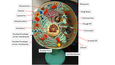
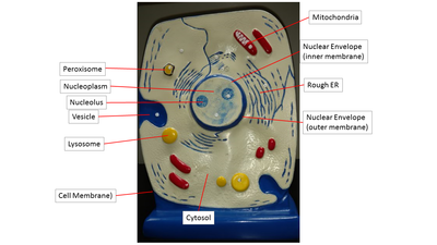
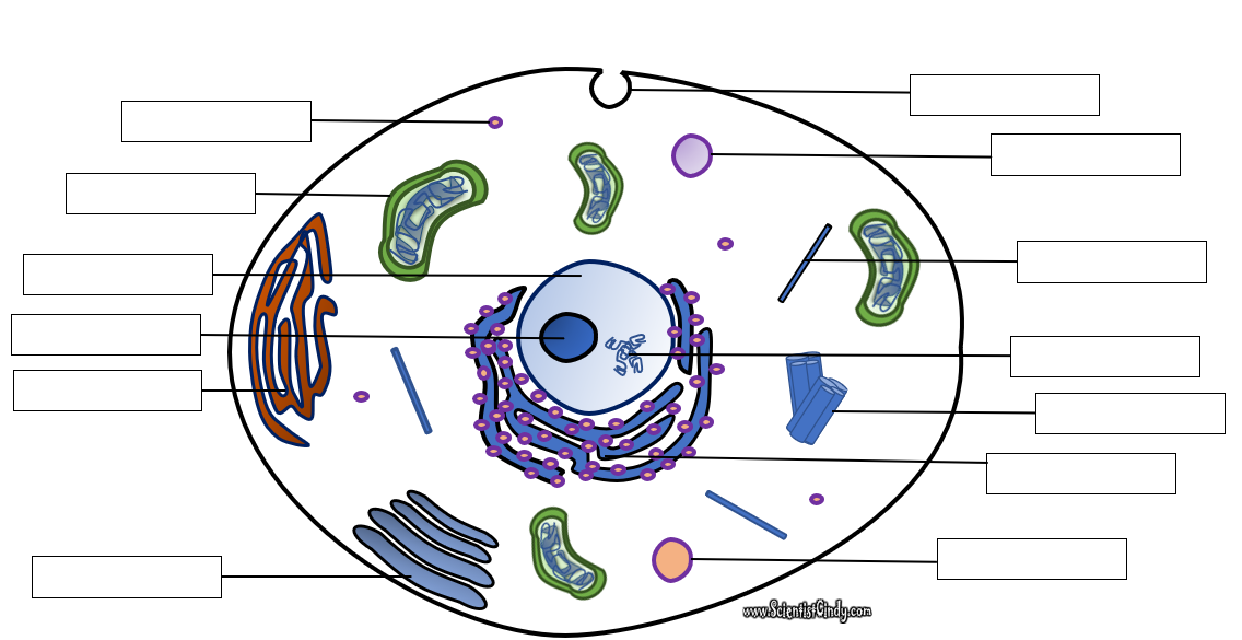
Anatomy of a Bacteria Cell
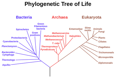
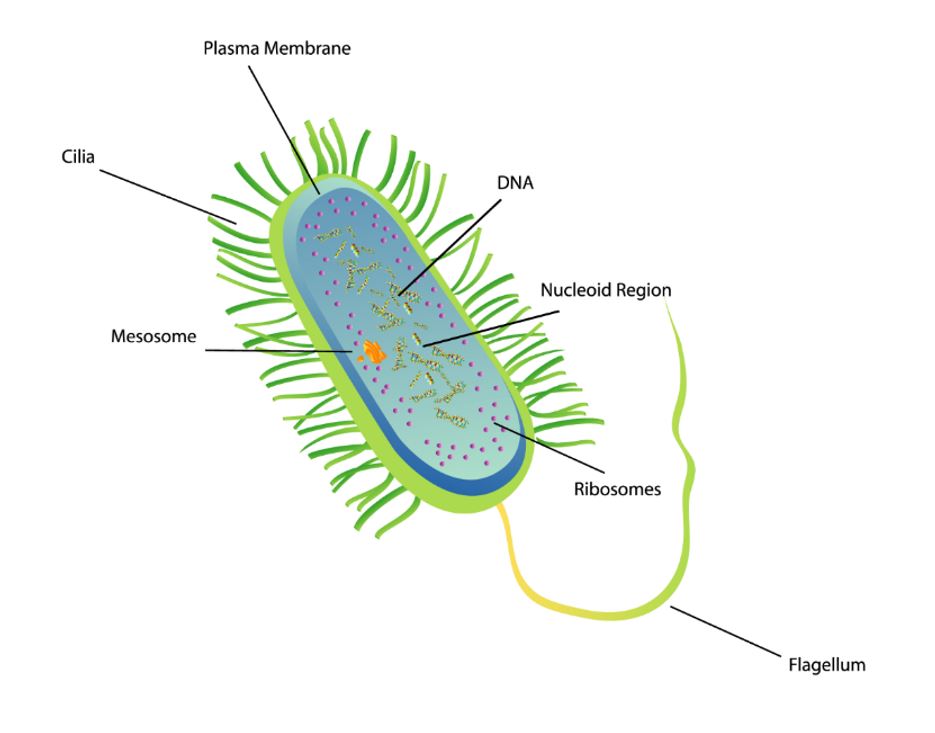
Prokaryotic organisms belong to the Archaea and Bacteria domains of life, whereas eukaryotic organisms belong to the Eukarya domain.
Prokaryotic cells existed from about one-billion years before eukaryotic single-celled organisms evolved. Prokaryotic cells are almost always smaller than eukaryotic cells. Prokaryotic cells lack membrane-bound organelles and do not have a nucleus. In contrast, eukaryotic cells do have a nucleus and do have membrane-bound organelles.
Prokaryotic cells existed from about one-billion years before eukaryotic single-celled organisms evolved. Prokaryotic cells are almost always smaller than eukaryotic cells. Prokaryotic cells lack membrane-bound organelles and do not have a nucleus. In contrast, eukaryotic cells do have a nucleus and do have membrane-bound organelles.
The Cell Membrane (Plasma Membrane)
- The Main Function of the Cell Membrane is TRANSPORT REGULATION -
The cell membrane is also called the plasma membrane. You can think of the membrane as the "skin" of the cell. Anything outside of the cell is considered "extracellular" and the contents inside the cell are considered "intracellular". The cell membrane protects the cell by creating a barrier between what is inside the cell and what is outside the cell.
In addition to this, the cell membrane does something our skin can’t do... It regulates what comes into the cell and what goes out of the cell. For this reason, we consider the cell membrane to be “SELECTIVELY PERMEABLE” which means that it allows some substances to enter or exit the cell, but not others. This is a very important function.
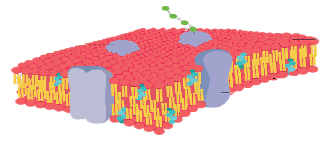
The Cell Membrane is selectively permeable due to its structure. The cell membrane is made up of a phospholipid bilayer.
The Cell Membrane is selectively permeable due to its structure. The cell membrane is made up of a phospholipid bilayer.
Phospholipid

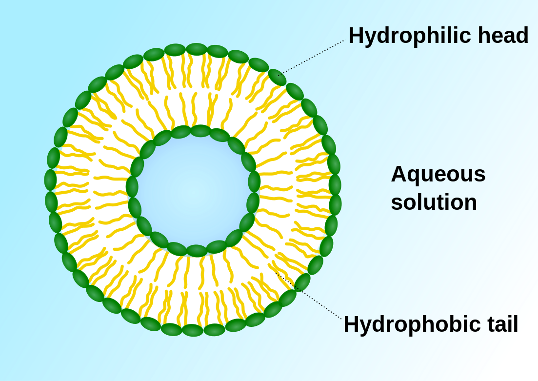
The phospholipid bilayer of the cell membrane has a unique structure. It is made up of an inner layer and an outer layer of phospholipids that are oriented with their 'tails' facing each other.
Phospholipids are considered amphiphilic, because they contain a polar, hydrophillic head that consists of a phosphate group and two nonpolar, hydrophobic fatty acid chains as 'tails'.
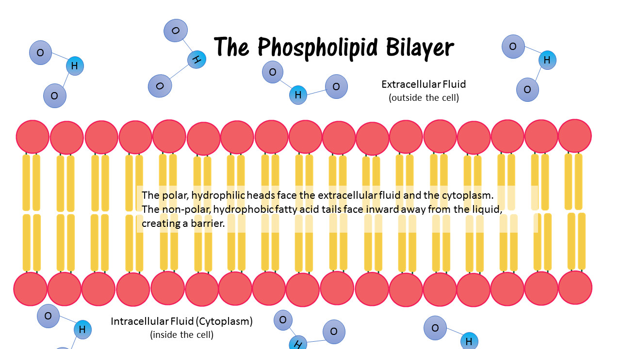
When the phospholipids form the cell membrane, the polar, hydrophillic (water-loving) heads are oriented towards the liquid outside the cells (extracellular fluid) and the liquid inside the cell (intracellular fluid). The tails of the phospholipids are oriented towards each other, away from the liquid, since they are made up of hydrophobic (water-fearing) fatty acid chains. This formation creates a barrier between the extracellular matrix and the intracellular fluid (cytoplasm).
Click Below to View a Video on the Mighty Membrane
Vesicular Transport
exocytosis = secretion and endocytosis = uptake
Endocytosis
|
Exocytosis
|
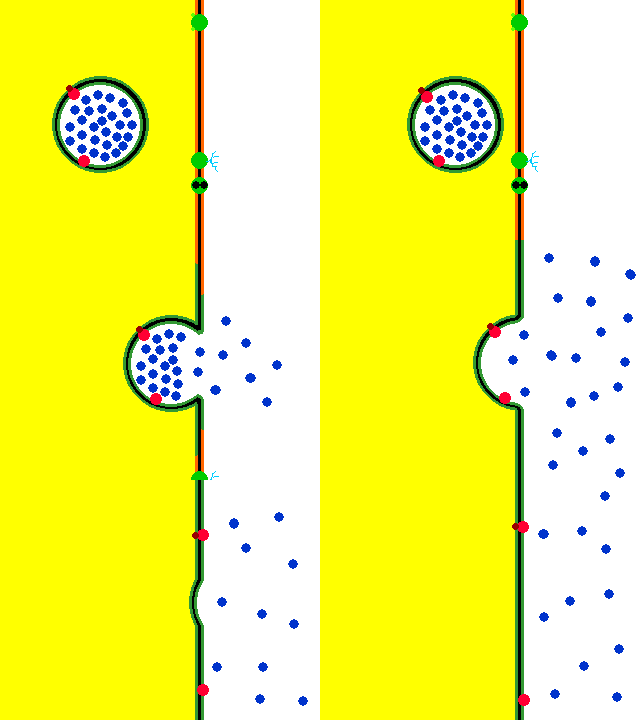
Vesicular transport is a vital part of the cell's function. Vesicles perform a variety of functions, including transport, metabolism, and temporary storage .
There are 3 different types of endocytosis.
- Phagocytosis – “cell eating” - In phagocytosis, one cell will engulf the target and then proceed to digest or destroy the contents of the vesicle. Phagocytosis is demonstrated by white blood cells that are appropriately named, "phagocytes".
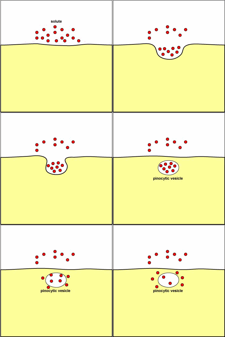
- Pinocytosis – “cell drinking” - Cells tend to sample small amounts of the extracellular fluid via pinocytosis. Pinocytosis is when the endocytosis results in a vesicle having only extracellular fluid as its contents.
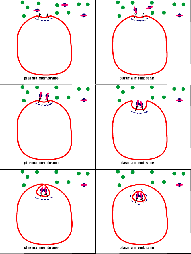
- Receptor-mediated endocytosis - This occurs when the plasma proteins bind only to certain molecules via a "lock and key" mechanism. The binding triggers the section of the membrane to "invaginate" and form a vesicle around the molecule that transports it into the cell from the extracellular fluid.
Receptor-Mediated EndocytosisGIF Courtesy of Source s10.lite.msu.edu
|
Endocytosis and Exocytosis |
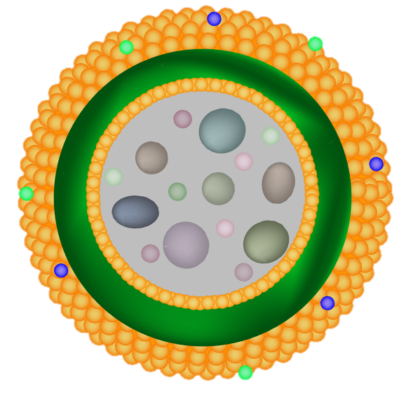
Vesicle
The Main Function of the Vesicle is Transport
By SuperManu - Own work, CC BY-SA 3.0, https://commons.wikimedia.org/w/index.php?curid=2918850

The vesicle consists of a small amount of fluid (and sometimes particles) surrounded by a phospholipid bilayer. This bilayer is made up of the same phospholipids that are found in the cell membrane of the plasma membrane. this is due to the fact that vesicles are actually made from the cell membrane itself!
.
.
Nucleus
The Main Function of the Nucleus is to Hold the DNA.
The nucleus is like the "brain" or the "command center" of the cell. This is due to the fact that the nucleus holds the DNA. The DNA acts as the cell's "instruction manual" which has the instructions for all of the functions of the cell, as well as the instructions for growth, development and cellular reproduction!

The structure of the nucleus - The nucleus is surrounded by the nuclear envelope, which is essentially a membrane (a phospholipid bilayer) that compartmentalizes (or separates) the nucleus from the rest of the cell. The liquid inside of the nucleus is called the nucleoplasm.
DNA stands for deoxyribonucleic acid. It is the "instruction manual" for the cell. The primary function of the DNA is to provide the instructions for protein synthesis to the cell through the processes of transcription and translation.
ENDOPLASMIC RETICULUM
There are 2 types of endoplasmic reticulum (ER); the rough endoplasmic reticulum and the smooth endoplasmic reticulum.
|
The Rough Endoplasmic Reticulum
(The Rough ER) The rough ER gets its name from its 'bumpy' or 'rough' appearance due to ribosomes that are attached to it. In the rough ER, ribosomes are assembled in the nucleolus and then exit the nucleus. Some of these ribosomes then attach themselves to the rough endoplasmic reticulum, where they will act as a "site of protein synthesis" for the cell.
|
The Smooth Endoplasmic Reticulum
(The Smooth ER) The smooth endoplasmic reticulum (Smooth ER) appears smooth in comparison to the rough ER. This is because the smooth ER does not have ribosomes bound to it. The smooth ER's main function is to make lipids.
|
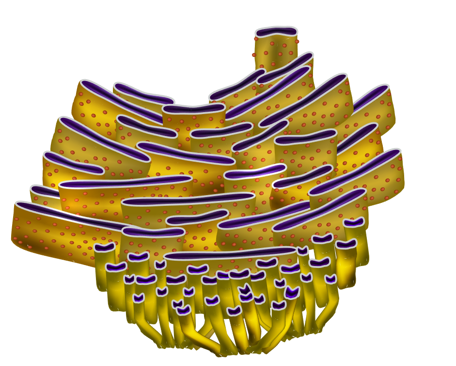
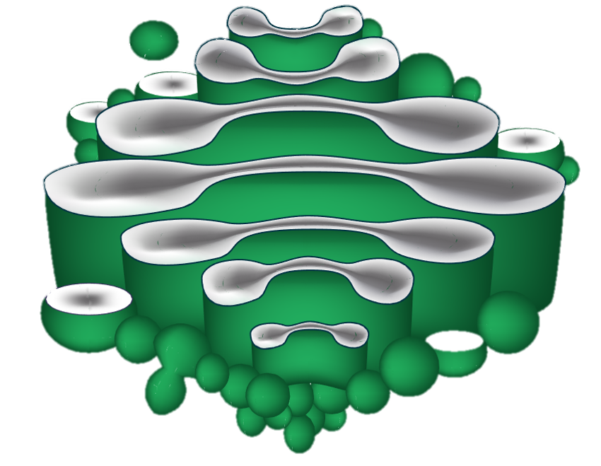
The Golgi Body
The Golgi Body
Lysozyme
Cell Wall
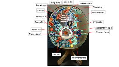
Summary of Structure and Function of Organelles
ORGANELLE or STRUCTURE |
FUNCTION |
SIMILAR TO |
NUCLEUS |
HOLDS THE DNA |
INSTRUCTION MANUAL |
NUCLEOLUS |
MAKES RIBOSOMES |
FACTORY |
MEMBRANE |
PROTECTION - REGULATION OF TRANSPORT |
SKIN |
GOLGI BODY |
MODIFIES, PACKAGES AND SHIPS PROTEINS |
POST OFFICE |
MITOCHONDRIA |
CREATES ENERGY (ATP) |
POWER PLANT |
ROUGH ENDOPLASMIC RETICULUM |
HAS RIBOSOMES |
|
SMOOTH ENDOPLASMIC RETICULUM |
LIPID PRODUCTION AND CALCIUM STORAGE |
|
PEROXISOME |
DETOXIFIES |
GARBAGE DISPOSAL |
LYSOSOME |
DIGESTS UNWANTED SUBSTANCES |
RECYCLING CENTER |
RIBOSOME |
THE SITE OF PROTEIN SYNTHESIS |
ASSEMBLY LINE |
NUCLEOPLASM |
LIQUID OF THE NUCLEUS |
|
NUCLEAR ENVELOPE |
MEMBRANE AROUND THE NUCLEUS |
"SKIN" OF NUCLEUS |
VESICLES |
TRANSPORTATION |
VEHICLES |
CYTOSOL |
LIQUID OF THE CELL THAT LIES INSIDE OF THE CELL MEMBRANE, AND OUTSIDE THE NUCLEUS |


