The Cardiovascular System
The cardiovascular system functions to circulate blood through the body. The blood carries vital nutrients, oxygen and hormones to the cells, and also picks up unwanted toxins, waste and carbon dioxide from the cells.
The cardiavascular system is extremely important in sustaining life. It’s proper functioning is responsible for the delivery of oxygen and nutrients to all cells, as well as the removal of carbon dioxide, waste products, maintenance of optimum pH, and the mobility of the
elements, proteins and cells, of the immune system. In developed countries, the two leading causes of death, myocardial infarction (heart attack) and stroke are each direct results of an arterial system that has been slowly and progressively compromised by years of deterioration.
elements, proteins and cells, of the immune system. In developed countries, the two leading causes of death, myocardial infarction (heart attack) and stroke are each direct results of an arterial system that has been slowly and progressively compromised by years of deterioration.
The 3 Components of the Cardiovascular System are...
1) The Heart - The function of the heart is to pump the blood around the body. It does this by creating a pressure gradient that forces the movement of the blood.
2) The Blood - The function of the blood is to deliver oxygen and nutrients to the cells of the body and to take away the unwanted waste products and carbon dioxide from the cells.
3) The Blood Vessels - The function of the blood vessels is to provide the network of passage ways that the blood can travel in. In short, the blood vessels allow the blood to travel through the body.
|
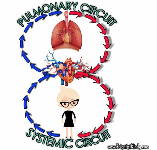
The cardiovascular system circulates blood around two loops.
|
In pulmonary circulation, deoxygenated blood travels from the heart to the lungs to gain oxygen and get rid of carbon dioxide before returning to the heart.
In contrast, in systemic circulation, oxygenated blood is pumped from the heart to the body and deoxygenated blood is returned back to the heart.
THE SYSTEMIC CIRCUIT
Systemic Circulation is the movement of blood from the heart through ALL OF THE PARTS OF THE BODY except THE LUNGS. As the blood travels around the body, the blood delivers oxygen and nutrients to the cells of the body and picks up the unwanted waste products and carbon dioxide from the cells. while bringing deoxygenated blood back to the heart.
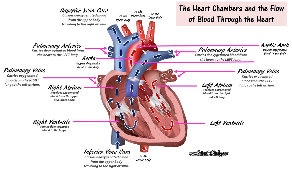
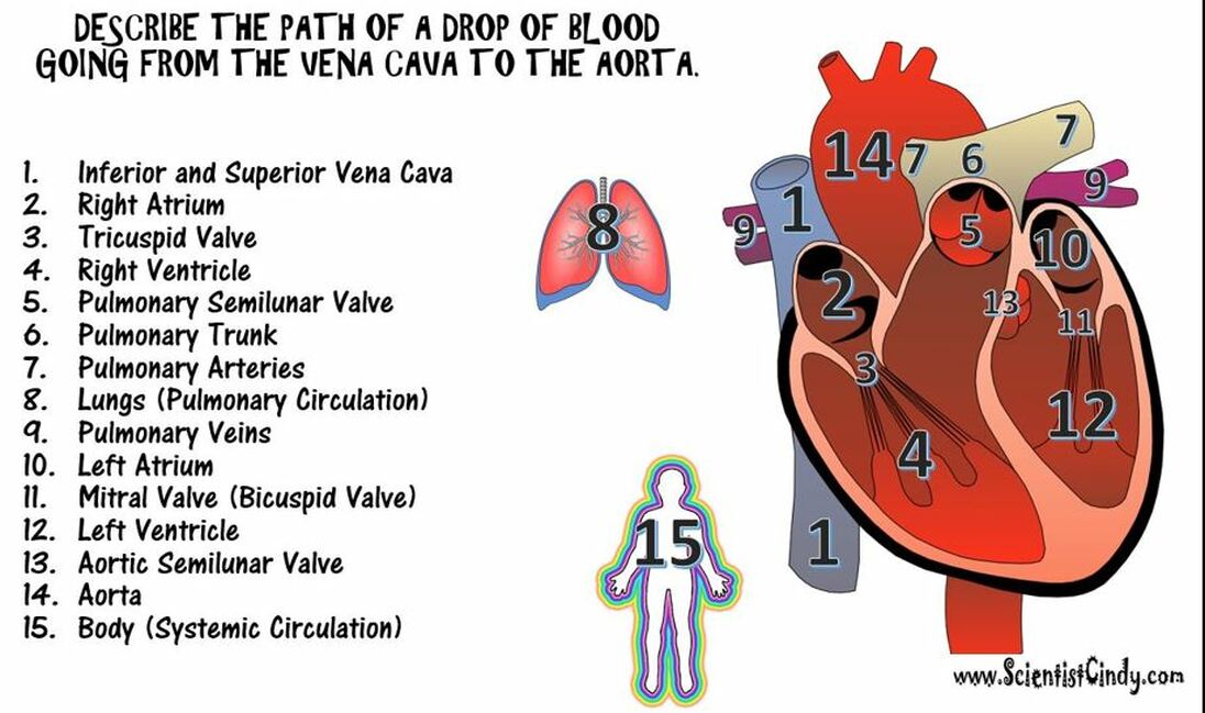
The systemic circuit begins when the oxygenated blood is pumped out of the left ventricle to the aorta (the largest artery in the body). The aorta branches into major arteries that travel to the upper and lower body. Once the blood has delivered the nutrients and oxygen to the and picked up the carbon dioxide and waste products from the cells, this deoxygenated blood returns to the heart through the superior and inferior vena cavae.
The systemic circuit supplies oxygenated blood to the organ system. Oxygenated blood from the lungs is returned to the left atrium, then the ventricle contracts and pumps blood into the aorta. Systemic arteries split from the aorta and direct blood into the capillaries. Cells consume the oxygen and nutrients and add carbon dioxide, wastes, enzymes and hormones. The veins drain the deoxygenated blood from the capillaries and return the blood to the right atrium.
The Systemic Circuit
|
The Pulmonary Circuit
|
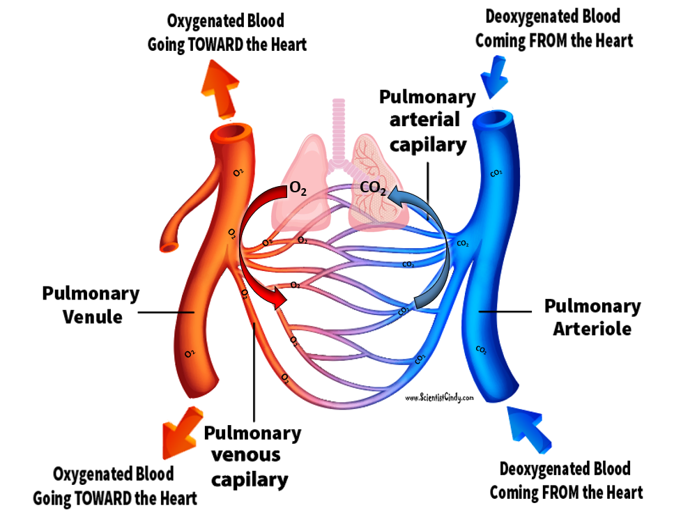
In the pulmonary circuit, blood is pumped to the lungs from the right ventricle of the heart. It is carried to the lungs via pulmonary arteries. At lungs, oxygen in the alveolae diffuses to the capillaries surrounding the alveolae and carbon dioxide inside the blood diffuses to the alveolae. As a result, blood is oxygenated which is then carried to the heart's left half -to the left atrium via pulmonary veins. Oxygen rich blood is prepared for the whole organs and tissues of the body. This is important because mitochondria inside the cells should use oxygen to produce energy from the organic compounds.
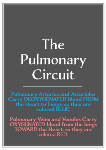
An artery is defined as any blood vessel that carries blood AWAY from the heart. POINTER: This is sometimes a point of confusion for students, because we usually think of arteries as carrying oxygenated blood, and the arteries are often colored in RED to illustrate this idea. HOWEVER, in the pulmonary circuit of the cardiovascular system, this IS NOT the case. In the pulmonary circuit of the cardiovascular system, the pulmonary arteries contain deoxygenated blood that travels from the heart to the lungs. When this blood passes by the lungs (the alveoli of the lungs), it gains oxygen molecules and releases carbon dioxide molecules. At this point, the blood becomes oxygenated and appears more reddish in color. This oxygenated blood then travels from the lungs back to the heart through the PULMONARY VEINS. A vein is any blood vessel that carries blood TO the heart. The blood vessels that carry blood to and from the lungs make up the pulmonary circuit (pulmonos = lung).
The Heart
The heart is a hollow, muscular organ about the size of a fist. It is responsible for pumping blood through the blood vessels by repeated, rhythmic contractions. The heart is composed of cardiac muscle, an involuntary muscle tissue that is found only within this organ. The term "cardiac" (as in cardiology) means "related to the heart” and comes from the Greek word kardia, for "heart." It has a four-chambered, double pump and is located in the thoracic cavity between the lungs. The cardiac muscle is self-exciting, meaning it has its own
conduction system. This is in contrast with skeletal muscle, which requires either conscious or reflex nervous stimuli. The heart's rhythmic contractions occur spontaneously, although
the frequency or heart rate can be changed by nervous or hormonal influence such as exercise or the perception of danger.
Heart Chambers
|
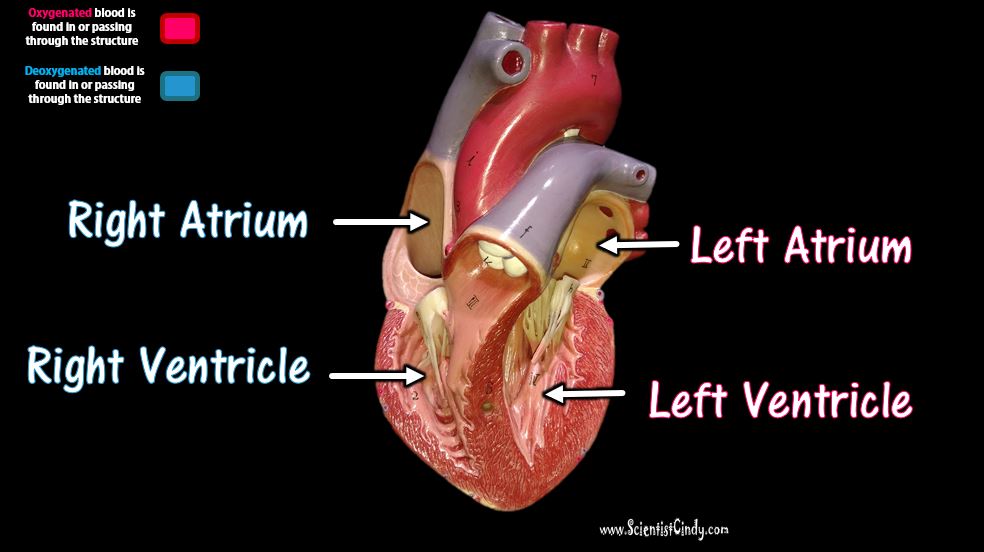
The heart has four chambers:
The two atria lie superiorly to the two ventricles. The atria are smaller with thin walls, while the ventricles are larger with thick muscular walls. |
|
Atria
The word "atrium" comes from the Latin word for "entrance". The atria (plural for atrium) of the heart function to RECEIVE blood. The word "ventricle" comes from the Latin word for "belly". This origin probably comes from the fact that the ventricles of the heart are the larger of the 4 chambers. They are muscular and perform the rhythmic contraction and relaxation of the cardiac muscle that creates the "pumping" action of the heart. The ventricles both SEND blood out of the heart.
There are two atria; the right atrium and the left atrium. The right atrium receives deoxygenated (oxygen-poor) blood from the body. The left atrium receives oxygenated (oxygen-rich) blood coming from the blood vessels surrounding the lungs. which has been oxygenated and is ready to be sent to the body. The right atrium receives deoxygenated blood from the upper portions of the body (the head, neck and arms) through the superior vena cava. The right atrium receives deoxygenated blood from the lower portions of the body (the torso and legs) through the inferior vena cava. The left atrium receives oxygenated blood from the left and right pulmonary veins wich carries blood coming from the blood vessels surrounding the lungs.
Ventricles
The ventricle is a heart chamber which collects blood from an atrium and pumps it out of the heart. There are two ventricles: the right ventricle pumps blood into the pulmonary circulation for the lungs, and the left ventricle pumps blood into the systemic circulation for the rest of the body. Ventricles have thicker walls than the atria, and thus can create the higher blood pressure. Comparing the left and right ventricle, the left ventricle has thicker walls because it needs to pump blood to the whole body.
The ventricle is a heart chamber which collects blood from an atrium and pumps it out of the heart. There are two ventricles: the right ventricle pumps blood into the pulmonary circulation for the lungs, and the left ventricle pumps blood into the systemic circulation for the rest of the body. Ventricles have thicker walls than the atria, and thus can create the higher blood pressure. Comparing the left and right ventricle, the left ventricle has thicker walls because it needs to pump blood to the whole body.
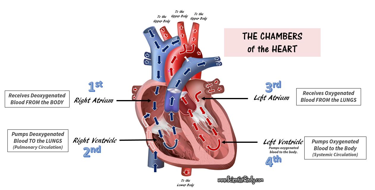
The Flow of Blood Through the Chambers of the Heart
The blood vessels that transport blood to and from all the body make up the SYSTEMIC CIRCUIT. So the 2 chambers of the heart that receive blood are the ATRIA (atria= plural; atrium= singular) and the two chambers of the heart that send blood are the VENTRICLES. The ventricles contract and relax rhythmically to create the pumping action of the heart.
The right atrium receives the deoxygenated blood that has traveled through the body. This blood has a low level of oxygen and a high level of carbon dioxide. This blood also has low pressure since it has traveled a good distance since it got its last "push" from the heart.
This deoxygenated blood from the lower and upper body travels to the right atrium, and then travels to the right ventricle. When the right ventricle contracts, it pushes the deoxygenated blood out of the heart and sends it to the lungs. (This is the pulmonary circuit). As this blood passes by the lungs, it picks up oxygen and gets rid of carbon dioxide.
The oxygenated blood from the lungs then travels back to the heart to get another "push" before it goes to the body (this is the systemic circuit).
The left atrium receives the oxygenated blood coming from the lungs. This blood enters the left atrium and then travels to the left ventricle. The left ventricle contracts to push the oxygenated blood out of the heart and to the body in order to deliver oxygen and nutrients to the cells and to pick up unwanted substances like waste and carbon dioxide.
The left atrium is the chamber of the heart that receives the oxygenated blood returning from the lungs through the PULMONARY VEINS. From the LEFT ATRIUM, this oxygenated blood travels to the LEFT VENTRICLE and then gets pumped to the body, to deliver oxygen and nutrients and to pick up carbon dioxide and waste products.
Heart Valves
The Valves of the Heart
Animated Heart Valve GIF Courtesy Of: By DrJanaOfficial - Own work, CC BY-SA 4.0, https://commons.wikimedia.org/w/index.php?curid=50477765
The function of the heart valves is to assure that blood flows in only one direction through the heart. When there is an increase in pressure of the blood, that pressure acts to push the valve open. The valves only open in one direction, which prevents back flow and guides the blood to flow in the proper direction.
The four main valves of the heart are as follows:
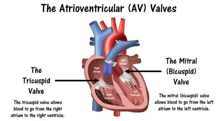
- The Tricuspid Valve is an atrioventricular (AV) valve that allows for the one-way movement of blood from the right atrium to the right ventricle.
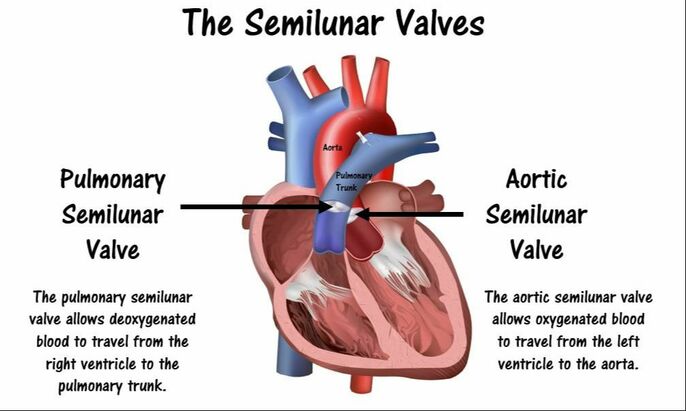
- The Pulmonary Semilunar Valve is a semilunar (SV) valve that allows for the one-way movement of blood from the right ventricle to the pulmonary arteries (which travel to the lungs).
- The Mitral (Bicuspid) Valve is an atrioventricular (AV) valve that allows for the one-way movement of blood from the left atrium to the left ventricle.
- The Aortic Semilunar Valve is a semilunar (SV) valve that allows for the one-way movement of blood from the left ventricle to the aorta (which travel to the body).
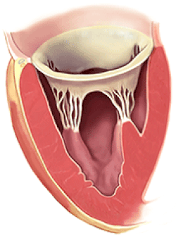
Valves
The two atrioventricular (AV) valves are one-way valves that ensure that blood flows from the atria to the ventricles, and not the other way. The two semilunar (SL) valves are present in the arteries leaving the heart; they prevent blood from flowing back into the ventricles. The sound heard in a heart beat is the heart valves shutting. The right AV valve is also called the tricuspid valve because it has three flaps. It is located between the right atrium and the right ventricle. The tricuspid valve allows blood to flow from the right atrium into the right ventricle when the heart is relaxed during diastole. When the heart begins to contract, the heart enters a phase called systole, and the atrium pushes blood into the ventricle. Then, the ventricle begins to contract and blood pressure inside the heart rises. When the ventricular pressure exceeds the pressure in the atrium, the tricuspid valve snaps shut. The left AV valve is also called the bicuspid valve because it has two flaps. It is also known as the mitral valve due to the resemblance to a bishop's mitre. This valve prevents blood in the left ventricle from flowing into the left atrium. As it is on the left side of the heart, it must withstand a great deal of strain and pressure; this is why it is made of only two cusps, as a simpler mechanism entails a reduced risk of malfunction.
The two atrioventricular (AV) valves are one-way valves that ensure that blood flows from the atria to the ventricles, and not the other way. The two semilunar (SL) valves are present in the arteries leaving the heart; they prevent blood from flowing back into the ventricles. The sound heard in a heart beat is the heart valves shutting. The right AV valve is also called the tricuspid valve because it has three flaps. It is located between the right atrium and the right ventricle. The tricuspid valve allows blood to flow from the right atrium into the right ventricle when the heart is relaxed during diastole. When the heart begins to contract, the heart enters a phase called systole, and the atrium pushes blood into the ventricle. Then, the ventricle begins to contract and blood pressure inside the heart rises. When the ventricular pressure exceeds the pressure in the atrium, the tricuspid valve snaps shut. The left AV valve is also called the bicuspid valve because it has two flaps. It is also known as the mitral valve due to the resemblance to a bishop's mitre. This valve prevents blood in the left ventricle from flowing into the left atrium. As it is on the left side of the heart, it must withstand a great deal of strain and pressure; this is why it is made of only two cusps, as a simpler mechanism entails a reduced risk of malfunction.
There are two remaining valves called the Semilunar Valves. They have flaps that resemble half moons. The pulmonary semilunar valve lies between the right ventricle and the pulmonary trunk. The aortic semilunar valve is located between the ventricle and the aorta.
Two of your heart valves are considered atrioventricular (AV) valves. The atrioventricular (AV) valves include the tricuspid valve and the mitral valve (also known as the bicuspid valve). The atrioventricular valves, as you might expect, lie between the atria and the ventricles of the heart. |
The other two heart valves are known as the the semilunar (SL) valves. The two semilunar (SL) valves include the pulmonary semilunar valve and the aortic semilunar valve.
|
The Flow of Blood Through the Heart INCLUDING THE VALVES
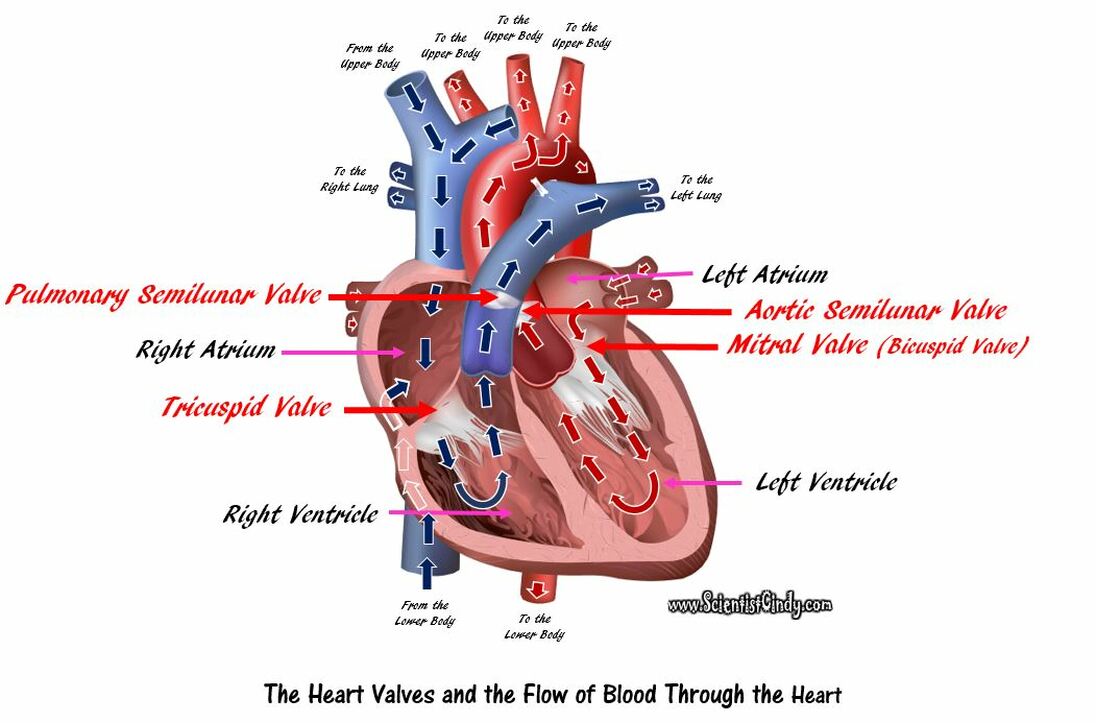
Deoxygenated blood comes from the body and goes to the right atrium. The blood then travels through the TRICUSPID VALVE to get to the right ventricle. Once the right ventricle contracts, this blood is then pumped through the PULMONARY SEMILUNAR VALVE to the pulmonary arteries which will carry the blood to the lungs.
Oxygenated blood comes from the lungs and enters the left atrium. The blood then travels through the MITRAL (BICUSPID) VALVE to the left ventricle. When the left ventricle contracts, the blood is pumped through the AORTIC SEMILUNAR VALVE to the aorta where it will travel to the body.
Oxygenated blood comes from the lungs and enters the left atrium. The blood then travels through the MITRAL (BICUSPID) VALVE to the left ventricle. When the left ventricle contracts, the blood is pumped through the AORTIC SEMILUNAR VALVE to the aorta where it will travel to the body.

THE BLOOD VESSELS
The cardiovascular system has specialized vessels that transport blood around the body.
These vessels fall into one of two broad categories; arteries and veins.
These vessels fall into one of two broad categories; arteries and veins.
|
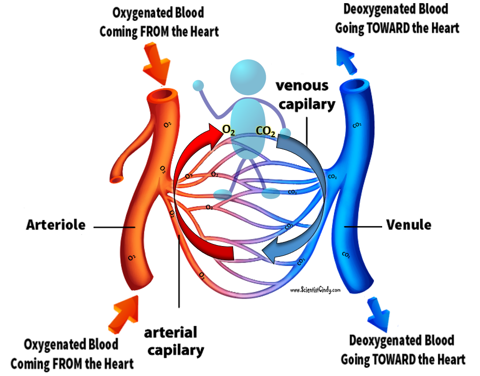
ARTERIES = Arteries are the blood vessels that are carrying blood AWAY FROM the heart. The arteries have high pressure.
VEINS = Veins are the blood vessels that are carrying blood TOWARDS the heart. The veins have low pressure since they are further away from the heart. Veins have one-way valves that prevent the blood from flowing backward. |
|
Inside the capillaries, exchange of oxygen and carbon dioxide takes place. Red blood cells inside the capillary releases their oxygen which passes through the wall and into the surrounding cells. No cells of the body can be more than just a few cells away from tissue. The
tissue then releases waste, such as carbon dioxide, which then passes through the wall and into the red blood cells.
ARTERIES
Arteries are muscular blood vessels that carry blood away from the heart, oxygenated and deoxygenated blood . The pulmonary arteries will carry deoxygenated blood to the lungs and the systemic arteries will carry oxygenated blood to the rest of the body. Arteries have a thick wall that consists of three layers. The inside layer is called the endothelial, the middle layer is mostly smooth muscle and the outside layer is connective tissue. The artery walls are thick so that when blood enters under pressure the walls can expand.
Arteries carry blood to smaller blood vessels called arterioles. From the arterioles, the blood then enters one or more capillaries. The capillaries are the sites of gas exchange and nutrient/waste exchange. The walls of capillaries are only one cell-layer thick. The walls of the capillaries must be extremely thin in order to allow for diffusion of gasses and other substances to occur. The capillaries are so thin that the blood cells can only pass in single file.
Arterioles
An arteriole is a small artery that extends and leads to capillaries. Arterioles have thick smooth muscular walls. These smooth muscles are able to contract (causing vessel constriction) and relax (causing vessel dilation). This contracting and relaxing affects blood pressure; the higher number of vessels dilated, the lower blood pressure will be. Arterioles are just visible to the naked eye.
Capillaries
Capillaries are the smallest of a body’s vessels; they connect arteries and veins, and most closely interact with tissues. They are very prevalent in the body; total surface area is about 6,300 square meters. Because of this, no cell is very far from a capillary, no more than 50 micrometers away. The walls of capillaries are composed of a single layer of cells, the endothelium, which is the inner lining of all the vessels. This layer is so thin that molecules such as oxygen, water and lipids can pass through them by diffusion and enter the tissues. Waste products such as carbon dioxide and urea can diffuse back into the blood to be carried away for removal from the body. The "capillary bed" is the network of capillaries present throughout the body. These beds are able to be “opened” and “closed” at any given time, according to need. This process is called autoregulation and capillary beds usually carry no more than 25% of the amount of blood it could hold at any time. The more metabolically active the cells, the more capillaries it will require to supply nutrients.
The Arteries
The main arteries of the heart include
1) The Aorta
2) The Pulmonary Trunk
3) The Pulmonary Arteries
4) The Coronary Arteries
The main arteries of the heart include
1) The Aorta
2) The Pulmonary Trunk
3) The Pulmonary Arteries
4) The Coronary Arteries
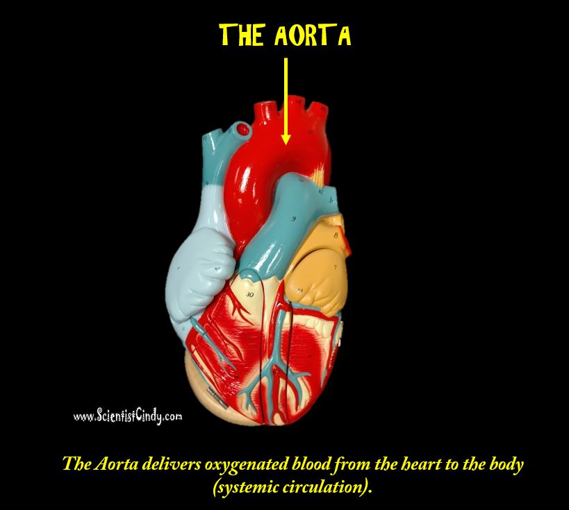
AORTA
|
The largest artery in the body is the AORTA. It is the largest and strongest artery in the body and it marks the beginning of the systemic circuit (HEART --> BODY --> HEART). Oxygenated blood gets pumped out of the left ventricle through the aortic semilunar valve, into the aorta. The aorta is the largest of the arteries in the systemic circuit. The blood is pumped from the left ventricle into the aorta and from there it branches to all parts of the body. The aorta is an elastic artery, and as such is able to distend. When the left ventricle contracts to force blood into the aorta, the aorta expands. This stretching gives the potential energy that will help maintain blood pressure during diastole, as during this time the aorta contracts passively.
|
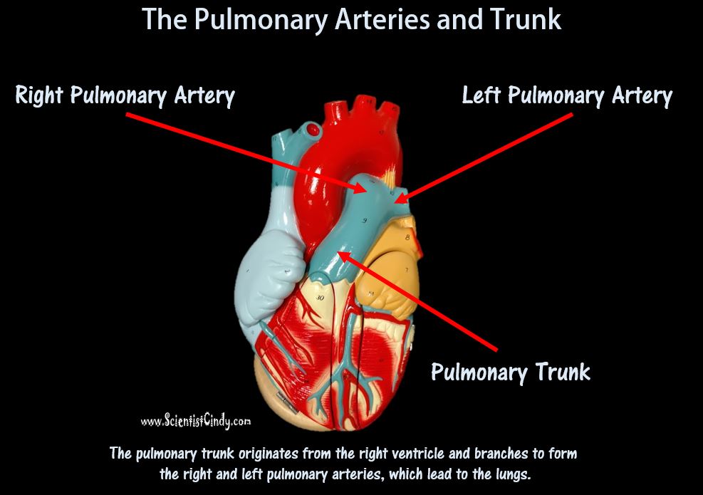
PULMONARY TRUNK AND PULMONARY ARTERIES
The second largest artery in the body is actually colored BLUE because it is carrying deoxygenated blood from the right ventricle to the lungs. This artery is called the pulmonary trunk. The pulmonary trunk branches off to form the right and left pulmonary arteries. The right pulmonary artery carries the blood to the right lung and the left pulmonary artery carries blood to the left lung.
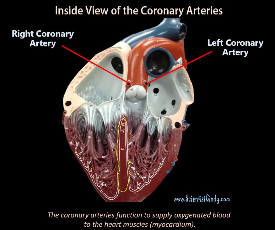
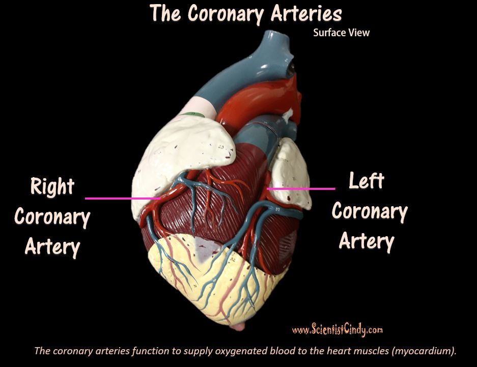
CORONARY ARTERIES
Although blood fills the chambers of the heart, the muscle tissue of the heart, or myocardium, is so thick that it requires coronary blood vessels to deliver blood deep into the myocardium. The vessels that supply blood high in oxygen to the myocardium are known as coronary arteries. The vessels that remove the deoxygenated blood from the heart muscle are known as cardiac veins. The coronary arteries that run on the surface of the heart are called epicardial coronary arteries. These relatively narrow vessels are commonly affected by atherosclerosis and can become blocked, causing angina (heart pain) or a myocardial infarction (heart attack). The coronary arteries are classified as "end circulation", since they represent the only source of blood supply to the myocardium.
|
There are two main coronary arteries:
• Right coronary artery • Left coronary artery Both of these arteries originate from the beginning (root) of the aorta, immediately above the aortic valve. |
|
The heart itself needs a supply of blood. There are two arteries that begin at the base of the aorta called the right and left coronary arteries. The right coronary artery supplies oxygenated blood to the cardiac muscles on the right side of the heart and the left coronary artery supplies oxygenated blood to the cardiac muscles on the left side of the heart.
|
|
THE VEINS
Systemic arteries split from the aorta and direct blood into the capillaries. Cells consume the oxygen and nutrients and add carbon dioxide, wastes, enzymes and hormones. The veins drain the deoxygenated blood from the capillaries and return the blood to the right atrium.
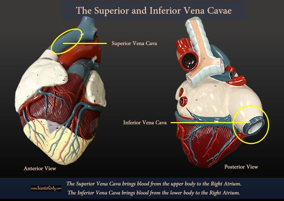
The largest veins of the body are the superior and inferior vena cava. The superior vena cava brings deoxygenated blood from the upper body to the right atrium. The inferior vena cava brings deoxygenated blood from the lower body to the right atrium.
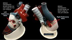
Oxygenated blood coming from the lungs returns to the heart through the right and left pulmonary veins. The right pulmonary veins bring oxygenated blood from the right lung to the left atrium of the heart. The left pulmonary veins bring oxygenated blood from the left lung to the left atrium of the heart.
VEINS
Veins carry blood to the heart. Most (~70%) of the blood in our bodies is found in veins (the venous system). The veins lack the smooth muscle that we see in arteries. Since veins are vessels that are headed back to the heart, it has been a long time (and distance) from its last "oush" from the heart. For this reason, veins have low blood pressure compared to arteries. Larger veins have one-way valves called venous valves to prevent the backflow of blood. Is a person is standing still for long periods or is bedridden, blood
can accumulates in veins and can cause varicose veins. Veins are used medically as points of access to the blood stream,
permitting the withdrawal of blood specimens (venipuncture) for testing purposes, and enabling the infusion of fluid, electrolytes, nutrition, and medications (intravenous delivery).
Venules
A venule is a small vein that allows deoxygenated blood to return from the capillary beds to the larger blood veins, except in the pulmonary circuit were the blood is oxygenated. Venules have three layers; they have the same makeup as arteries with less smooth muscle, making them thinner.
TISSUE LAYERS OF THE HEART
|
Pericardium
The pericardium (lining of the pericardial cavity) is the thick, membranous sac that surrounds the heart. It protects and lubricates the heart. There are two layers to the pericardium: the fibrous pericardium and the serous pericardium. The serous pericardium is divided into two layers; in between these two layers there is a space called the pericardial cavity. The walls of the heart have 3 layers.
|
|
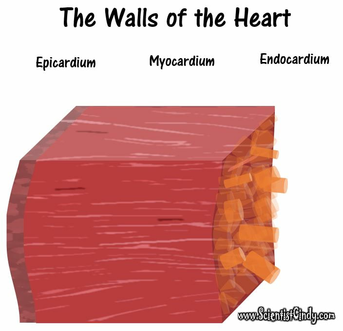
Endocardium
The endocardium is the innermost lining of the heart which consists of the endothelial cells forming a smooth membrane in places, and a pocked and tribeculated surface in others (mainly the ventricles, or lower pumping chambers). |
Epicardium
The outer most layer next to the myocardium is known as the Epicardium. This is the outer layer after endocardium and myocardium that consists of a thin layer of connective tissue and fat. Myocardium
The myocardium is the muscular tissue of the heart. The myocardium is composed of specialized cardiac muscle cells with an ability not possessed by muscle tissue elsewhere in the body. Cardiac muscle, like other muscles, can contract, but it can also conduct electricity, like nerves. The blood to the myocardium is supplied by the coronary arteries. If these arteries are occluded by atherosclerosis and/or thrombosis, this can lead to angina pectoris or myocardial infarction due to ischemia (lack of oxygen). Failure of the heart to contract properly (for various reasons) is termed heart failure, generally leading to fluid retention, edema, pulmonary edema, renal insufficiency, hepatomegaly, a shortened life expectancy and decreased quality of life. |
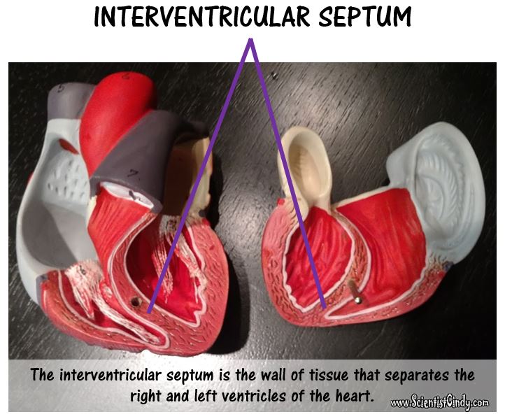
The interventricular septum (ventricular septum) is the thick wall separating the lower chambers (the ventricles) of the heart from one another. The ventricular septum is directed backward and to the right, and is curved toward the right ventricle. The greater portion of it is thick and muscular and constitutes the muscular ventricular septum. Its upper and posterior part, which separates the aortic vestibule from the lower part of the right atrium and upper part of the right ventricle, is thin and fibrous, and is termed the membranous ventricular septum.
The Heart Beat
|
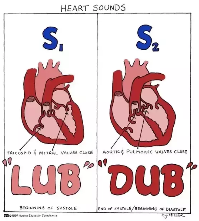
S1 - 1st sound of the heart beat (LUB)
|
|
The “lub” is the sound of the mitral and tricuspid valves closing as the heart starts to contract; the “dub” is the sound of the aortic and pulmonary valves closing at the end of the contraction.
|
|
|
Blood pressure (BP), the force per unit area exerted on a vessel wall by the contained blood, is expressed in millimeters of
mercury (mm Hg). For example, a blood pressure of 120 mm Hg is equal to the pressure exerted by a column of mercury
120 mm high. Unless stated otherwise, the term blood pressure means systemic arterial blood pressure in the largest arteries near the heart. The pressure gradient—the differences in blood pressure within the vascular system—provides the driving force that keeps blood moving, always from an area of higher pressure to an area of lower pressure, through the body.
The terms diastole and systole refer to when the heart muscles relax and contract. The balance between diastole and systole determines a person's blood pressure.
Cardiac Cycle
The cardiac cycle is the sequence of events that occurs when the heart beats. As the heart beats, it circulates blood through pulmonary and systemic circuits of the body.
There are two phases of the cardiac cycle:
SYSTOLE and DIASTOLE
The cardiac cycle is the sequence of events that occurs when the heart beats. As the heart beats, it circulates blood through pulmonary and systemic circuits of the body.
There are two phases of the cardiac cycle:
SYSTOLE and DIASTOLE
What are diastole and systole?
Diastole is when the heart muscle relaxes and systole is when the heart muscle contracts.Diastole is defined by the following characteristics:
Diastole is when the heart muscle relaxes and systole is when the heart muscle contracts.Diastole is defined by the following characteristics:
- Diastole is when the heart muscle relaxes.
- When the heart relaxes, the chambers of the heart fill with blood, and a person's blood pressure decreases.
- Systole is when the heart muscle contracts.
- When the heart contracts, it pushes the blood out of the heart and into the large blood vessels of the circulatory system. From here, the blood goes to all of the organs and tissues of the body.
- During systole, a person's blood pressure increases.
Every single 'beat' of the heart involves three major stages:
Throughout the cardiac cycle, the blood pressure increases and decreases. As ventricles contract the pressure rise, causing the AV valves to slam shut.
- atrial systole
- ventricular systole
- complete cardiac diastole
Throughout the cardiac cycle, the blood pressure increases and decreases. As ventricles contract the pressure rise, causing the AV valves to slam shut.
Cardiac Cycle
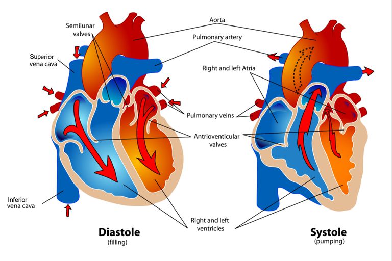
- Ventricles begin contraction, pressure rises, and AV valves close (lub).
- Pressure builds, semilunar valves open, and blood is ejected into arteries.
- Pressure in ventricles falls; semilunar valves close (dub).
- Pressure in ventricles falls below that of atria, and AV valve opens. Ventricles fill.
- Atria contract, sending last of blood to ventricles.
Cardiac cycle is the term used to describe the relaxation and contraction that occur, as a heart works to pump blood through the body.
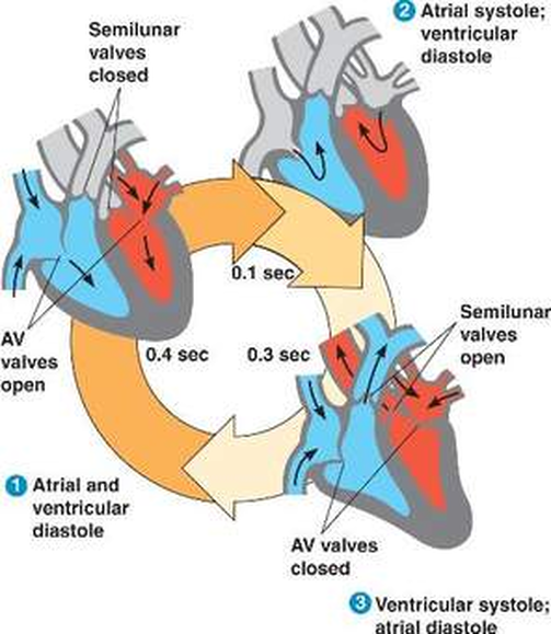
There are 3 phases involved in cardiac cycle;
- Atrial systole [systole= contraction]
- Ventricular systole
- Complete cardiac diastole [cardiac= atria and ventricle] [diastole= rest].
1) Atrial systole = both the right and left atria CONTRACT
The first phase of the cardiac cycle is ATRIAL SYSTOLE. This is when both atria contract. During atrial systole, the contraction of both atria forces blood from the atria to travels through the atrioventricular valves and enter into the ventricles. As this blood travels from the atria to the ventricles, the atria quickly fill back up with blood coming into the heart. The right atrium is filled with deoxygenated blood coming from the body through the superior and inferior vena cava. The left atrium is filled with oxygenated blood from the lungs traveling through the right and left pulmonary veins.
2) Ventricular systole = both the right and left ventricles CONTRACT
The second phase of the cardiac cycle is VENTRICLE SYSTOLE. This is when both ventricles contract. During ventricular systole, the contraction of both ventricles to force blood into the pulmonary [lungs for oxygenation] and systemic [throughout the body] circulation, it takes place for about 0.3s.
The impulse sent by SA node is delayed for 0.1s before reaching the AV node, which results in contraction of the atria before the ventricles. As the AV node receives the impulse of contraction, it sends it inferiorly to the bundle of HIS, which continues downwards to the apex of the heart through the right and left AV bundle. The impulse is conducted superior-laterally to the level of the ventricles, then the right and left AV bundle together with the purkinje fibres initiate the impulse of ventricular myocardial contraction, hereby forcing the blood into the systemic circulation.
3) Cardiac diastole = All 4 chambers of the heart are relaxed.
Cardiac diastole is the time in which the entire heart is relaxed. This means that we have ventricular diastole in which the ventricles are relaxed, as well as atrial diastole in which the atria are relaxed.
* Note that atrial systole and ventricular systole DO NOT OCCUR AT THE SAME TIME. In atrial systole, both the right and left atria contract at the same time. In ventricular systole, both the right and left ventricles contract at the same time. In contrast, atrial diastole and ventricular diastole occur simultaneously (at the same time), such that all 4 chambers are relaxed.
Cardiac Cycle
KEEP IN MIND: Blood flows from higher to lower pressure.
Contraction of the heart muscle in heart chambers is called SYSTOLE. The contraction of the heart muscles functions to increase the pressure within that chamber, which forces the blood out of that chamber through the appropriate one-way valve. Atrial systole occurs just before ventricular systole. Both atria contract at about the same time and both ventricles contract at about the same time.
SYSTOLE
Let's look at what happens in each chamber during the SYSTOLIC PHASE of the cardiac cycle.
- When the right atrium contracts (during atrial systole), this contraction increases the pressure of the blood in that chamber. This contraction (or increased force) forces blood out of the right atrium and into the right ventricle through the one-way valve (tricuspid valve).
- When the right ventricle contracts (during ventricular systole), this contraction increases the pressure of the blood in that chamber. This contraction (or increased force) forces blood out of the right ventricle, through the one-way valve (pulmonary semilunar valve) and into the pulmonary trunk. You may recall that the pulmonary trunk branches off to form the right and left pulmonary arteries which lead to the lungs.
- When the left atrium contracts (during atrial systole), this contraction increases the pressure of the blood in that chamber. This contraction (or increased force) forces blood out of the left atrium and into the left ventricle through the one-way valve (bicuspid or mitral valve).
- When the left ventricle contracts (during ventricular systole), this contraction increases the pressure of the blood in that chamber. This contraction (or increased force) forces blood out of the left ventricle, through the one-way valve (aortic semilunar valve) and into the aorta. You may recall that the aorta branches off to provide all areas of the body (except the lungs) with oxygen-rich blood.
DIASTOLE
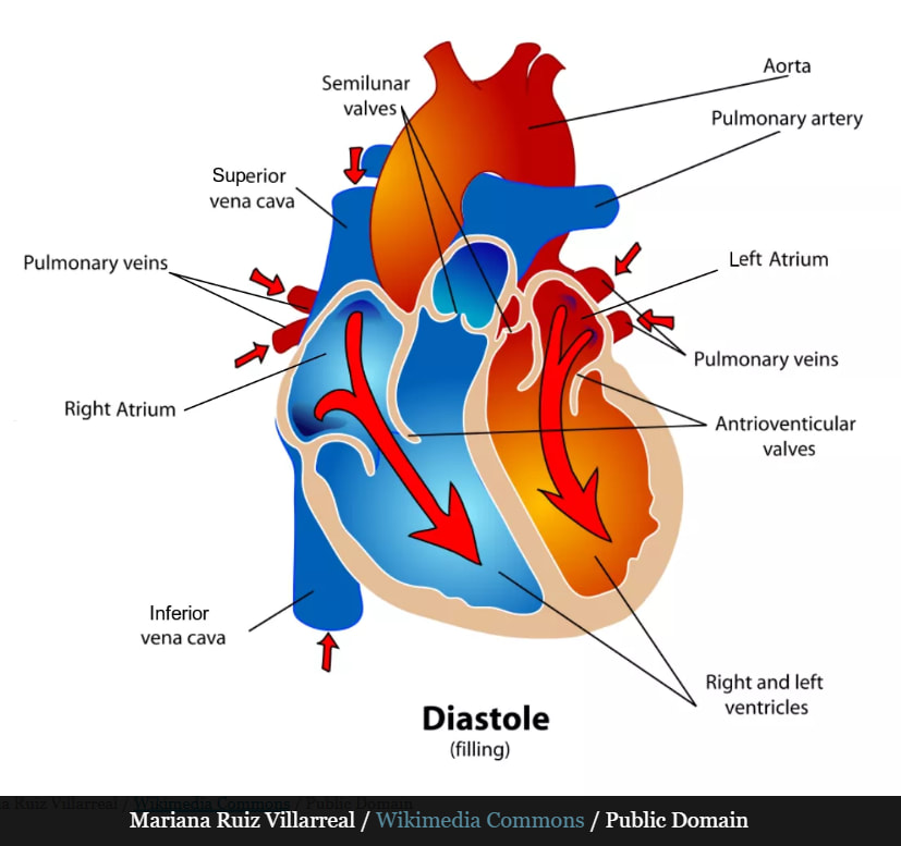
Relaxation of the heart muscles around the chambers of the heart is called DIASTOLE. When a chamber becomes relaxed (after it has contracted during SYSTOLE) the pressure in those now relaxed chambers drops. Since blood travels from high pressure to low pressure, the chambers will quickly fill back up with blood during diastole. All 4 heart chambers undergo diastole at the same time.
Let's look at what happens during diastole in each chamber.
- The right atrium will fill up with blood from the superior and inferior venae cavae. Since the atria contract a little bit before the ventricles do (atrial systole occurs just prior to ventricular systole), the pressure in the right atrium will be higher that the pressure in the right ventricle.
- When the right ventricle relaxes (during ventricular diastole) high pressure blood from the right atrium passes through the one-way valve (tricuspid valve) and fills the right ventricle.
- The left atrium will fill up with blood from the right and left pulmonary veins. Since the atria contract a little bit before the ventricles do (atrial systole occurs just prior to ventricular systole), the pressure in the left atrium will be higher that the pressure in the left ventricle.
- When the left ventricle relaxes (during ventricular diastole) high pressure blood from the left atrium passes through the one-way valve (bicuspid or mitral valve) and fills the left ventricle.
2nd Distole Period
In the second diastole period, the semilunar valves close and the atrioventricular valves open. Oxygenated blood from the pulmonary veins fills the left atrium (blood from the venae cavae is also filling the right atrium at this time). The SA node contracts again triggering both atria to contract. Atrial contraction causes the left atrium to empty its contents into the left ventricle (the right atrium is also emptying blood into the right ventricle at this time). The mitral valve, located between the left atrium and left ventricle, prevents oxygenated blood from flowing back into the left atrium.
2nd Systole Period
During the second systole period, the atrioventricular valves close and the semilunar valves open. The ventricles receive impulses and contract. Oxygenated blood in the left ventricle is pumped to the aorta and the aortic valve prevents the oxygenated blood from flowing back into the left ventricle (oxygen-depleted blood is also being pumped from the right ventricle to the pulmonary artery at this time). The aorta branches out to provide oxygenated blood to all parts of the body through systemic circulation. After its tour through the body, oxygen-depleted blood is returned to the heart via the venae cavae.
Heart rate is a term used to describe the frequency of the cardiac cycle and is calculated as the number of contractions (heart beats) of the heart in one minute or simply "beats per minute" (bpm).
When resting, the adult human heart beats at about 70 bpm (males) and 75 bpm (females).
A heart rate below 60 bpm is bradycardia.
A heart rate over 100 bpm is tachycardia.
Resting heart rates can be significantly lower in athletes, and significantly higher in the obese. The body can increase the heart rate in response to a wide variety of conditions in order to increase the cardiac output (the amount of blood ejected by the heart per unit time). Exercise, environmental stressors or psychological stress can cause the heart rate to increase above the resting rate. The pulse is the most straightforward way of measuring the heart rate, but it can be deceptive when some strokes do not lead to much cardiac output. In these cases (as happens in some arrhythmias), the heart rate may be considerably higher than the pulse.
Cardiac Muscle TissueCardiac muscle is striated muscle that is present only in the heart. Cardiac muscle fibers have a single nucleus, are branched, and joined to one another by intercalated discs that contain gap junctions for depolarization between cells and desmosomes to hold the fibers together when the heart contracts. Contraction in each cardiac muscle fiber is triggered by Ca++ ions in a similar manner as skeletal muscle, but here the Ca++ ions come from SR and through voltage-gated calcium channels in the sarcolemma. Pacemaker cells stimulate the spontaneous contraction of cardiac muscle as a functional unit, called a syncytium.
Cardiac muscle tissue is only found in the heart. Highly coordinated contractions of cardiac muscle pump blood into the vessels of the circulatory system. Similar to skeletal muscle, cardiac muscle is striated and organized into sarcomeres, possessing the same banding organization as skeletal muscle (Figure 1). However, cardiac muscle fibers are shorter than skeletal muscle fibers and usually contain only one nucleus, which is located in the central region of the cell. Cardiac muscle fibers also possess many mitochondria and myoglobin, as ATP is produced primarily through aerobic metabolism. Cardiac muscle fibers cells also are extensively branched and are connected to one another at their ends by intercalated discs. An intercalated disc allows the cardiac muscle cells to contract in a wave-like pattern so that the heart can work as a pump.
Intercalated discs are part of the sarcolemma and contain two structures important in cardiac muscle contraction:
gap junctions and desmosomes.
- A gap junction forms channels between adjacent cardiac muscle fibers that allow the depolarizing current produced by cations to flow from one cardiac muscle cell to the next. This joining is called electric coupling, and in cardiac muscle it allows the quick transmission of action potentials and the coordinated contraction of the entire heart. This network of electrically connected cardiac muscle cells creates a functional unit of contraction called a syncytium.
- The remainder of the intercalated disc is composed of desmosomes. A desmosome is a cell structure that anchors the ends of cardiac mContractions of the heart (heartbeats) are controlled by specialized cardiac muscle cells called pacemaker cells that directly control heart rate. Although cardiac muscle cannot be consciously controlled, the pacemaker cells respond to signals from the autonomic nervous system (ANS) to speed up or slow down the heart rate. The pacemaker cells can also respond to various hormones that modulate heart rate to control blood pressure.
The wave of contraction that allows the heart to work as a unit, called a functional syncytium, begins with the pacemaker cells. This group of cells is self-excitable and able to depolarize to threshold and fire action potentials on their own, a feature called autorhythmicity; they do this at set intervals which determine heart rate. Because they are connected with gap junctions to surrounding muscle fibers and the specialized fibers of the heart’s conduction system, the pacemaker cells are able to transfer the depolarization to the other cardiac muscle fibers in a manner that allows the heart to contract in a coordinated manner.
Another feature of cardiac muscle is its relatively long action potentials in its fibers, having a sustained depolarization “plateau.” The plateau is produced by Ca++ entry though voltage-gated calcium channels in the sarcolemma of cardiac muscle fibers. This sustained depolarization (and Ca++ entry) provides for a longer contraction than is produced by an action potential in skeletal muscle. Unlike skeletal muscle, a large percentage of the Ca++ that initiates contraction in cardiac muscles comes from outside the cell rather than from the SR.
Another feature of cardiac muscle is its relatively long action potentials in its fibers, having a sustained depolarization
uscle fibers together so the cells do not pull apart during the stress of individual fibers contracting
Summary
As with all of the body systems, the cardiovascular system plays a part in maintaining
homeostasis. The nervous system regulates the functioning of the heart based on what the
heart is supposed to do. The pumping of the heart maintains normal blood pressure and
proper oxygenation of tissues. The vascular system forms passageways for the blood, but
they aren't simply just a pipeline system. The vessels are not passive tubes, but rather
active contributors to homeostasis. The arteries and veins help maintain blood pressure,
and the capillaries provide sites for the necessary exchanges of materials between the blood
and the tissues.
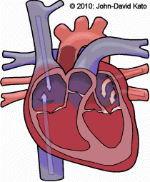
Structure Function
- AORTA - LARGEST ARTERY - BRINGS OXYGENATED BLOOD FROM THE LEFT VENTRICLE TO THE BODY
- SUPERIOR VENA CAVA - BRINGS DEOXYGENATED BLOOD FROM THE UPPER BODY TO THE RIGHT ATRIUM
- INFERIOR VENA CAVA (NOT SHOWN) - BRINGS DEOXYGENATED BLOOD FROM THE LOWER BODY TO THE RIGHT ATRIUM
- AORTIC ARCH - HOLDS THE 3 ARTERIES THAT BRANCH UPWARD TO SEND BLOOD TO UPPER BODY (the brachiocephalic trunk, the left common carotid artery and the left subclavian artery)
- DESCENDING AORTA (NOT SHOWN) - THIS IS THE PORTION OFTHE AORTA THAT COMES AFTER THE AORTIC ARCH AND TURNS INFERIORLY TO SUPPLY OXYGENATED BLOOD TO THE LOWER BODY.
- PULMONARY TRUNK - BRINGS DEOXYGENATED BLOOD FROM THE RIGHT VENTRICLE TO THE RIGHT AND LEFT PULMONARY ARTERIES.
- LEFT PULMONARY ARTERIES - BRINGS DEOXYGENATED BLOOD TO THE LEFT LUNG
- RIGHT PULMONARY ARTERIES (NOT SHOWN) - BRINGS DEOXYGENATED BLOOD TO RIGHT LUNG
- LEFT PULMONARY VEINS - BRINGS OXYGENATED BLOOD FROM THE LEFT LUNG TO THE LEFT ATRIUM
- RIGHT PULMONARY VEINS (NOT SHOWN) - BRINGS OXYGENATED BLOOD FROM THE RIGHT LUNG TO THE LEFT AT