Chapter 9 Study Guide
The Central Nervous System
9.1 Emergent Properties of Neural Networks
Neurons in the nervous system link together to form circuits that have specific functions. The most complex circuits are those of
the brain, in which billions of neurons are linked into intricate networks that converge and diverge, creating an infinite number of
possible pathways. Signaling within these pathways creates thinking, language, feeling, learning, and memory—the complex behaviors
that make us human. Some neuroscientists have proposed thatthe functional unit of the nervous system be changed from the
individual neuron to neural networks because even the most basic functions require circuits of neurons.
9.2 Evolution of Nervous Systems
All animals have the ability to sense and respond to changes in their environment. Even single-cell organisms such as Paramecium
are able to carry out the basic tasks of life: finding food, avoiding becoming food, finding a mate. Yet, these unicellular organisms
have no obvious brain or integrating center. They use the resting membrane potential that exists in living cells and many of the
same ion channels as more complex animals to coordinate their daily activities.
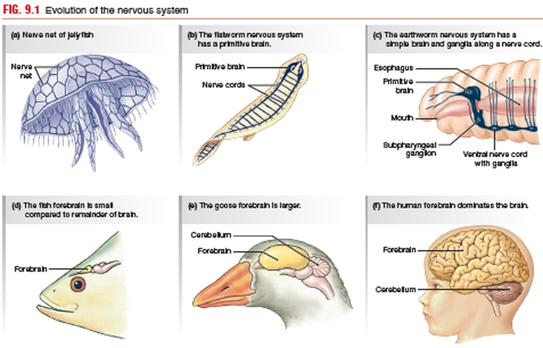
Some of the first multicellular animals to develop neurons were members of the phylum Cnidaria, the jellyfish and sea anemones.
Their nervous system is a nerve net composed of sensory neurons, connective interneurons, and motor neurons that innervate
muscles and glands (FIG. 9.1a). These animals respond to stimuli with complex behaviors, yet without input from an identifiable
control center. If you watch a jellyfish swim or a sea anemone maneuver a piece of shrimp into its mouth, it is hard to imagine
how a diffuse network of neurons can create such complex coordinated movements. However, the same basic principles of
neural communication apply to jellyfish and humans. Electrical signals in the form of action potentials, and chemical signals passing
across synapses, are the same in all animals. It is only in the number and organization of the neurons that one species differs
from another.
In the primitive flatworms, we see the beginnings of a nervous system as we know it in higher animals, although in flatworms the
distinction between central nervous system (CNS) and peripheral nervous system is not clear. Flatworms have a rudimentary brain consisting of a cluster of nerve cell bodies concentrated in the head, or cephalic region. Two large nerves called nerve cords come off the primitive brain and lead to a nerve network that innervates distal regions of the flatworm body (Fig. 9.1b).
The segmented worms, or annelids, such as the earthworm,
have a more advanced central nervous system (Fig. 9.1c). Clusters of cell bodies are no longer restricted to the head region, as they
are in flatworms, but also occur in fused pairs, called ganglia (singular ganglion) [p. 232], along a nerve cord. Because each segment
of the worm contains a ganglion, simple reflexes can be integrated within a segment without input from the brain. Reflexes that do not require integration in the brain also occur in higher animals and are called spinal reflexes in humans and other vertebrates.
Annelids and higher invertebrates have complex reflexes controlled through neural networks.
Nerve cell bodies clustered into brains persist throughout the more advanced phyla and become increasingly more complex.
One advantage to cephalic brains is that in most animals, the head is the part of the body that first contacts the environment as the
animal moves. For this reason, as brains evolved, they became associated with specialized cephalic receptors, such as eyes for vision
and chemoreceptors for smell and taste. In the higher arthropods, such as insects, specific regions of the brain are associated with particular functions. More complex brains are associated with complex behaviors, such as the ability of social insects like ants and bees to organize themselves into colonies, divide labor, and communicate with one another. The octopus (a cephalopod mollusk) has the most sophisticated brain development among the invertebrates, as well as the most sophisticated behavior.
seen in the forebrain region {fore, in front}, which includes the cerebrum
{cerebrum, brain; adjective cerebral}.
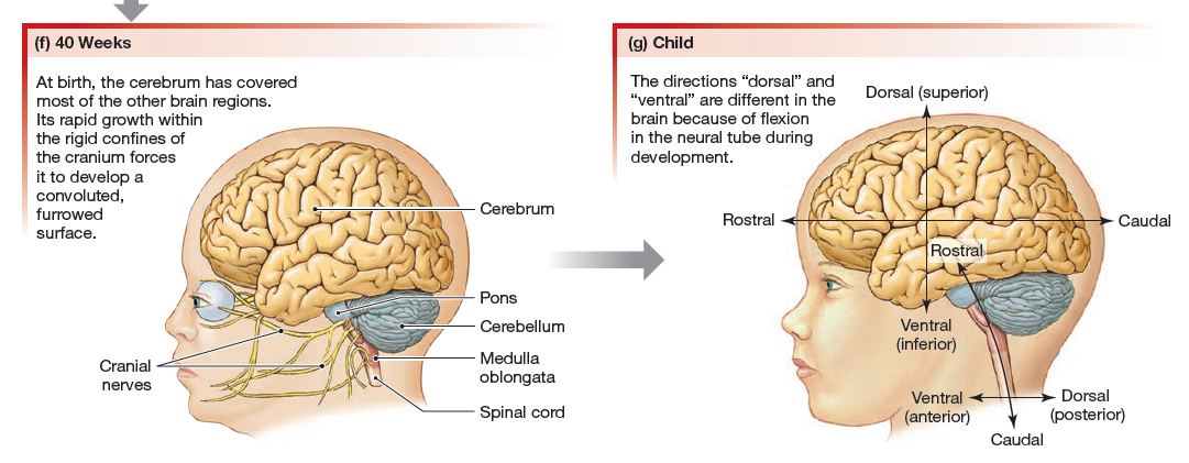
In fish, the forebrain is a small bulge dedicated mainly to processing olfactory information about odors in the environment (Fig. 9.1d). In birds and rodents, part of the forebrain has enlarged into a cerebrum with a smooth surface (Fig. 9.1e).
In humans, the cerebrum is the largest and most distinctive part of the brain, with deep grooves and folds (Fig. 9.1f). More than
anything else, the cerebrum is what makes us human. All evidence indicates that it is the part of the brain that allows reasoning and
cognition. The other brain structure whose evolution is obvious in the vertebrates is the cerebellum, a region of the hindbrain devoted to coordinating movement and balance. Birds (Fig. 9.1e) and humans (Fig. 9.1f) both have well-developed cerebellar structures. The cerebellum, like the cerebrum, is readily identifiable in these animals by its grooves and folds.
The vertebrate CNS consists of the brain and the spinal cord. As you learned in the previous section, brains increase in complexity
and degree of specialization as we move up the phylogenetic tree from fish to humans. However, if we look at the
vertebrate nervous system during development, a basic anatomical pattern emerges. In all vertebrates, the CNS consists of
layers of neural tissue surrounding a fluid-filled central cavity lined with epithelium.
9.3 Anatomy of the Central
Nervous System
The Embryologic Perspective
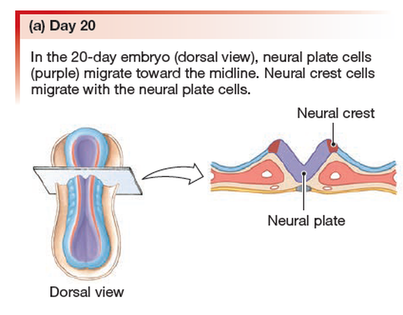
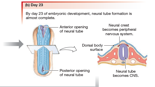
The development of the nervous system starts early in embryonic development. The outer layer of the embryo, the ectoderm, gives rise to the skin and the nervous system. A specialized region of this layer, the neuroectoderm, becomes a groove that folds in and becomes the neural tube beneath the dorsal surface of the embryo. The anterior end of the neural tube develops into the brain, and the posterior region becomes the spinal cord. Tissues at the edges of the neural groove, when it closes off, are called the neural crest and migrate through the embryo to give rise to PNS structures as well as some non-nervous tissues.
The development of the nervous system starts early in embryonic development. The outer layer of the embryo, the ectoderm, gives rise to the skin and the nervous system. A specialized region of this layer, the neuroectoderm, becomes a groove that folds in and becomes the neural tube beneath the dorsal surface of the embryo. The anterior end of the neural tube develops into the brain, and the posterior region becomes the spinal cord. Tissues at the edges of the neural groove, when it closes off, are called the neural crest and migrate through the embryo to give rise to PNS structures as well as some non-nervous tissues.
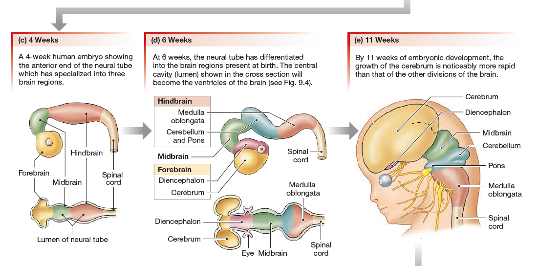
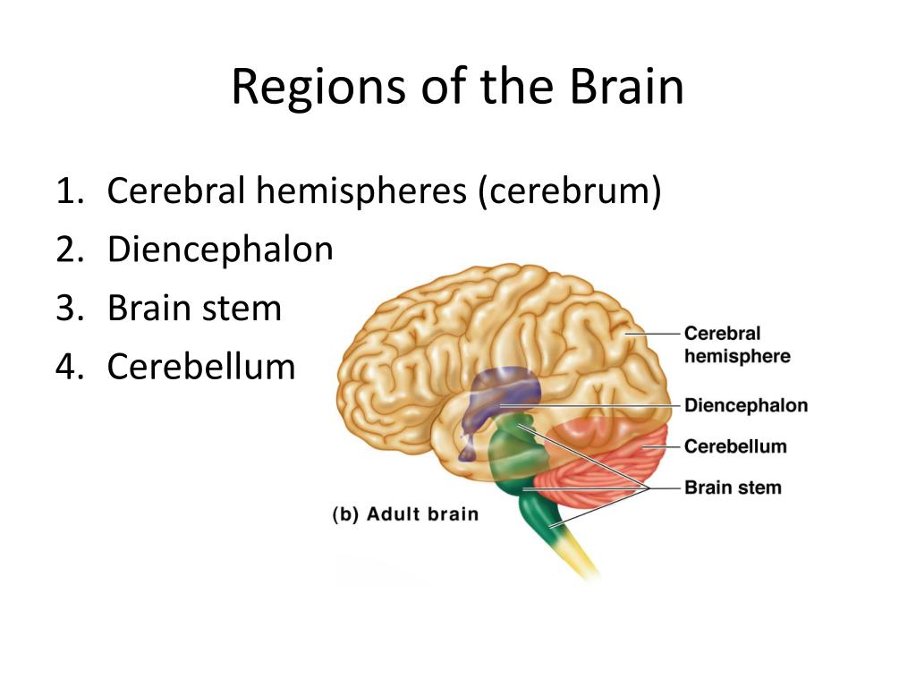
The brain develops from this early tube structure and gives rise to specific regions of the adult brain. As the neural tube grows and differentiates, it enlarges into three vesicles that correspond to the forebrain, midbrain, and hindbrain regions of the adult brain. Later in development, two of these three vesicles differentiate further, resulting in five vesicles. Those five vesicles can be aligned with the four major regions of the adult brain. The cerebrum is formed directly from the telencephalon. The diencephalon is the only region that keeps its embryonic name. The mesencephalon, metencephalon, and myelencephalon become the brain stem. The cerebellum also develops from the metencephalon and is a separate region of the adult brain.
The spinal cord develops out of the rest of the neural tube and retains the tube structure, with the nervous tissue thickening and the hollow center becoming a very small central canal through the cord. The rest of the hollow center of the neural tube corresponds to open spaces within the brain called the ventricles, where cerebrospinal fluid is found.
The spinal cord develops out of the rest of the neural tube and retains the tube structure, with the nervous tissue thickening and the hollow center becoming a very small central canal through the cord. The rest of the hollow center of the neural tube corresponds to open spaces within the brain called the ventricles, where cerebrospinal fluid is found.
The Central Nervous System
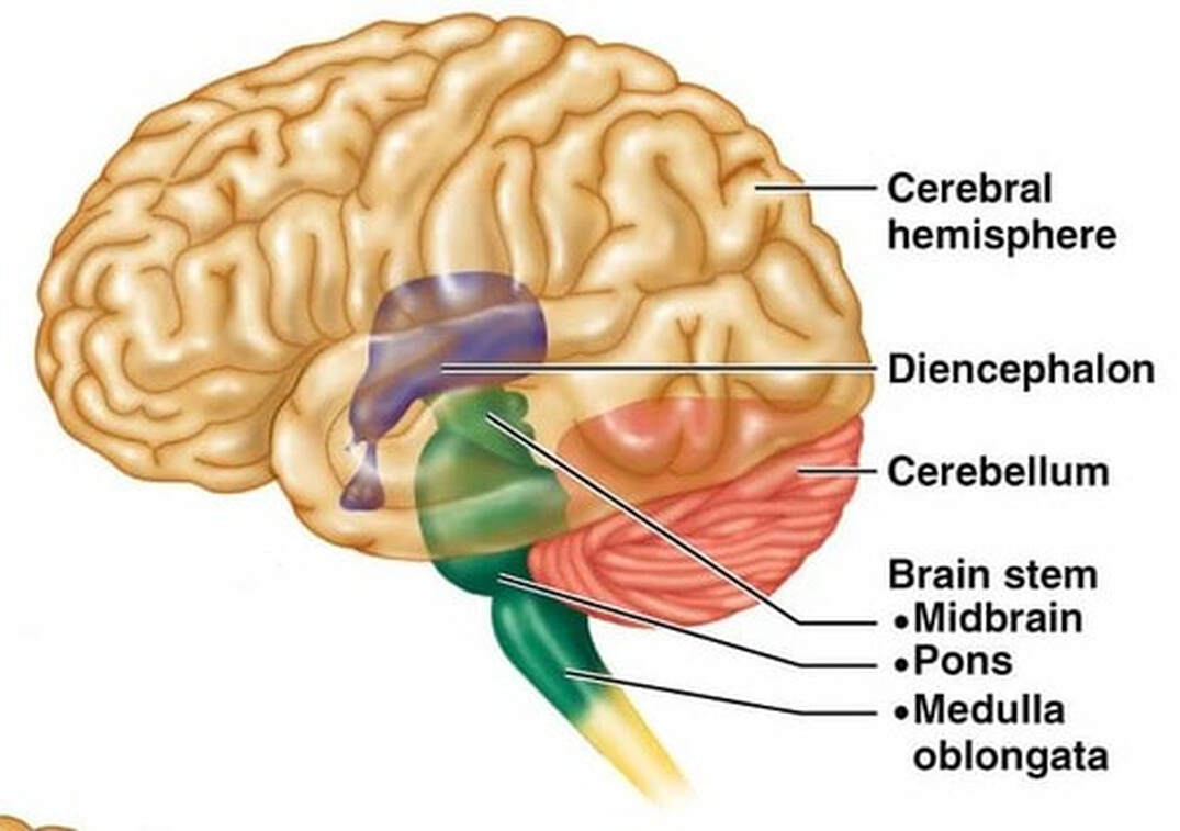
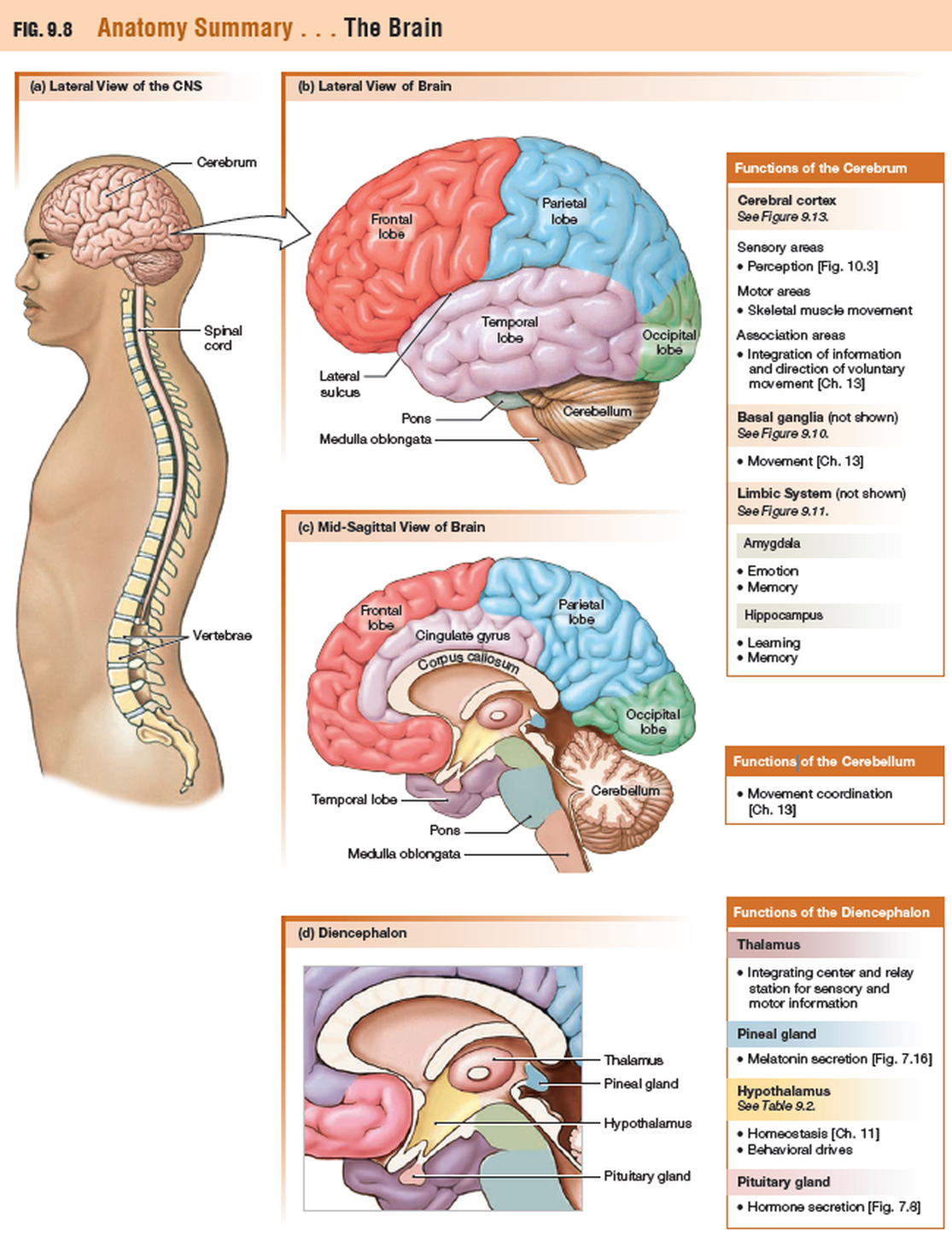
The adult brain is separated into four major regions: the cerebrum, the diencephalon, the brain stem, and the cerebellum. The cerebrum is the largest portion and contains the cerebral cortex and subcortical nuclei. It is divided into two halves by the longitudinal fissure.
The adult brain is separated into four major regions: the cerebrum, the diencephalon, the brain stem, and the cerebellum. The cerebrum is the largest portion and contains the cerebral cortex and subcortical nuclei. It is divided into two halves by the longitudinal fissure.
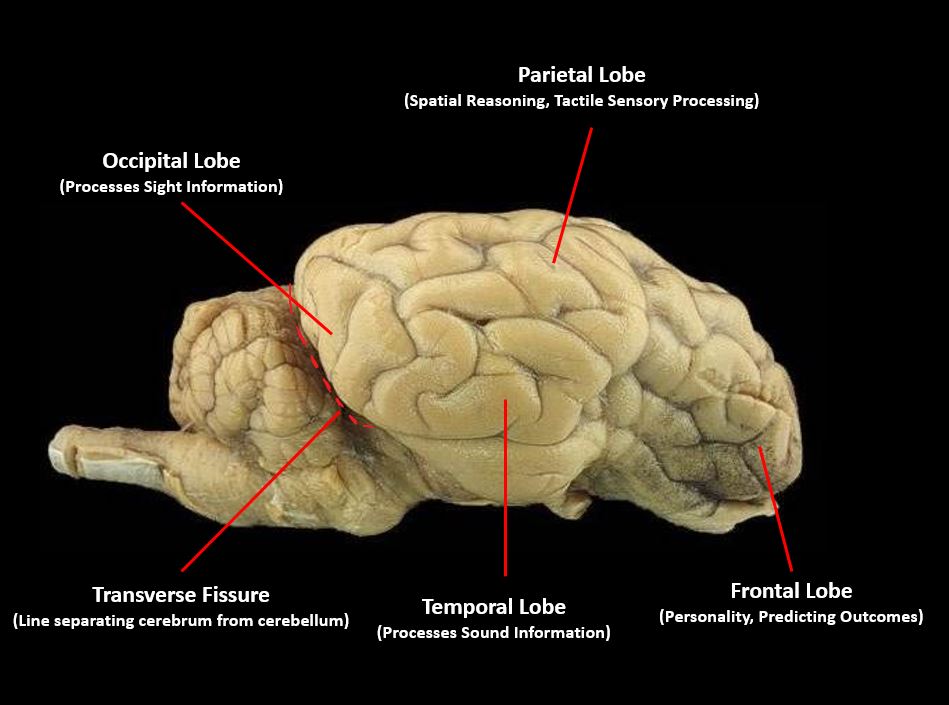
The cortex is separated into the frontal, parietal, temporal, and occipital lobes. The frontal lobe is responsible for motor functions, from planning movements through executing commands to be sent to the spinal cord and periphery. The most anterior portion of the frontal lobe is the prefrontal cortex, which is associated with aspects of personality through its influence on motor responses in decision-making.
The other lobes are responsible for sensory functions. The parietal lobe is where somatosensation is processed. The occipital lobe is where visual processing begins, although the other parts of the brain can contribute to visual function. The temporal lobe contains the cortical area for auditory processing, but also has regions crucial for memory formation.
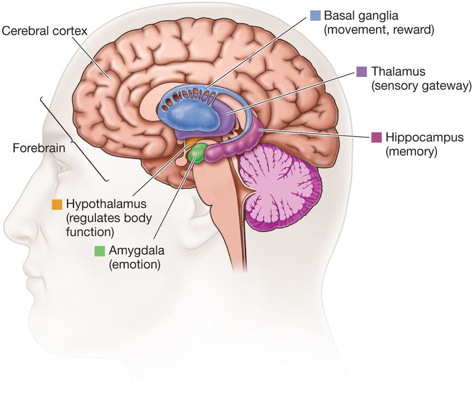
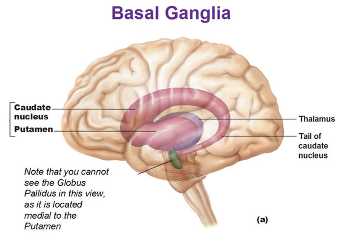
Nuclei beneath the cerebral cortex, known as the subcortical nuclei, are responsible for augmenting cortical functions. The basal nuclei receive input from cortical areas and compare it with the general state of the individual through the activity of a dopamine-releasing nucleus. The output influences the activity of part of the thalamus that can then increase or decrease cortical activity that often results in changes to motor commands. The basal forebrain is responsible for modulating cortical activity in attention and memory. The limbic system includes deep cerebral nuclei that are responsible for emotion and memory.
The diencephalon includes the thalamus and the hypothalamus, along with some other structures. The thalamus is a relay between the cerebrum and the rest of the nervous system. The hypothalamus coordinates homeostatic functions through the autonomic and endocrine systems.
The brain stem is composed of the midbrain, pons, and medulla. It controls the head and neck region of the body through the cranial nerves. There are control centers in the brain stem that regulate the cardiovascular and respiratory systems.
The cerebellum is connected to the brain stem, primarily at the pons, where it receives a copy of the descending input from the cerebrum to the spinal cord. It can compare this with sensory feedback input through the medulla and send output through the midbrain that can correct motor commands for coordination.
The meninges
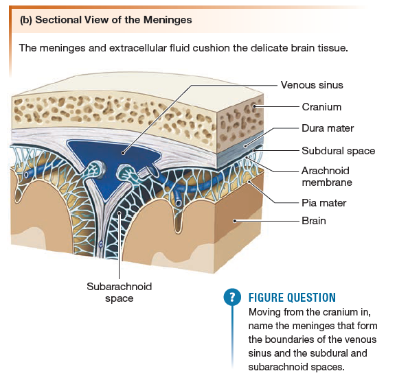
The meninges are membranes that help stabilize the neural tissue and protect it from bruising against the bones of the skeleton. Starting from the bones and moving toward the neural tissue, the membranes are (1) the dura mater, (2) the arachnoid
membrane, and (3) the pia mater
membrane, and (3) the pia mater
- The dura mater -The dura mater {durare, to last + mater, mother} is the thickest of the three membranes (think durable). It is associated with veins that drain blood from the brain through vessels or cavities called sinuses.
- The arachnoid membrane - The middle layer, the arachnoid {arachnoides, cobweblike} membrane, is loosely tied to the inner membrane, leaving a subarachnoid space between the two layers.
- The pia mater - The inner membrane, the pia mater {pius, pious + mater, mother}, is a thin membrane that adheres to the surface of the brain and spinal cord. Arteries that supply blood to the brain are associated with this layer.
Circulation and the Central Nervous System
The CNS has a privileged blood supply established by the blood-brain barrier. Establishing this barrier are anatomical structures that help to protect and isolate the CNS. The arterial blood to the brain comes from the internal carotid and vertebral arteries, which both contribute to the unique circle of Willis that provides constant perfusion of the brain even if one of the blood vessels is blocked or narrowed. That blood is eventually filtered to make a separate medium, the CSF, that circulates within the spaces of the brain and then into the surrounding space defined by the meninges, the protective covering of the brain and spinal cord.
The blood that nourishes the brain and spinal cord is behind the glial-cell–enforced blood-brain barrier, which limits the exchange of material from blood vessels with the interstitial fluid of the nervous tissue. Thus, metabolic wastes are collected in cerebrospinal fluid that circulates through the CNS. This fluid is produced by filtering blood at the choroid plexuses in the four ventricles of the brain. It then circulates through the ventricles and into the subarachnoid space, between the pia mater and the arachnoid mater. From the arachnoid granulations, CSF is reabsorbed into the blood, removing the waste from the privileged central nervous tissue.
The blood, now with the reabsorbed CSF, drains out of the cranium through the dural sinuses. The dura mater is the tough outer covering of the CNS, which is anchored to the inner surface of the cranial and vertebral cavities. It surrounds the venous space known as the dural sinuses, which connect to the jugular veins, where blood drains from the head and neck.
9.4 The Spinal Cord
The spinal cord is the major pathway for information flowing back and forth between the brain and the skin, joints, and muscles of
the body. In addition, the spinal cord contains neural networks responsible for locomotion. If the spinal cord is severed, there is
loss of sensation from the skin and muscles as well as paralysis, loss of the ability to voluntarily control muscles.
The spinal cord is divided into four regions: cervical, thoracic, lumbar, and sacral, named to correspond to the adjacent vertebrae.
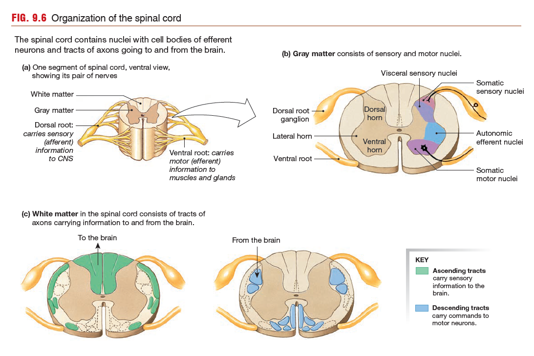
Each spinal region is subdivided into segments, and each segment gives rise to a bilateral pair of spinal nerves. Just before a spinal nerve joins the spinal cord, it divides into two branches called roots.
The white matter of the spinal cord can be divided into a number of columns composed of tracts of axons that transfer information up and down the cord.
Each spinal region is subdivided into segments, and each segment gives rise to a bilateral pair of spinal nerves. Just before a spinal nerve joins the spinal cord, it divides into two branches called roots.
- The dorsal root of each spinal nerve is specialized to carry incoming sensory information.
- Sensory fibers from the dorsal roots synapse with interneurons in the dorsal horns of the gray matter. The dorsal horn cell bodies are
organized into two distinct nuclei, one for somatic information and one for visceral information.
- Sensory fibers from the dorsal roots synapse with interneurons in the dorsal horns of the gray matter. The dorsal horn cell bodies are
- The ventral root carries information from the CNS to muscles and glands.
- The ventral horns of the gray matter contain cell bodies of motor neurons that carry efferent signals to muscles and glands. The ventral horns are organized into somatic motor and autonomic nuclei. Efferent fibers leave the spinal cord via the ventral root.
The white matter of the spinal cord can be divided into a number of columns composed of tracts of axons that transfer information up and down the cord.
- Ascending tracts take sensory information to the brain. They occupy the dorsal and external lateral portions of the spinal cord. Descending tracts carry mostly efferent (motor) signals from the brain to the cord. They occupy the ventral and interior lateral portions of the white matter.
9.5 The Brain
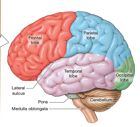
The Brain Stem Is the Oldest Part of the Brain
The brain stem is the oldest and most primitive region of the brain and consists of structures that derive from the embryonic midbrain and hindbrain. The brain stem can be divided into white matter and gray matter, and in some ways, its anatomy is similar to that of the spinal cord. Some ascending tracts from the spinal cord pass through the brain stem, while other ascending tracts synapse there. Descending tracts from higher brain centers also travel through the brain stem on their way to the spinal cord.
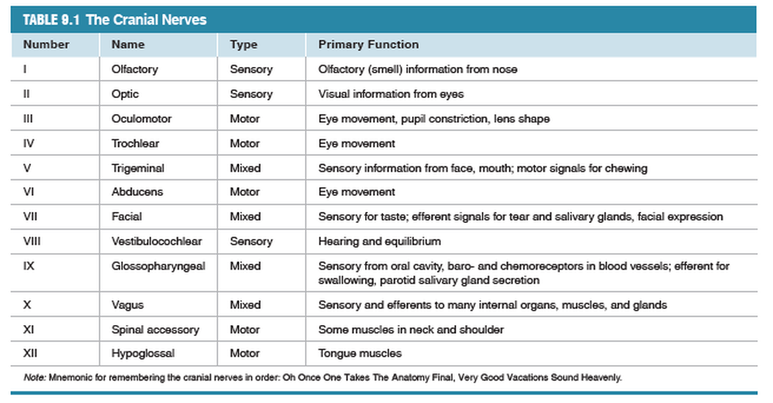
Pairs of peripheral nerves branch off the brain stem, similar to spinal nerves along the spinal cord. Eleven of the 12 cranial nerves (numbers II–XII) originate along the brain stem. (The first cranial nerve, the olfactory nerve, enters the forebrain.)
Cranial nerves carry sensory and motor information for the head and neck.
The cranial nerves are described according to whether they include sensory fibers, efferent fibers, or both (mixed nerves).
The brain stem contains numerous discrete groups of nerve cell bodies, or nuclei. Many of these nuclei are associated with the
reticular formation, a diffuse collection of neurons that extends throughout the brain stem.
Starting at the spinal cord and moving toward the top of the skull, the brain stem consists of the medulla oblongata, the pons, and the midbrain.
The Medulla
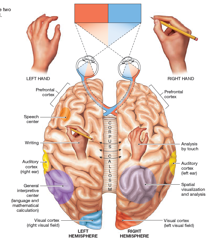
The medulla oblongata, frequently just called the medulla {medulla, marrow; adjective medullary}, is the transition from the spinal cord into the brain proper (Fig. 9.8f). Its white matter includes ascending somatosensory tracts {soma, body} that bring sensory information to the brain, and descending corticospinal tracts that convey information from the cerebrum to the spinal cord. About 90% of corticospinal tracts cross the midline to the opposite side of the body in a region of the medulla known as the pyramids. As a result of this crossover, each side of the brain controls the opposite side of the body. Gray matter in the medulla includes nuclei that control many involuntary functions, such as blood pressure, breathing, swallowing, and
vomiting.
The Pons
The pons {pons, bridge; adjective pontine} is a bulbous protrusion on the ventral side of the brain stem above the medulla
and below the midbrain. Because its primary function is to act as a relay station for information transfer between the cerebellum
and cerebrum, the pons is often grouped with the cerebellum. The pons also coordinates the control of breathing along with centers
in the medulla.
and below the midbrain. Because its primary function is to act as a relay station for information transfer between the cerebellum
and cerebrum, the pons is often grouped with the cerebellum. The pons also coordinates the control of breathing along with centers
in the medulla.
The Midbrain
The third region of the brain stem, the midbrain, or mesencephalon {mesos, middle}, is a relatively small area that lies
between the lower brain stem and the diencephalon. The primary function of the midbrain is control of eye movement, but it also
relays signals for auditory and visual reflexes.
between the lower brain stem and the diencephalon. The primary function of the midbrain is control of eye movement, but it also
relays signals for auditory and visual reflexes.
The Cerebellum
The Cerebellum Coordinates Movement. The cerebellum is the second largest structure in the brain. The name cerebellum means “little brain,” and, indeed, most of the nerve cells in the brain are in the cerebellum. The specialized function of the cerebellum is to process sensory information and coordinate the execution of movement.
The Diencephalon
The Diencephalon Contains the Centers for Homeostasis. The diencephalon is composed of two main sections, the
thalamus and the hypothalamus, and two endocrine structures, the pituitary and pineal glands.
thalamus and the hypothalamus, and two endocrine structures, the pituitary and pineal glands.
The Thalamus
Most of the diencephalon is occupied by many small nuclei that make up the thalamus. The thalamus receives sensory fibers from the optic tract, ears, and spinal cord as well as motor information from the cerebellum. It projects fibers to the cerebrum, where the information is processed. The thalamus is often described as a relay station because almost all sensory information from lower parts of the CNS passes through it. Like the spinal cord, the thalamus can modify information passing through it, making it an integrating center as well as a relay station.
The Hypothalamus
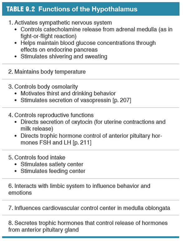
The hypothalamus lies beneath the thalamus. It is the
center for homeostasis and contains centers for various behavioral drives, such as hunger and thirst. Output from the hypothalamus also influences many functions of the autonomic division of the nervous system, as well as a variety of endocrine functions. The hypothalamus receives input from multiple sources, including the cerebrum, the reticular formation, and various sensory receptors. Output from the hypothalamus goes first to the thalamus and eventually to multiple effector pathways.
The Pituitary Gland
The posterior pituitary (neurohypophysis) is a down-growth of the hypothalamus and secretes neurohormones that are synthesized in
hypothalamic nuclei. The anterior pituitary (adenohypophysis) is a true endocrine gland. Its hormones are regulated by hypothalamic
neurohormones secreted into the hypothalamic- hypophyseal portal system.
The Cerebrum
The Cerebrum Is the Site of Higher Brain Functions. It is the largest and most distinctive part of the human brain and fills most of the
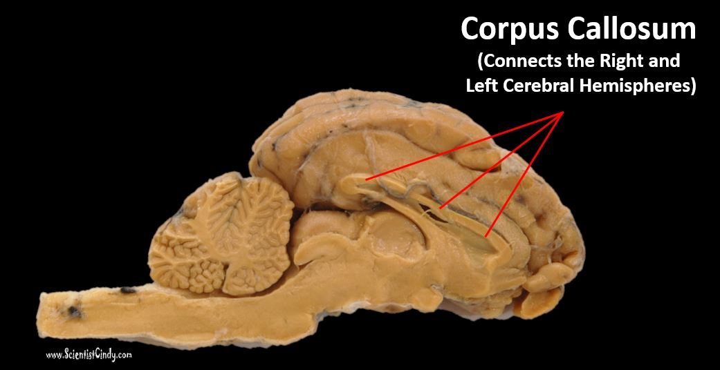
cranial cavity. It is composed of two hemispheres connected primarily at the corpus callosum, a distinct structure formed by axons passing from one side of the brain to the other. This connection ensures that the two hemispheres communicate and cooperate with each other.
cranial cavity. It is composed of two hemispheres connected primarily at the corpus callosum, a distinct structure formed by axons passing from one side of the brain to the other. This connection ensures that the two hemispheres communicate and cooperate with each other.
Each cerebral hemisphere is divided into four lobes, named for the bones of the skull under which they are located: frontal, parietal, temporal, and occipital.
Sulci and Gyri
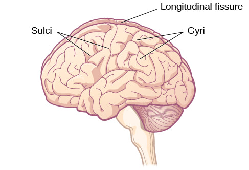
The surface of the cerebrum in humans and other primates has a furrowed, walnut-like appearance, with grooves called sulci
dividing convolutions called gyri.
dividing convolutions called gyri.
The Cerebral Cortex
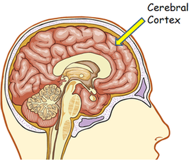
Neurons of he cerebral cortex are arranged in anatomically distinct vertical columns and horizontal layers. It is within these layers
that our higher brain functions arise.
that our higher brain functions arise.
The Basal Ganglia
The basal ganglia is a group of neurons which are involved in the control of movement. The basal ganglia are also called the basal nuclei.
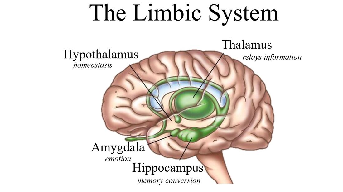
The Limbic System
The limbic system acts as the link between higher cognitive functions, such as reasoning, and more primitive emotional responses, such as fear.
The major areas of the limbic system are the amygdala and cingulate gyrus, which are linked to emotion and memory,
and the hippocampus, which is associated with learning and memory.
The major areas of the limbic system are the amygdala and cingulate gyrus, which are linked to emotion and memory,
and the hippocampus, which is associated with learning and memory.
9.6 Brain Function
The brain receives sensory input from the internal and external environments, integrates and processes the information,
and, if appropriate, creates a response. What makes the brain more complicated than this simple reflex pathway, however,
is its ability to generate information and output signals in the absence of external input. Modeling this intrinsic input requires a more
complex diagram.
The three systems that influence output by the motor systems of the body:
(1) the sensory system, which monitors the internal and external environments and initiates reflex responses
(2) a cognitive system that resides in the cerebral cortex and is able to initiate voluntary responses
(3) a behavioral state system, which also resides in the brain and governs sleep-wake cycles and other intrinsic behaviors.
The Sensory System
The sensory system monitors the internal and external environments and sends information to neural integrating centers, which
in turn initiate appropriate responses.
The primary somatic sensory cortex (also called the somatosensory cortex) in the parietal lobe is the termination point of pathways from the skin, musculoskeletal system, and viscera. The somatosensory pathways carry information about touch, temperature, pain, itch, and body position.
Damage to this part of the brain leads to reduced sensitivity of the skin onthe opposite side of the body because sensory fibers cross to the opposite side of the midline as they ascend through the spine or medulla.
The special senses of vision, hearing, taste, and olfaction (smell) each have different brain regions devoted to processing
their sensory input (Fig. 9.13). The visual cortex, located in the occipital lobe, receives information from the eyes. The auditory
cortex, located in the temporal lobe, receives information from the ears. The olfactory cortex, a small region in the temporal lobe, receives input from chemoreceptors in the nose. The gustatory cortex, deeper in the brain near the edge of the frontal lobe,
receives sensory information from the taste buds.
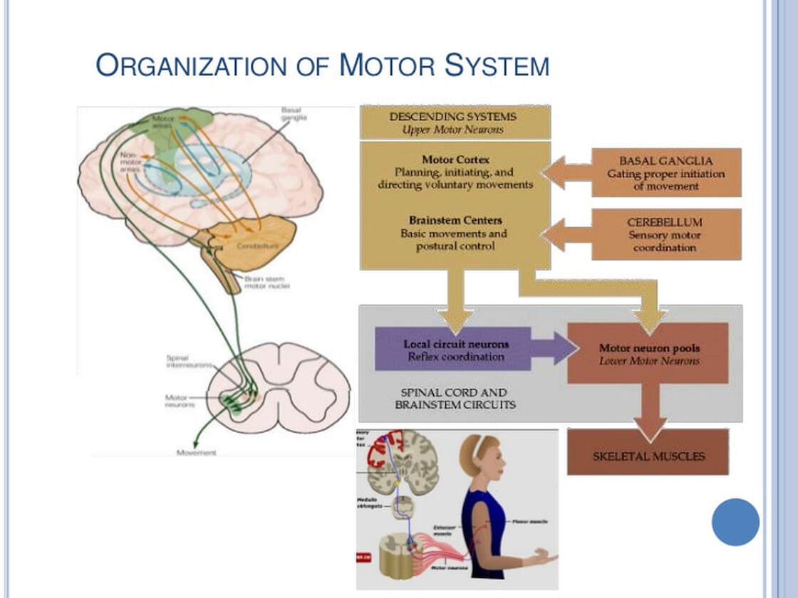
The Motor System
The Motor System Governs Output from the CNS. The motor output component of the nervous system is associated with the efferent division of the nervous system.
Motor output can be divided into three major types:
(1) skeletal muscle movement, controlled by the somatic motor division;
(2) neuroendocrine signals, which are neurohormones secreted into the blood by neurons located primarily in the hypothalamus and adrenal medulla
(3) visceral responses, the actions of smooth and cardiac muscle or endocrine and exocrine glands.

