|
|
Practice Identifying Your Tissue With This Slide Show!
Then try the quiz at the bottom of this page!
The 4 Types of Connective Tissues
Connective tissues include a wide variety of tissue types with a wide variety of functions, only one of those functions is to connect tissues and organs together. For example, your bones and your cartilage are connective tissues that function as the structural support of the body. Fat tissue and blood are connective tissues that function to store and carry nutrients. Connective tissue proper acts to surround delicate vessels and nerves and contains specialized cells that fight infection.
|
The four broad categories of connective tissue are classified according to the characteristics of their ground substance and the types of fibers found within the matrix.
The 4 Types of Connective Tissues Include: 1) Connective Tissue Proper 2) Cartilage 3) Bone 4) Blood |
|
Connective Tissue Proper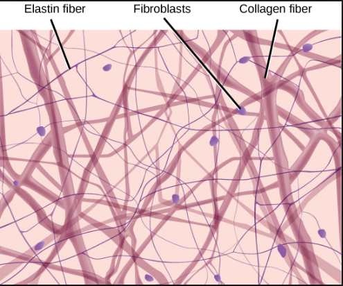
|
Cartilage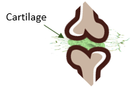
|
Bone
|
Blood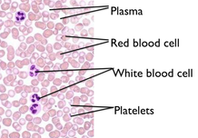
|
There are 3 characteristics that are shared by all of the different types of connective tissue.
- All connective tissue contains relatively few cells with large space between them.
- All connective tissue contains a large amount of extracellular matrix (ground substances and protein fibers).
- All connective tissues have the same embryonic origin; the mesenchyme (comes from mesoderm germ layer of embryo).
- Special cell that secrete extracellular matrix proteins
•fibroblasts for areolar tissue and reticular C.T.,
•adipocytes for adipose
•chondroblasts for cartilage
•osteoblasts for bone
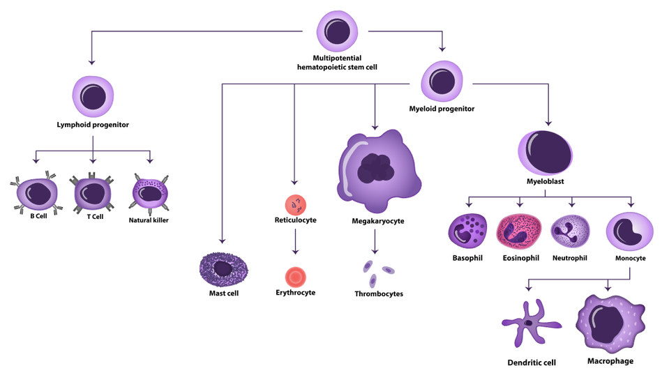
•hematopoietic stem cell for blood
Connective Tissue
- The Matrix is REAL -
Yes, the matrix is real... and it's in your connective tissue!
Yes, the matrix is real! Connective tissues are composed of not only of specific cell types, but also the protein fibers and ground substance that make up the surrounding extracellular matrix. Connective tissue cells are interspersed within the unique extracellular matrix of the tissue. The composition and density of proteins and molecules that make up the matrix varies with the different types of connective tissues. It is the specific composition of the cells, ground substance and the protein fibers that make up the connective tissue that gives it the ability to provide its specific function within the body. This is the complementarity between structure and function in connective tissues.
What is the "Ground Substance"? The ground substance refers to the substance that surrounds the cells and fibers of the extracellular matrix. The ground substance is a thick or jelly-like substance in connective tissues, except for in bone and cartilage in which the ground substance is calcified.
What are the "Protein Fibers"? The matrix has a scaffolding made up of fibrous proteins. that provide support for the connective tissue. There are 3 types of protein fibers that can be found in connective tissues: 1) collagen fibers, 2) reticular fibers, and 3) elastic fibers. The types, density, and distribution of the protein fibers is unique in different connective tissue types.
What are the "Protein Fibers"? The matrix has a scaffolding made up of fibrous proteins. that provide support for the connective tissue. There are 3 types of protein fibers that can be found in connective tissues: 1) collagen fibers, 2) reticular fibers, and 3) elastic fibers. The types, density, and distribution of the protein fibers is unique in different connective tissue types.
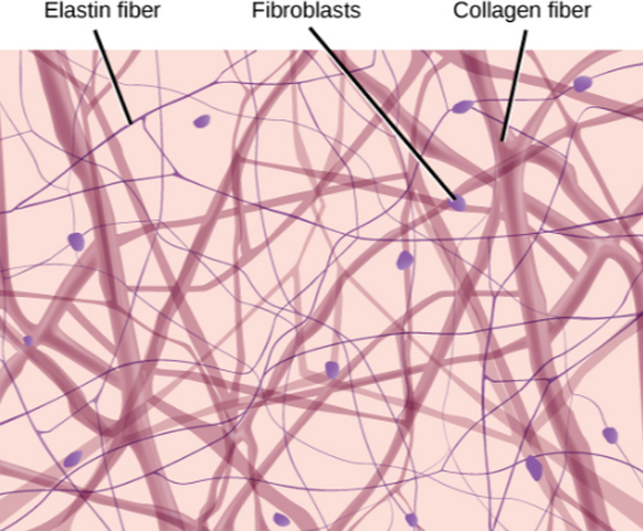
The matrix of connective tissues is made up of three types of fibers. The amounts and density of collagen fibers, reticular fibers, and elastic fibers.
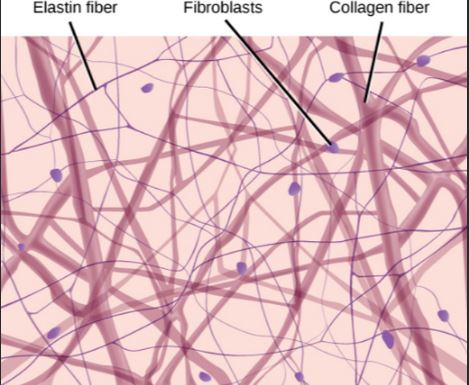
Collagen fibers are the strongest, most visible (under the microscope) and most abundant type of fiber in connective tissues. They are bundles of fibers that are cross-linked to each other. They are resistant to being stretched and they add strength to the connective tissue.
Reticular fibers are thin collagen fibrils that form a scaffolding network of fibers. The word reticulum means "network". Reticular fibers allow a lot more flexibility in being moved or stretched due to the lack of cross-links that would hinder their movement.
Elastic fibers contain the protein elastin, which allows for a great deal of stretching, while being able to return to its original non-stretched position.
IMAGE Courtesy of BOUNDLESS figures.boundless-cdn.com/19479/full/figure-33-02-06.jpeg
The Common Origin of Connective Tissue
It may be hard to believe that these very different tissue types are related, but it is true!
The cells that make up the different connective tissue types all have the same embryonic origin.
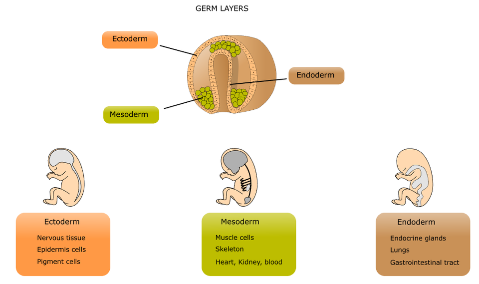
During the early stages of development, the embryo form 3 distinct layers; the ectoderm (outer layer), the mesoderm (middle layer, and endoderm (inner layer) and ).
The cells of the mesoderm have mesenchymal stem cells that can differentiate into many different cell types.
During the early stages of development, the embryo form 3 distinct layers; the ectoderm (outer layer), the mesoderm (middle layer, and endoderm (inner layer) and ).
The cells of the mesoderm have mesenchymal stem cells that can differentiate into many different cell types.
The Origin of Connective Tissue Cell Types: The mesenchymal stem cells give rise to the different cell types needed to form all of the different connective tissue types. Mesenchymal stem cells give rise to the osteoblasts which form your bones, the chondrocytes that form your cartilage, the fibrocytes that form your connective tissue proper, and the adipocytes that form your adipose (fat) cells.
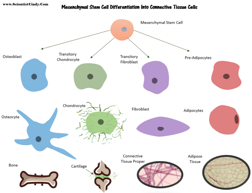
The origin of blood cells - Blood cells are also considered connective tissue, but they are derived from hematopoietic stem cells that are made in your bone marrow which was derived from mesenchymal stem cells. Hematopoietic stem cells give rise to the various types of white blood cells, red blood cells and platelets.
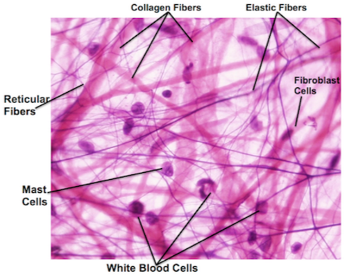
Defense cells of the connective tissue proper. The blood usually takes all of the glory when people think of the cells that fight infection, but you have many types of immune cells that are at work in your connective tissue proper screening and destroying invaders. Your immune cells include phagocytes (neutrophils, monocytes, and macrophages), inflammatory mediators (basophils, mast cells, and eosinophils), and natural killer cells (NK cells).
Connective Tissue Proper
The first category of connective tissue we will explore is called the connective tissue proper. The main function of connective tissue proper is to bind tissues, and to resist stress and taring due to stretching and tension placed on the tissue. Connective tissue proper, found in most organs, is characterized by a predominance of fibers (mainly type I collagen) in the extracellular matrix. Its varied functions are chiefly related to binding cells and tissues into organs and organ systems. Its sub-classes are based on the type, density, and orientation of its fibers.
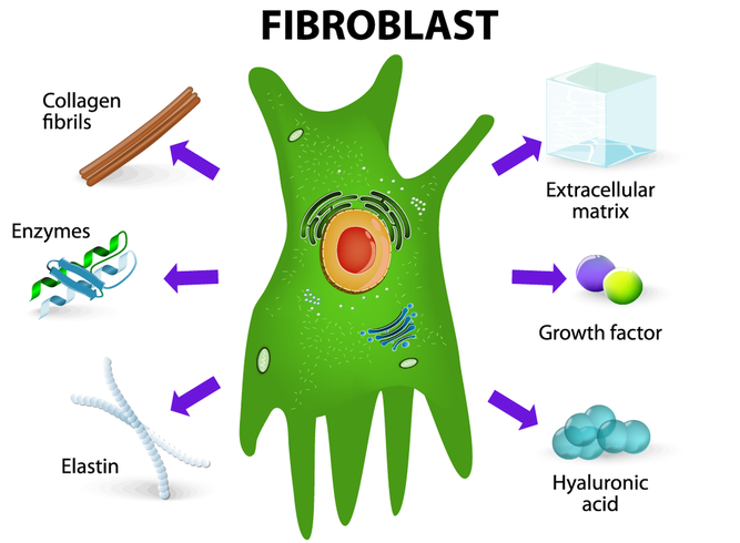
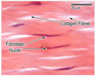
The primary cell type in connective tissue proper is the fibroblast.
The fibroblast produces components for the extracellular matrix of the connective tissue proper, which includes collagen and elastin. The fibroblast means “fiber formers”, and they live up to their name. Fibroblasts manufacture proteins and secrete them (via exocytosis) into the extracellular matrix where they act as building blocks for the matrix.
Connective tissue proper includes a few other cell types in addition to its primary cell type, the fibroblast. These additional cells include fibrocytes, defense cells, and adipose (fat) cells. The ground substance in connective tissue proper is the consistency of jelly and is composed of collagen and elastin.
What Tissue Types Are Considered "Connective Tissue Proper"?
Connective tissue proper contains tissues that fall into 2 different categories.
1) Loose Connective Tissues
2) Dense Connective Tissues
There are 3 types of Loose Connective Tissue.
1) Areolar Connective Tissue
2) Adipose (Fat) Tissue
3) Reticular Tissue
There are 3 types of Dense Connective Tissue.
1) Dense Regular Connective Tissue
2) Dense Irregular Connective Tissue
3) Elastic Connective Tissue
Areolar Connective Tissue
A Type of Loose Connective Tissue
A Type of Loose Connective Tissue
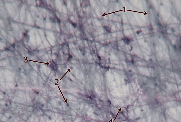
Here is an illustration of areolar connective tissue. Areloar connective tissue is very 'loose'. Areolar tissue is composed of a lot of matrix and relatively few cells. The primary cell of areolar connective tissue is the flibroblast. The fibroblast function in the areloar tissue to form the fibers of the matrix and the surrounding ground substance.
Areolar connective tissue is a loose connective tissue.The appearance of areolar connective tissue makes me think of "paint brush strokes".
Areolar tissue appears as a disorganized network of fibers with lots of space between them and a small number of spread out cells. The ground substance of the areolar tissue is a viscous liquid that surrounds the cells and fibers. Areolar tissue is strong, yet flexible and elastic and resists taring. This tissue surrounds blood vessels and nerves, is found in and around most of your organs, and is an important component for your skin where it acts to support the epithelia, and to anchor your skin to underlying muscle tissues.
|
The areolar tissue surrounds the organs provides flexible structure and support. The areolar tissue allows a high degree of movement between adjacent body parts.
The key functions of areolar tissue can be summarized as providing:
|
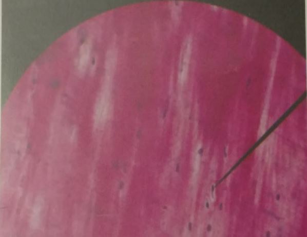
Know THIS Slide for the Practical!
Areolar Connective Tissue Slide
|
Areolar Connective Tissue Slide
|
Areolar Connective Tissue Slide
|
Adipose Connective Tissue
A Type of Loose Connective Tissue

Adipose (fat) tissue function to store energy and produce heat.
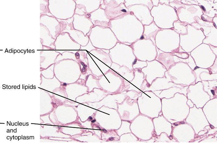
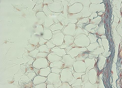
Adipose (fat) cells, is considered a loose connective tissue of the connective tissue proper. There are actually two different types of fat tissue; white fat and brown fat. Your white fat stores energy (in the form of a lipid droplet, while brown fat functions to generate body heat. Infants have a lot more brown fat than adults do. This is one of the reasons that infants have a higher normal temperature than adults.
Adipose (fat) cells, is considered a loose connective tissue of the connective tissue proper. There are actually two different types of fat tissue; white fat and brown fat. Your white fat stores energy (in the form of a lipid droplet, while brown fat functions to generate body heat. Infants have a lot more brown fat than adults do. This is one of the reasons that infants have a higher normal temperature than adults.
|
Adipose Connective Tissue Slides Know THIS Slide for the Practical!
|
|

|
|
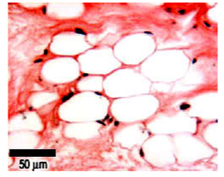
Under the microscope, adipose cells (adipocytes) appear as large ovals with the nucleus off to one side. This odd appearance is due to the presence of a large lipid (fat) droplet that flattens the nucleus and pushes the cytoplasm to one end of the cell. The lipid droplet that takes of most of the area within these cells is hydrophobic, so it does not take up any of the applied stain. So, a slide containing adipose cells is going to appear as mostly "white space". However, it is important to locate the cell membrane and the nuclei to ensure that the slide is of adipose cells and not just out of focus.
Visceral Fat vs. Subcutaneous Fat
Visceral fat is different from subcutaneous fat underneath the skin, and intramuscular fat interspersed in skeletal muscles. You may have heard that people who have a large accumulation of adipose tissue in the abdomen region are at a higher risk diabetes, heart disease, cancer and stroke. This is true, but why?
"Belly fat" is an indicator of "visceral fat". Visceral fat surrounds these visceral organs and can effect their function. Remember that your visceral organs include all of the organs contained within your ventral cavity. Men are more likely to have their excess adipose tissue stored in the abdomen due to male sex hormones. Women, on the other hand, tend to carry their excess adipose tissue on the buttocks, thighs, and hips in women due to female sex hormones. When women reach menopause, the decrease in estrogen production by the ovaries induces a change in the location of fat store from the thighs, hips and buttocks to the abdomen where it is more detrimental to health.
Subcutaneous fat is found just below the skin in a region called the hypodermis and is not related to the obesity-related diseases, cancer and stroke.
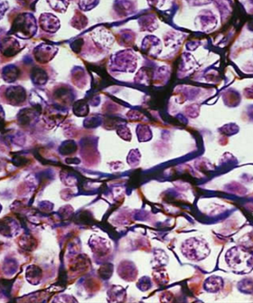
Reticuluar Connective Tissue
A Type of Loose Connective Tissue
Reticular Connective Tissue. |
|
|
Reticular connective tissue is a type of loose connective tissue that contains a network of reticular fibers. The word "recticulum" means (net or network). Reticular fibers are very noticeable in these tissues. These reticular fibers are synthesized by fibroblasts (also called reticular cells). Reticular connective tissue is found in bone marrow and surrounding the kidneys, the spleen, and the lymph nodes. Reticular connective tissue forms a flexible 'skeleton' or internal framework, that can support many free blood cells (largely lymphocytes) in lymph nodes, the spleen, and red bone marrow. |
Slides of reticular connective tissue.
Know THIS Slide for the Practical! |
|
|
|
There are 3 types of Dense Connective Tissue:
Dense Regular Connective Tissue - has a rope-like arrangement of fiber bundles
Dense Irregular connective tissue - has a wavy, woven fabric-like arrangement
Elastic Connective Tissue - has stacked swiggly wavy lines
Dense Regular Connective Tissue - has a rope-like arrangement of fiber bundles
Dense Irregular connective tissue - has a wavy, woven fabric-like arrangement
Elastic Connective Tissue - has stacked swiggly wavy lines
Dense Regular Connective Tissue
A Type of Dense Connective Tissue
Dense regular connective tissue
The fibers of this tissue are tightly packed into parallel bundles, between which are a few highly attenuated, spindle-shaped fibroblasts. The small, cigar-shaped nuclei of the fibroblasts are oriented parallel to the fibers; the cytoplasm is difficult to distinguish with the light microscope. There is little room for the ground substance, which nevertheless permeates the tissue.
The fibers of this tissue are tightly packed into parallel bundles, between which are a few highly attenuated, spindle-shaped fibroblasts. The small, cigar-shaped nuclei of the fibroblasts are oriented parallel to the fibers; the cytoplasm is difficult to distinguish with the light microscope. There is little room for the ground substance, which nevertheless permeates the tissue.
|
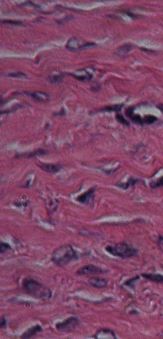
Know THIS Slide for the Practical!

Slide of tendon
|
Know THIS Slide for the Practical!

|
|
Dense regular connective tissue is packed with collagen fibers. This makes them very strong and resistant to stretch. This unique property allows them to transit mechanical force over long distances using a minimum of material and space, while resisting mechanical forces from other directions. This tissue therefore serves to transmit the force of muscle contraction, to attach bones to one another, and to protect other tissues and organs.
Dense regular connective tissue is found in tendons, ligaments, periosteum, perichondrium, deep fascia, and some organ capsules. You are already familiar with these tissues, even if you don't already know it. In some cuts of meat, you can see these tissues as the white stringy substance that has a harder consistency. In anatomy, a ligament is the dense regular connective tissue that connects bones to other bones. and a tendon is the dense regular connective tissue that connects bones to muscle (for example, the hamstring.)
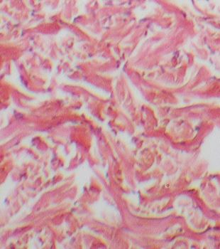
Dense Irregular Connective Tissue
- WE DO NOT HAVE DENSE IRREGULAR TISSUE SLIDE FOR PRACTICAL -

|
|
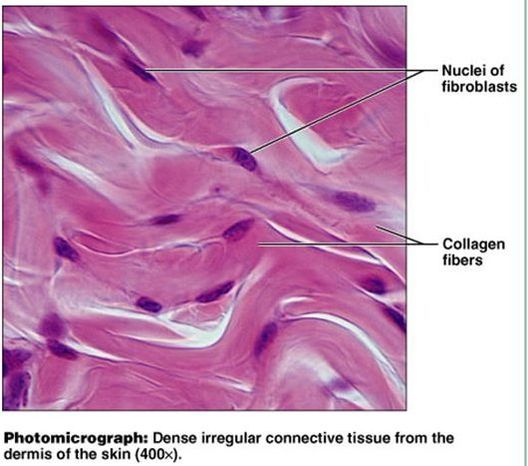
Dense irregular connective tissue consists of tightly packer collagen fibers that appear less organized and less uniform that dense regular connective tissue tissue. The nuclei in this tissue are stretch long and the tissue may appear wavy under a microscope. The major cell on dense irregular connective tissue is the fibroblasts.
Dense irregular connective tissue is able to withstand tension from multiple directions. It provides support and strength. This tissue is found in areas that experience stretching from multiple angles, such as the dermis of the skin, the submucosa of the digestive tract and the fibrous capsules of organs and joints.
Dense irregular connective tissue is able to withstand tension from multiple directions. It provides support and strength. This tissue is found in areas that experience stretching from multiple angles, such as the dermis of the skin, the submucosa of the digestive tract and the fibrous capsules of organs and joints.
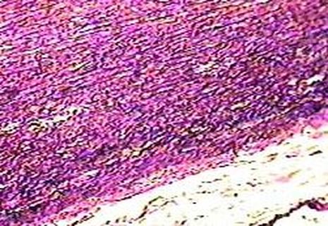
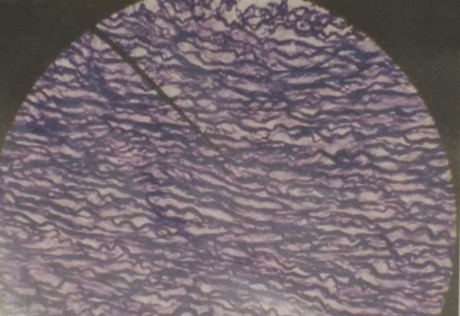
Elastic Connective Tissue
A Type of Dense Connective Tissue
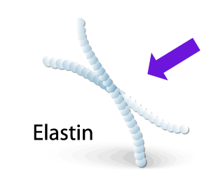
Elastic fibers are present in different concentrations in all of the connective tissues proper. Elastic fibers are made with elastin that has the ability to stretch and then return to its original shape after being stretched. Connective tissues that have a very large amount of elastin are referred to as elastic connective tissue.
Elastic connective tissues are present in relatively high concentration in several organs, including the largest arteries in the body.
Elastic connective tissues are present in relatively high concentration in several organs, including the largest arteries in the body.
|
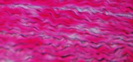
Know THIS Slide for the Practical!
|
Fibroblasts or fibrocytes are the primary cell in elastic connective tissue. The fibroblast cells make he elastin necessary for the elastic fibers and also provide other proteins and molecules that contribute to the matrix.
Cartilage
WHAT IS CARTILAGE?
Cartilage is a supportive connective tissue that functions in the body to provide structural support, protection of soft tissues and increases strength. You are born with a lot more cartilage than you have as an adult. As you grow, much of this cartilage is used to guide the growth of bones. For example, the human skull is composed of separated bones at birth and then fuses during development to form the skull. Once the bones have fused, the cartilage is broken down by the body and not replaced. This is why infants are born with a "soft spot" at the top of their head that disappears.
Cartilage tissue consists of a dense collection of collagen and elastin fibers. The primary cell in cartilage are chondrocytes. The fibers and the ground substance that makes up the matrix of cartilage is made from chrondroblasts (instead of fibroblasts as seen in connective tissue proper) which are immature chondrocytes.
Cartilage is usually closely associated with a dense irregular connective tissue (called perichondrium). This configuration is very important for the cartilage. Cartilage does not have blood or lymph vessels and does not have nerves (in not innervated) . The cartilage gets is nutrients from dense irregular tissue of the perichondrium via diffusion through the matrix. Whenever there is an injury that occurs in an area that contains a lot of cartilage (like the Achilles heel ) the amount of time necessary for healing is greatly increased. The fact that cartilage has no nerves, means that cartilage is not sensitive to pain or pressure,
Cartilage is closely related to bone. Whenever cartilage gets calcified, the chondrocytes of the cartilage die and the cartilage will be replaced by bone tissue. Cartilage has more flexibility than bone, due to a flexible material called chondroitin which exists in the matrix. chondroitin is sometimes taken as a supplement for joint pain. .
|
There are 3 types of cartilage.
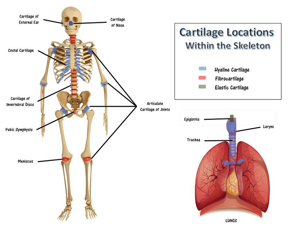
1) Hyaline Cartilage 2) Fibrocartilage 3) Elastic Cartilage. |
|
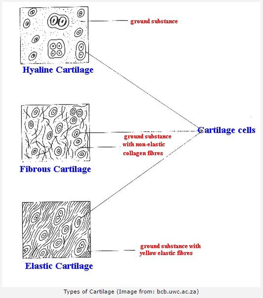
Hyaline Cartilage
|
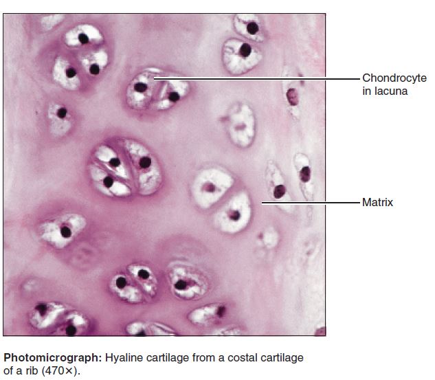
Hyaline cartilage is the most abundant type of cartilage found in our body. Your entire skeleton actually started out as hyaline cartilage when you were developing in the womb. As an adult, you have hyaline cartilage covering the surfaces of your bones at each of your joints. Your ribs also contain hyaline cartilage at the anterior ends. You have hyaline cartilage in your bronchi, your bronchial tubes, nose and trachea. You even have hyaline cartilage that forms your vocal cords!
|
|
|
Hyaline cartilage tissue appears as a shiny bluish-white, material that contains collagen fibrils and chondrocytes. Hyaline Cartilage functions is the body to allow for the gliding motion of adjacent structure by providing a smooth surface. It also provides flexible support. Hyaline cartilage also acts to connect the ribs to the sternum (the costal region).
Know THIS Slide for the Practical!
|
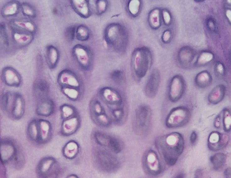
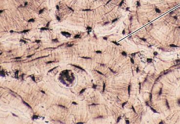
Hyaline cartilage means "glass. It gets its name from its clear glass-like appearance when observed with the naked eye (this quality is not visible on stained slides). When viewed under the light microscope, hyaline cartilage has clearly defined cells (the chondrocytes) that seem round. The cells are surrounded by a blank spaces called lacunae. In histology, a lacuna is a small space containing an osteocyte in bone or chondrocyte in cartilage.The fiber portion of the matrix of hyaline cartilage contains only thin fibrils of collagen that are too thin to be seen with a light microscope. The ground substance has a jelly-like consistency and holds large amounts of water. The structural properties of hyaline cartilage given it the ability to resists compression and provide strength and flexibility.
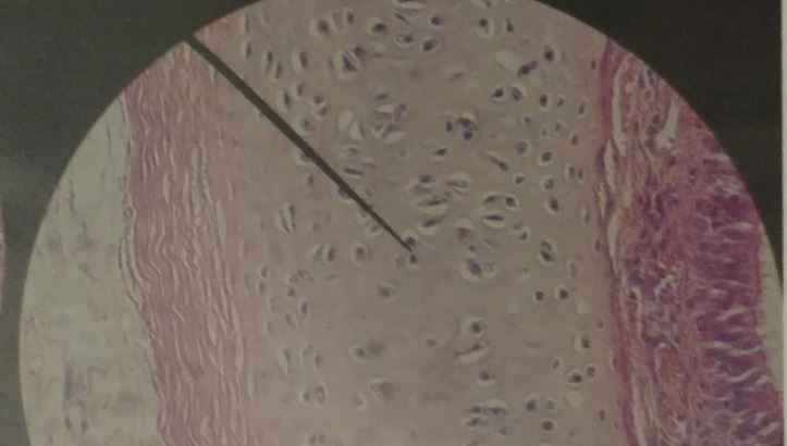
Fibrocartilage (Fibrous Cartilage)
Know THIS Slide for the Practical!
|
|
|
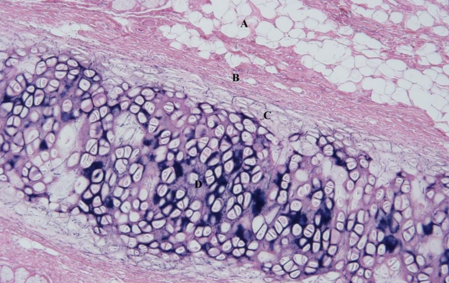
Fibrocartilage is a tough form of cartilage that consists of chondrocytes scattered among clearly visible dense bundles of collagen fibers within the matrix. Fibrocartilage is found in portions of your joints, your intevertebral discs, your knee (as the meniscus) and your pubic symphysis which joins the anterior portions of your hip bones together Fibrocartilage provides structure and support. It is also the strongest type of cartilage.
Fibrocartilage has the ability to resist strong compression and strong tension (pushing and pulling) forces. The areas of the body that have this type of cartilage are subjected to a lot of push and pull (compression and tension) forces. its consistency lies somewhere between hyaline cartilage and dense regular connective tissue. Fibrocartilage has thick collagen fibers which makes it somewhat similar to dense regular connective tissue, however the cartilage includes chondrocytes within lacunae. In histology, a lacuna is a small space containing an osteocyte in bone or chondrocyte in cartilage.
Fibrocartilage has the ability to resist strong compression and strong tension (pushing and pulling) forces. The areas of the body that have this type of cartilage are subjected to a lot of push and pull (compression and tension) forces. its consistency lies somewhere between hyaline cartilage and dense regular connective tissue. Fibrocartilage has thick collagen fibers which makes it somewhat similar to dense regular connective tissue, however the cartilage includes chondrocytes within lacunae. In histology, a lacuna is a small space containing an osteocyte in bone or chondrocyte in cartilage.
Elastic Cartilage
|
Know THIS Slide for the Practical! |
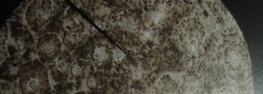
Elastic cartilage contains a lot of elastic fibers with thin collagen fiberils in its matrix. As you would expect, this cartilage is more elastic
than hyaline cartilage. This exatra elasticity allows for better tolerance of repeated bending. The body has elastic cartilage tissue in the eustacian tubes which regulate the inner ear pressure, it form the structure of the outer ear (the auricle) and it forms the epiglottis. The epiglottis, which bends down to cover the glottis (opening) of the larynx each time we swallow, is made of elastic cartilage, as is the highly bendable cartilage in the outer ear. Elastic cartilage contains chondrocytes that are dispersed within a a threadlike network of elastic fibrers. Elastic cartilage functions to provides support. It also provides structure to the area. .
than hyaline cartilage. This exatra elasticity allows for better tolerance of repeated bending. The body has elastic cartilage tissue in the eustacian tubes which regulate the inner ear pressure, it form the structure of the outer ear (the auricle) and it forms the epiglottis. The epiglottis, which bends down to cover the glottis (opening) of the larynx each time we swallow, is made of elastic cartilage, as is the highly bendable cartilage in the outer ear. Elastic cartilage contains chondrocytes that are dispersed within a a threadlike network of elastic fibrers. Elastic cartilage functions to provides support. It also provides structure to the area. .
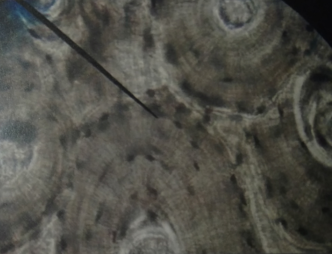
Bone (Osseous Tissue)
Bone is a tissue, also known as osseous tissue. Bone is alive and filled with blood vessels. It has incredible stregthe due to inorganic calcium salts that form a rigid ground substance of the matrix. Bone functions in the body to provide structural support and protection to internal body structures. Bone is able to withstand a great deal of compression and tension (pushing force and pulling force). The primary cell of bone tissue is the osteoblast. Osteoblastss secrete the collagen fibers and the materials needed for the ground substance. These materials are then calcified by the addition of calcium salts which form the rigid ground substance of the bone. When bone is mature, the matrix consists of collagen fibers surrounded by a calcified rigid ground substance. The osteoblasts mature into the osteocytes. The osteocytes reside in the lacunae filling up the space.

Bone tissue is a supportive type of connective tissue. It acts to shield the internal organs and to provide structure and support. Bone tissue includes compact bone and spongy bone. In bone, the matrix is rigid and described as calcified because of the deposited calcium salts.
|
|
Blood
Animated GIF Courtesy of Source scinerds.tumblr.com
Hematopoiesis
|
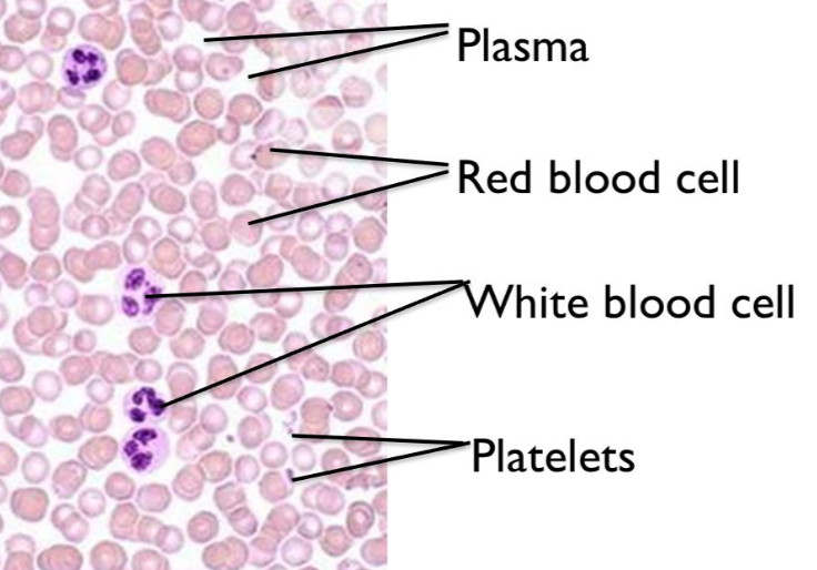
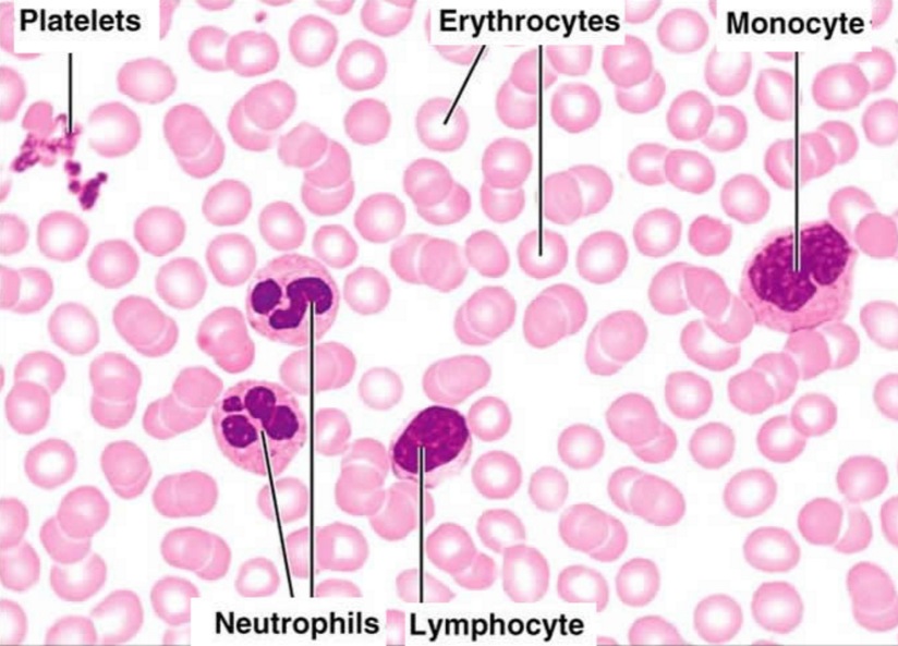
Fluid connective tissue includes lymph and blood. Various specialized cells circulate in a watery fluid containing salts, nutrients, and dissolved proteins. Red blood cells do not have a nucleus! We don't see purple in the red blood cells, because there is no nucleus to take up the purple nuclear stain. Blood is pretty different from the other connective tissue types. It is liquid and it does not bind things
together or provide any mechanical or structural support. However, it develops from the mesenchyme like the other connective tissue types. Blood consists of blood cells surrounded by a liquid nonliving matrix called blood plasma. Its cells and matrix are very different from those in other connective tissues. Blood functions as the transport vehicle for the cardiovascular system, carrying defense cells, nutrients, wastes, respiratory gases, and many other substances throughout the body. |
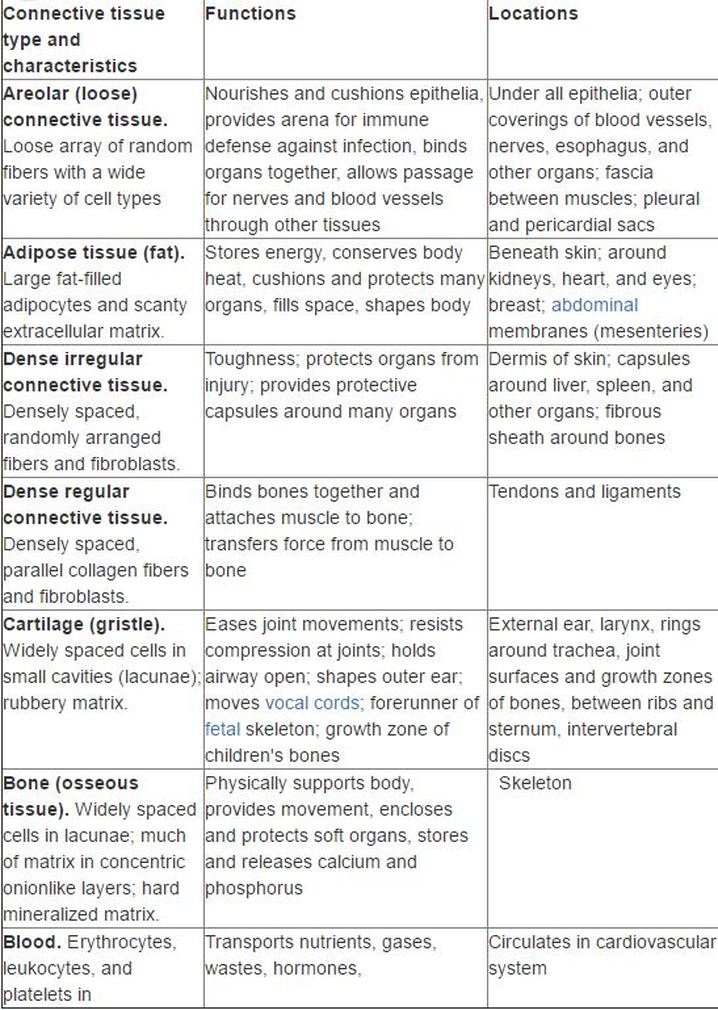
Summary Table