THE DERMIS
Looking Beneath the the Surface
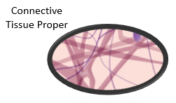
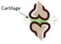
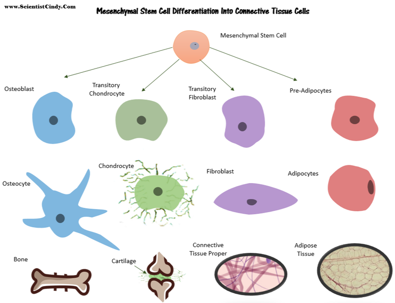
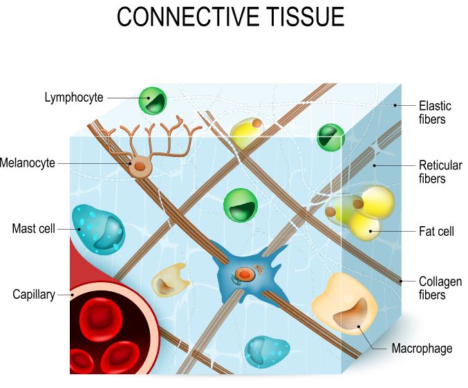
Connective tissues include a wide variety of tissue types with a wide variety of functions, only one of those functions is to connect tissues and organs together. For example, your bones and your cartilage are connective tissues that function as the structural support of the body. Fat tissue and blood are connective tissues that function to store and carry nutrients. Connective tissue proper acts to surround delicate vessels and nerves and contains specialized cells that fight infection.
The four broad categories of connective tissue are classified according to the characteristics of their ground substance and the types of fibers found within the matrix.
The four broad categories of connective tissue are classified according to the characteristics of their ground substance and the types of fibers found within the matrix.
There are 3 characteristics that are shared by all of the different types of connective tissue.
- All connective tissue contains relatively few cells with large spaces between them.
- All connective tissue contains a large amount of extracellular matrix (ground substances and protein fibers).
- All connective tissues have the same embryonic origin; the mesenchyme (comes from mesoderm germ layer of embryo).
- Special cell that secrete extracellular matrix proteins
•fibroblasts for areolar tissue and reticular C.T.,
•adipocytes for adipose
•chondroblasts for cartilage
•osteoblasts for bone
•hematopoietic stem cell for blood
A quick word about
CONNECTIVE TISSUES
before we dive in.
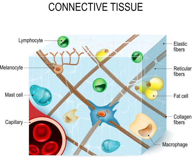
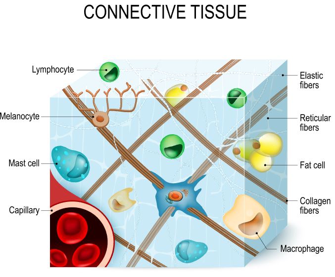
Connective tissues are composed of not only of specific cell types, but also the protein fibers and ground substance that make up the surrounding extracellular matrix. Connective tissue cells are interspersed within the unique extracellular matrix of the tissue. The composition and density of proteins and molecules that make up the matrix varies with the different types of connective tissues. It is the specific composition of the cells, ground substance and the protein fibers that make up the connective tissue that gives it the ability to provide its specific function within the body. This is the complementarity between structure and function in connective tissues.
THE MATRIX IS REAL!
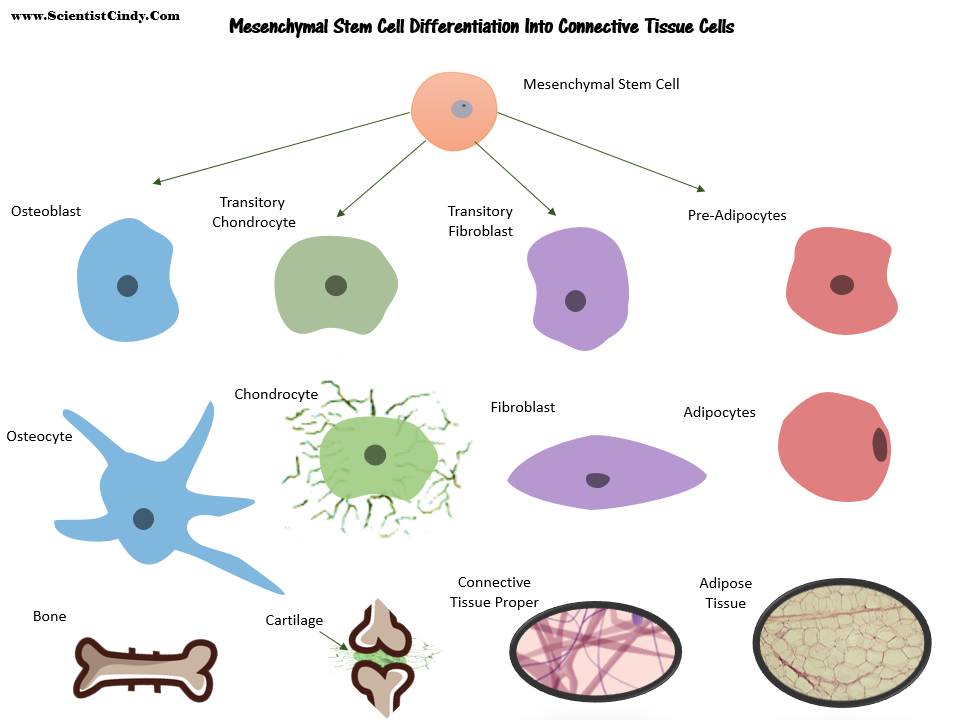
Connective Tissues Share the Same Embryonic Origin, Coming From the Mesenchymal Stem Cells
The connective tissues (connective tissue proper, bone, cartilage and blood) have the same embryonic origin. They all originate from mesenchymal stem cells, except for the blood. Blood originates from hematopoietic stem cells of the red bone marrow within bone tissue.
|
|
What is the "Ground Substance"? The ground substance refers to the substance that surrounds the cells and fibers of the extracellular matrix. The ground substance is a thick or jelly-like substance in connective tissues, except for in bone and cartilage in which the ground substance is calcified.
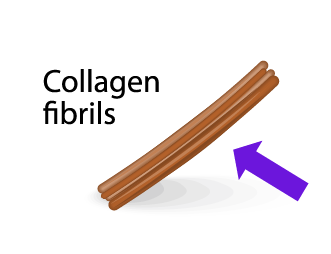
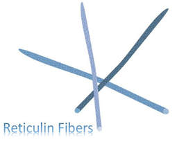
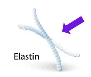
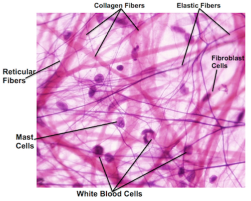
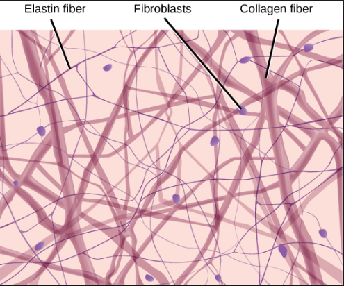
What are the "Protein Fibers"? The matrix has a scaffolding made up of fibrous proteins. that provide support for the connective tissue. There are 3 types of protein fibers that can be found in connective tissues: 1) collagen fibers, 2) reticular fibers, and 3) elastic fibers. The types, density, and distribution of the protein fibers is unique in different connective tissue types.
|
The 3 Types of Protein Fibers Found in
Connective Tissues
The Dermis
|
The dermis is the larger, more durable portion of our skin which lies deep to the epithelium. It is primarily composed of connective tissue proper.
Connective tissue proper contains the following types of cells:
|
|
|
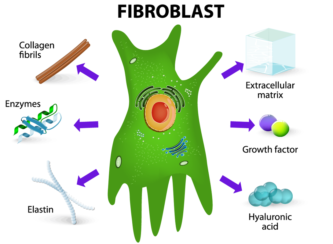
Fibroblasts...
|
The Layers of the Dermis
|
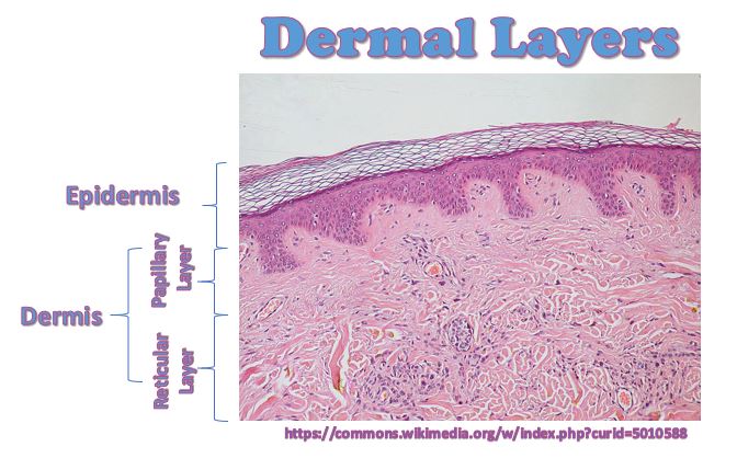
The thin, superficial layer of the dermis is called the papillary layer. The papillary layer is composed of areolar connective tissue which has 1) collagen fibers, 2) reticular fibers and 3) elastic fibers. Elastic fibers provide the stretch-recoil properties of skin, and collagen hydrates the skin by holding onto water molecules.
|
|
|
The key functions of areolar tissue can be summarized as providing:
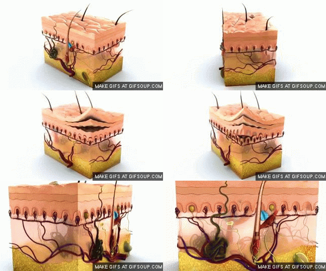
Elastic fibers provide the stretch-recoil properties of skin.
|
It is important that the papillary layer is composed of LOOSE connective tissue, so that the phagocytes and other defensive cells (mast cells and white blood cells) can travel around and "patrol" the area looking for any foreign invaders that may have penetrated the protective barrier of the epidermis.
|
The Papillary Layer of the Dermis
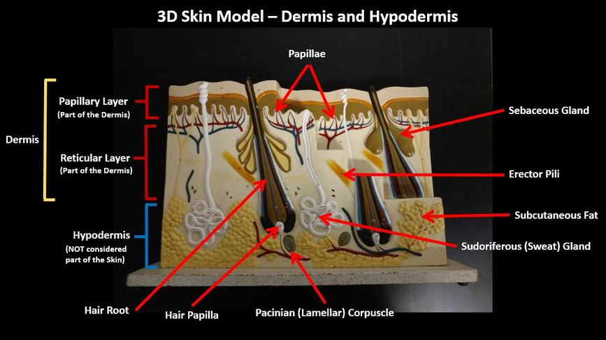
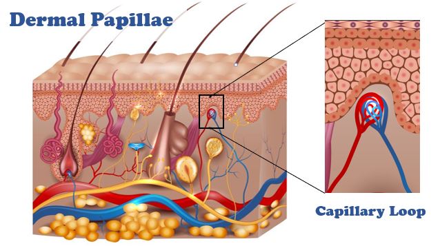
The papillary layer of the dermis gets its name from the structures called the "dermal papillae". These are the ridges that lie just below the epidermis.
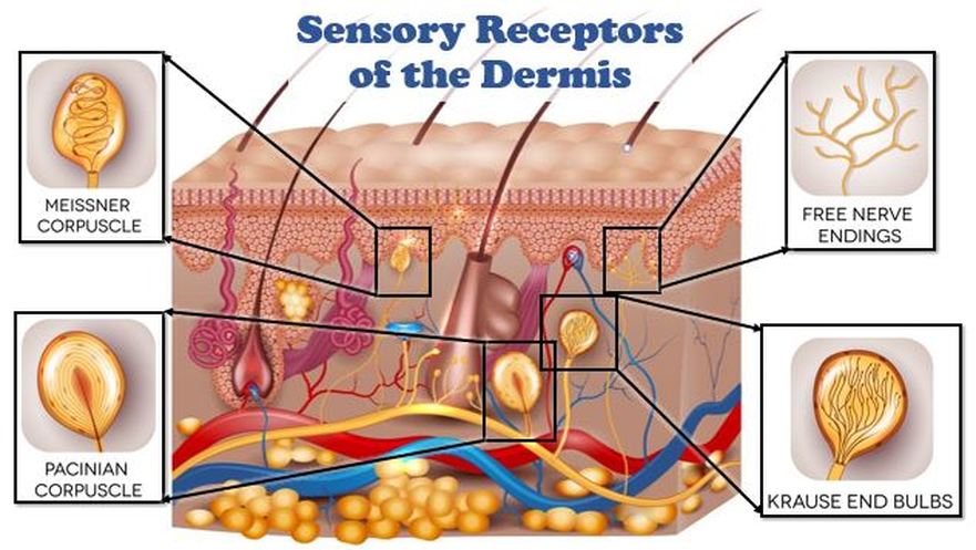
The papillae and the papillary layer as a whole, contain many specialized features and structures that enable the dermis to have various functions.
Capillary Loops
The blood vessels make a looped structure within many of the papillae called a capillary loop. Since the epidermis does not have its own blood supply, it gets its nutrients and oxygen from the tiny blood vessels that travel through the nearby papillae.
The Reticular Layer of the Dermis
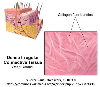
The reticular layer of the dermis lies beneath (or deep to) the papillary layer of the dermis and above (or superficial to) the hypodermis. The reticular layer is much larger than the papillary layer and is composed of dense irregular connective tissue.
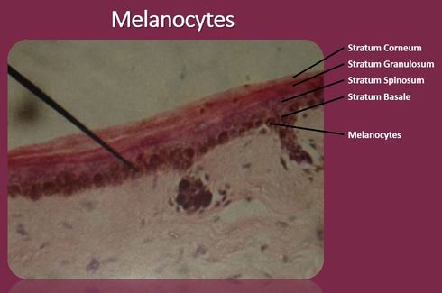
Melanin is produced in the skin and comes in various colors and types. Exposure to UV radiation stimulates melanogenesis which causes the skin to darken. Melanin filters out UV radiation and protects the skin from UVB radiation damage which can lead to sun burns, premature aging and melanoma (skin cancer).
Clinical Observations of Abnormal Skin Coloration
- BLUE SKIN (cyanosis) - When the blood is severely lacking in oxygen, people with a light complexion can appear to turn blue. This is because you are able to see the color of the blood through their skin. Cyanosis is not evident in people with darker complexions. Cyanosis is a indication of severe distress and help should be sought immediately. Possible causes of cyanosis include heart failure or severe respiratory distress.
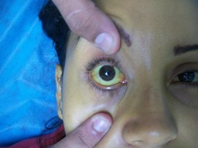
- YELLOW SKIN (Jaundice) - Jaundice is a severe condition in which medical attention should be sought immediately. The white portion of the eyes (sclera) becomes yellow along with a yellowing of the skin. This condition is usually a sign of a failing liver and the yellowing of the tissues is due to the build up of the excess yellow bile stored in the tissues.
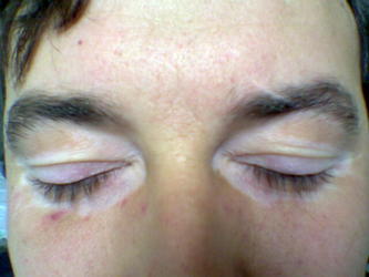
- BRONZE SKIN (Bronzing): A sudden unexplained bronze, metallic-like appearance of the skin can be an indication of one of the following:
- Addisons disease - Addisons disease is marked by an underactive adrenal cortex and therefore, a reduction in the release of steroid hormones.
- Pituitary gland - a pituitary tumor could be releasing too much melanocyte stimulating hormone (MSH).
- BLACK and/or BLUE SKIN (bruises and hematomas) - Bruises and hematomas are created from broken blood vessels. The blood the leaks out of the broken blood vessels creating a patch of discoloration. When larger amounts of blood are released from broken blood vessels, the blood forms a blood clot under the skin called a hematoma. The word "hemotoma" is derived from the Latin words for “blood swelling”

