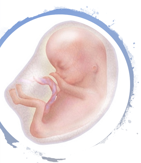
Nervous System Development
Week 1-3 of Embryonic Development
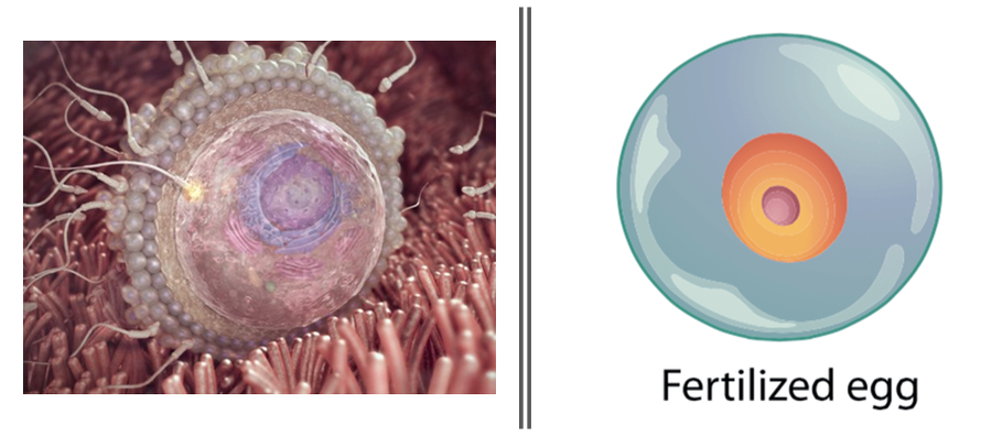
We begin life as a fertilized egg called a zygote.
We begin life as a fertilized egg called a zygote.
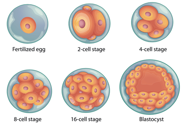
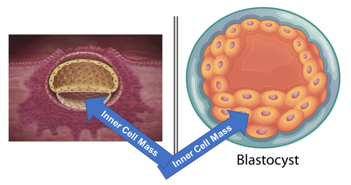
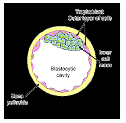
This fertilized egg rapidly divides forming 2 cells, 4 cells, 8 cells, 16 cells, and then at about the 32 cell stage of development, the beginnings of a structure known as a BLASTOCYST Forms. The blastocyst contains a fluid-filled sac and cells that congregate to one side. The cells that congregate to one side of the blastocyst are called the INNER CELL MASS.
Week 3
Around the 3rd week of embryonic development, the Inner Cell Mass of the Blastocyst
differentiates into 2 distincy populations of cells called the Epiblast and the Hypoblast.
Around the 3rd week of embryonic development, the Inner Cell Mass of the Blastocyst
differentiates into 2 distincy populations of cells called the Epiblast and the Hypoblast.
- Epiblast Cells: From inner cell mass, will ultimately give rise to the three primary germ layers and the entire embryo.
- Hypoblast Cells: These cells are the first to migrate and eventually disintegrate.
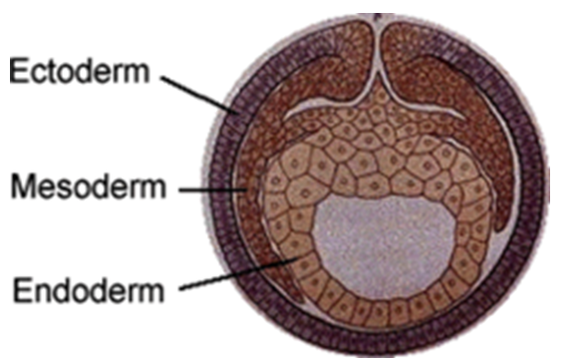
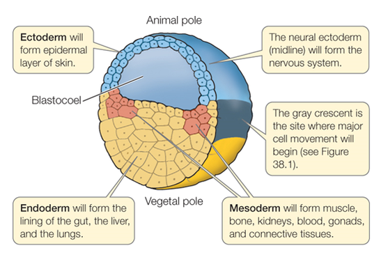
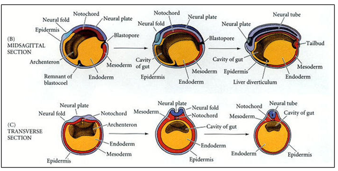
About week 4 of embryonic developmemt, the EPIBLAST gives rise to the 3 primary germ layers. The cross-section of the blastocyst reveals the positions of these three germ layers which are:
•The 3 Primary Germ Layers of the blastocyst undergo many interactions in order to evolve into organ, bone, muscle, skin or neural tissue that make up the developing embryo.
- The ectoderm
- The mesoderm
- The endoderm
•The 3 Primary Germ Layers of the blastocyst undergo many interactions in order to evolve into organ, bone, muscle, skin or neural tissue that make up the developing embryo.
|
ECTODERM
- The outside layer is the ectoderm. This becomes the epidermal (skin) layers, the brain and the spinal cord. MESODERM - The middle layer is the mesoderm. This becomes the connective tissues including the bones and the blood, as well as the gonads and the kidneys. ENDODERM - Inner layer is the endoderm. This becomes the digestive systems and the respiratory system. |
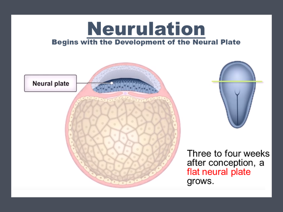
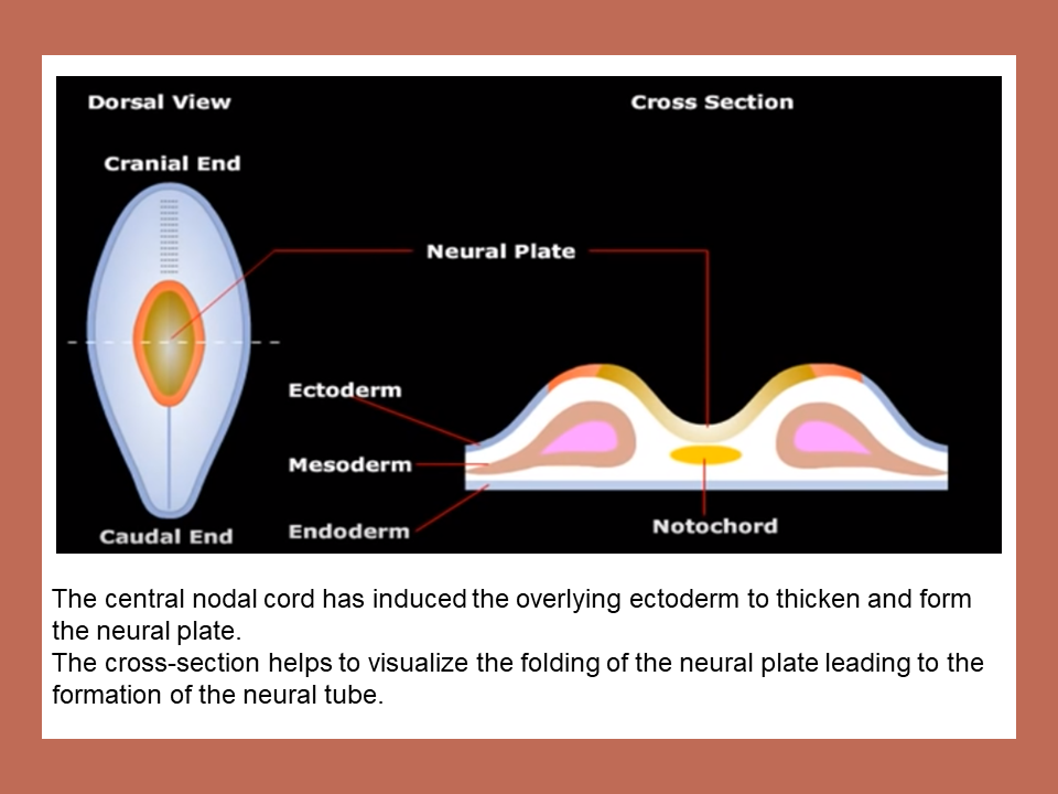
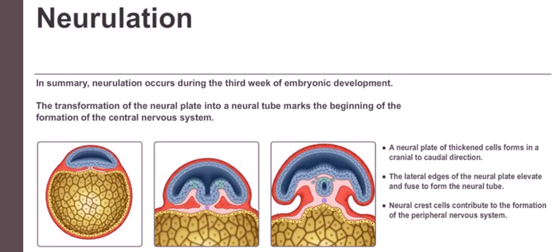
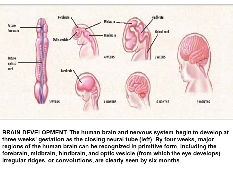
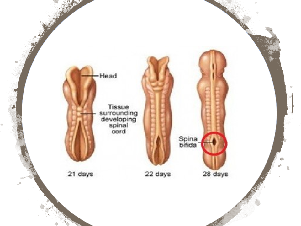
About 3-4 weeks after conception, the process of neurulation begins. Neurulation is the process of neural tube formation and its development into the spinal cord and the brain. Any complication during this complex process can result in neural tube defects.
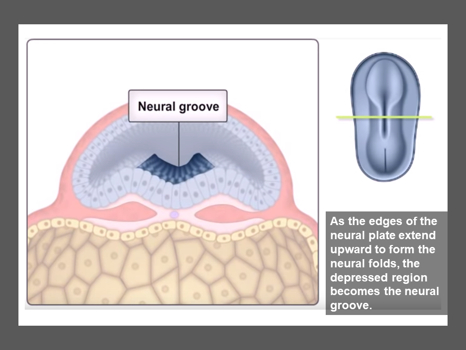
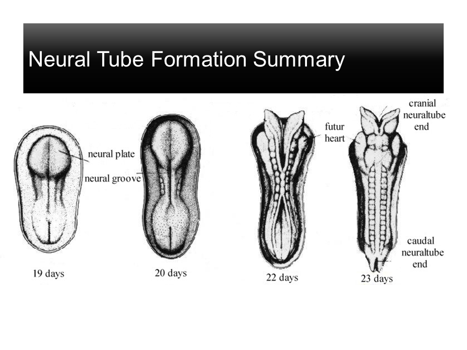
The process of neurulation begins with the formation of the NEURAL PLATE.
The process of neurulation begins with the formation of the NEURAL PLATE.
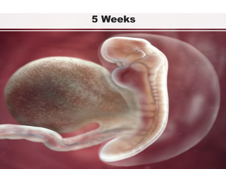
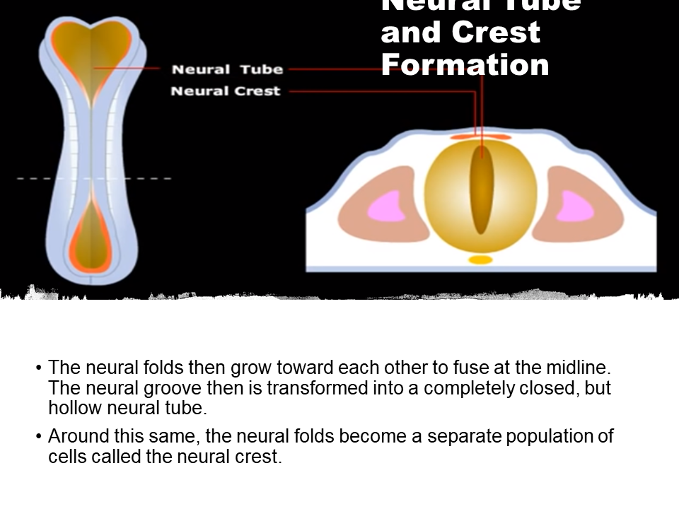
Around 5 weeks of development, the process of neurulation becomes more evident through the formation of the neural tube and neural crest.
|
5 Weeks - Neurulation (Neural Tube and Neural Crest Formation)
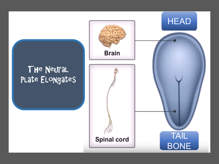
The neural plate elongates. There is a morphological difference between the rostral end (head) and the caudal end (tail bone) of the structure at this point. The neural crest cells migrate throughout the body to form a wide variety of cell types including Supporting cells of the nervous system – Glial Cells The coverings of the nervous system – The Meninges Neural Tube and Crest Formation
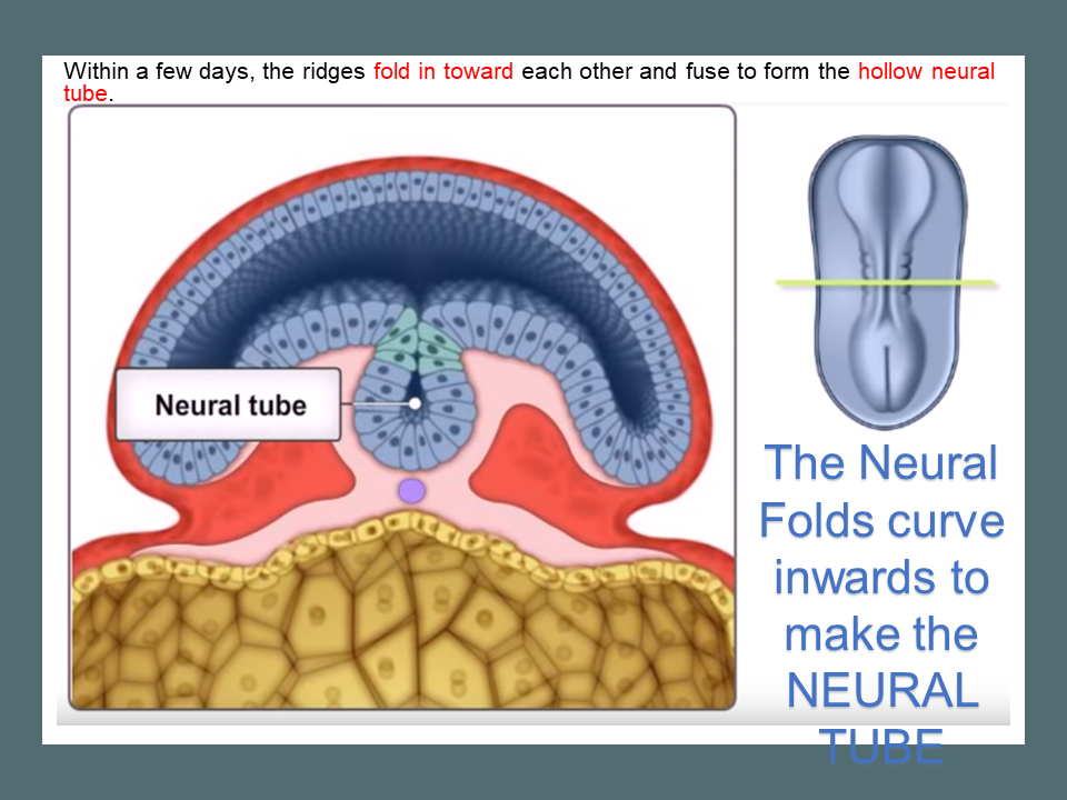
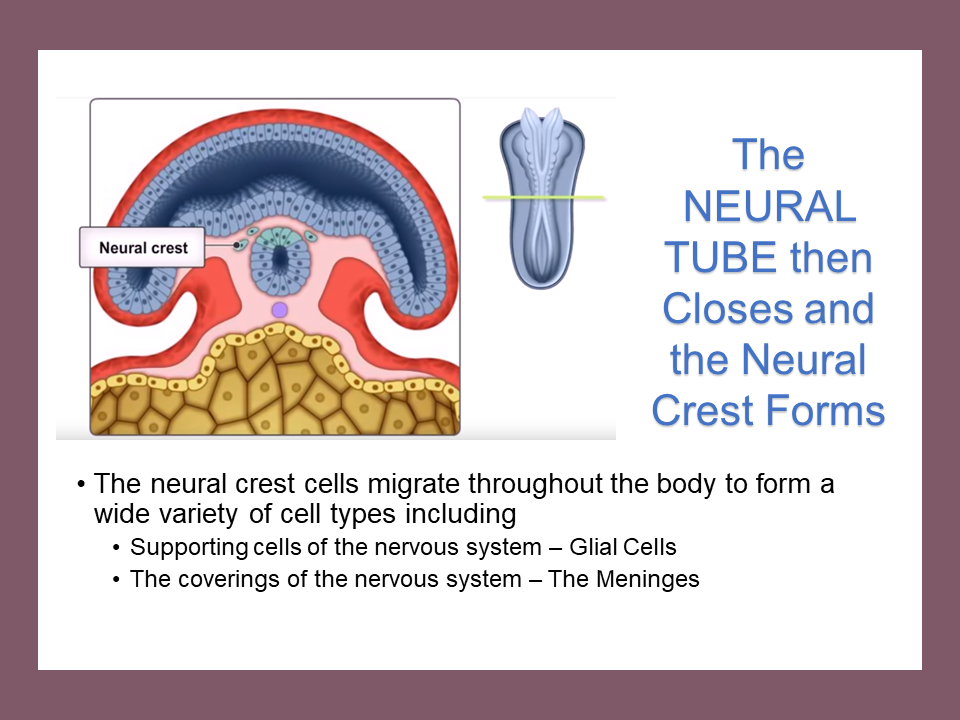
The neural folds then grow toward each other to fuse at the midline. The neural groove then is transformed into a completely closed, but hollow neural tube. Around this same, the neural folds become a separate population of cells called the neural crest. • 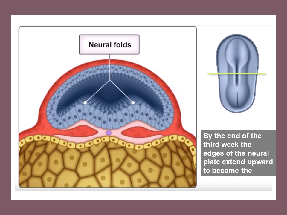
|
|
|
|
The neural crest cells migrate throughout the body to form a wide variety of cell types including
- Supporting cells of the nervous system – Glial Cells
- The coverings of the nervous system – The Meninges
|
|
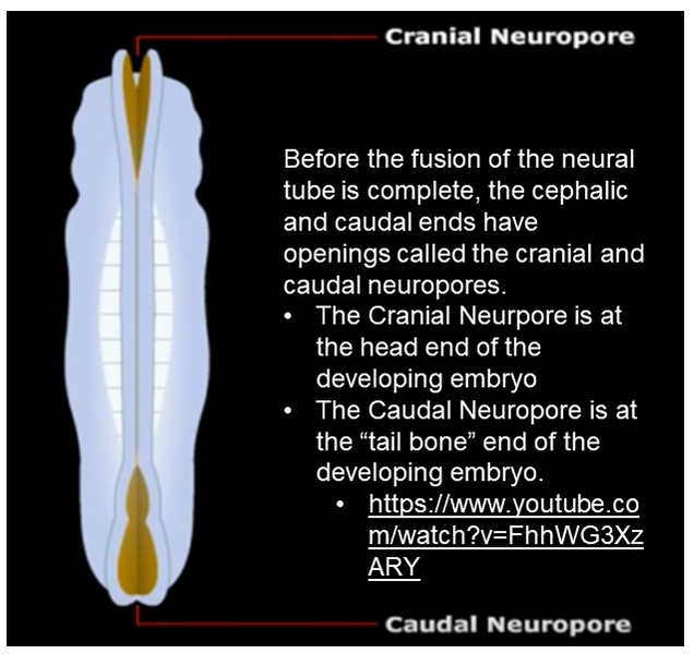
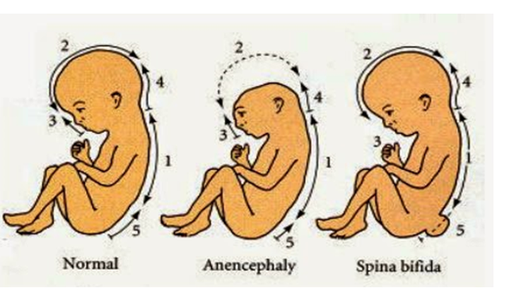
Before the fusion of the neural tube is complete, the cephalic and caudal ends have openings called the cranial and caudal neuropores.
•The Cranial Neurpore is at the head end of the developing embryo
•The Caudal Neuropore is at the “tail bone” end of the developing embryo.
•The Cranial Neurpore is at the head end of the developing embryo
•The Caudal Neuropore is at the “tail bone” end of the developing embryo.
Sonic hedgehog
Sonic hedgehog is a protein that in humans is encoded by the SHH ("sonic hedgehog") gene. Sonic hedgehog is one of three proteins in the mammalian signaling pathway family called hedgehog, the others being desert hedgehog (DHH) and Indian hedgehog (IHH).
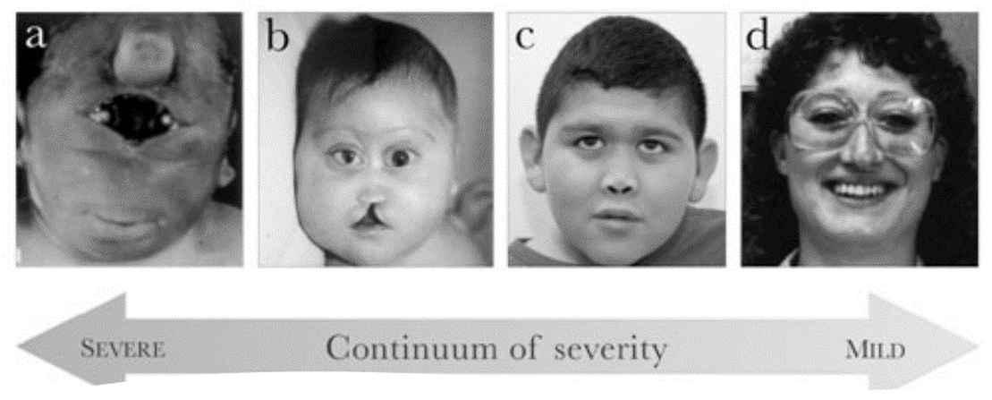
Sonic Hedgehog Disorders More than 100 mutations in the SHH gene have been found to cause nonsyndromic holoprosencephaly. Nonsyndromic holoprosencephaly is a condition that occurs when the brain fails to divide into two hemispheres during early development. SHH gene mutations are the most common cause of nonsyndromic holoprosencephaly. These mutations reduce or eliminate the activity of Sonic Hedgehog. The eyes will not form normally and the brain does not separate into two hemispheres. The development of other parts of the face is affected if the eyes do not move to their proper position. The signs and symptoms of nonsyndromic holoprosencephaly are caused by abnormal development of the brain and face.Sonic hedgehog is a protein that in humans is encoded by the SHH ("sonic hedgehog") gene. Sonic hedgehog is one of three proteins in the mammalian signaling pathway family called hedgehog, the others being desert hedgehog (DHH) and Indian hedgehog (IHH).
Sonic hedgehog is a protein that in humans is encoded by the SHH ("sonic hedgehog") gene. Sonic hedgehog is one of three proteins in the mammalian signaling pathway family called hedgehog, the others being desert hedgehog (DHH) and Indian hedgehog (IHH).
Sonic Hedgehog Disorders More than 100 mutations in the SHH gene have been found to cause nonsyndromic holoprosencephaly. Nonsyndromic holoprosencephaly is a condition that occurs when the brain fails to divide into two hemispheres during early development. SHH gene mutations are the most common cause of nonsyndromic holoprosencephaly. These mutations reduce or eliminate the activity of Sonic Hedgehog. The eyes will not form normally and the brain does not separate into two hemispheres. The development of other parts of the face is affected if the eyes do not move to their proper position. The signs and symptoms of nonsyndromic holoprosencephaly are caused by abnormal development of the brain and face.Sonic hedgehog is a protein that in humans is encoded by the SHH ("sonic hedgehog") gene. Sonic hedgehog is one of three proteins in the mammalian signaling pathway family called hedgehog, the others being desert hedgehog (DHH) and Indian hedgehog (IHH).
Sonic Hedgehog Disorders
More than 100 mutations in the SHH gene have been found to cause nonsyndromic holoprosencephaly. Nonsyndromic holoprosencephaly is a condition that occurs when the brain fails to divide into two hemispheres during early development. SHH gene mutations are the most common cause of nonsyndromic holoprosencephaly. These mutations reduce or eliminate the activity of Sonic Hedgehog. The eyes will not form normally and the brain does not separate into two hemispheres. The development of other parts of the face is affected if the eyes do not move to their proper position. The signs and symptoms of nonsyndromic holoprosencephaly are caused by abnormal development of the brain and face.
More than 100 mutations in the SHH gene have been found to cause nonsyndromic holoprosencephaly. Nonsyndromic holoprosencephaly is a condition that occurs when the brain fails to divide into two hemispheres during early development. SHH gene mutations are the most common cause of nonsyndromic holoprosencephaly. These mutations reduce or eliminate the activity of Sonic Hedgehog. The eyes will not form normally and the brain does not separate into two hemispheres. The development of other parts of the face is affected if the eyes do not move to their proper position. The signs and symptoms of nonsyndromic holoprosencephaly are caused by abnormal development of the brain and face.
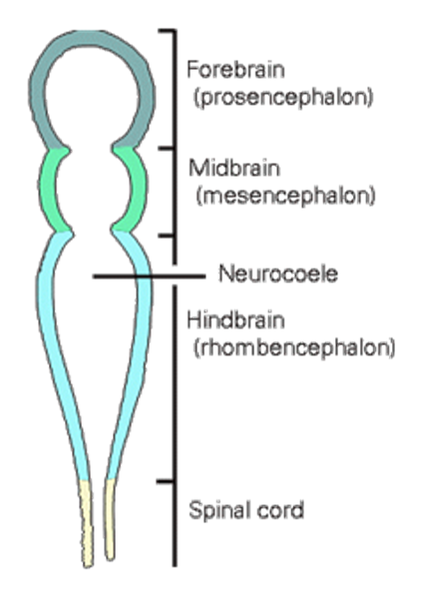
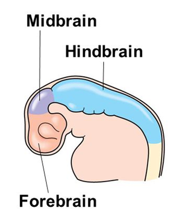
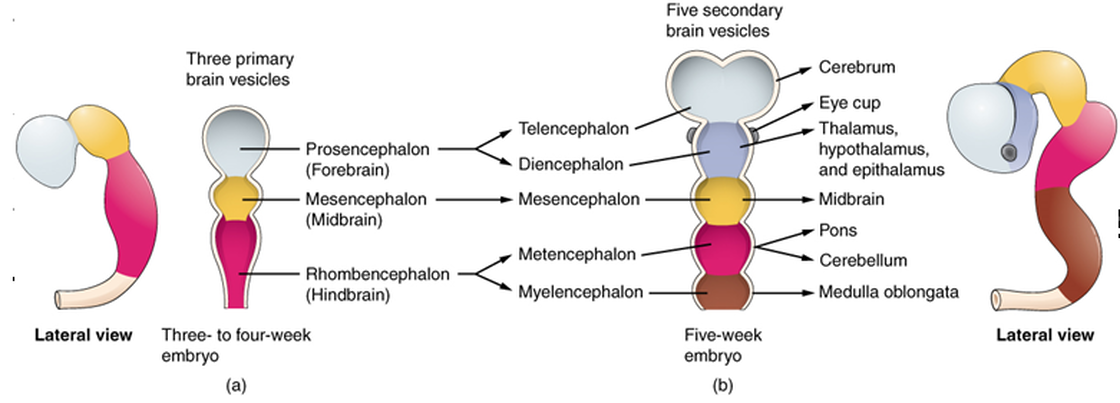
The Three Primary Vesicles
|
3 primary vesicles of the Neural Tube
The top of the tube thickens into three bulges that form the three primary vesicles. The 3 primary vesicles are:
|
|
|
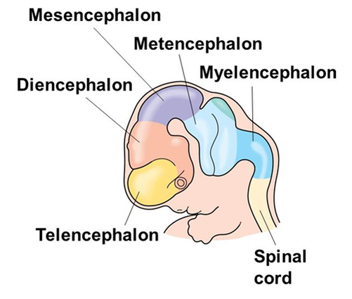
The Five Secondary Vesicles
|
5 secondary vesicles of the Neural Tube
These three develop later into five secondary vesicles:
|
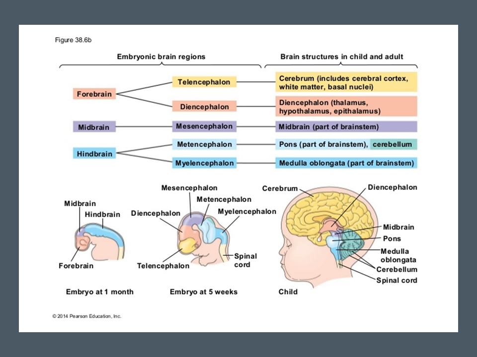
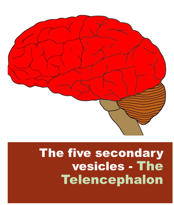
The Telencephalon
Telencephalon is also known as the cerebrum. The telencephalon refers to the region of the brain that includes the cerebral cortex and the hippocampus and basal ganglia. The five secondary vesicles- |
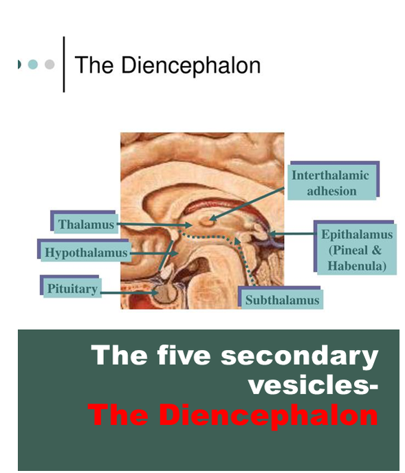
The Diencephalon
These three develop later into five secondary vesicles the forebrain develops into The prosencephalon (forebrain) develops into The Telencephalon The Diencephalon Becomes structures for regulating growth, hormones, sleep/wake cycles, and more. • |
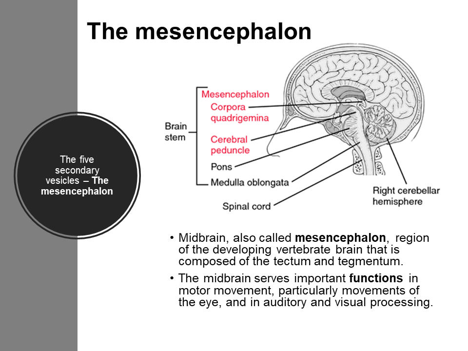
The mesencephalon
Midbrain, also called mesencephalon, region of the developing vertebrate brain that is composed of the tectum and tegmentum. The midbrain serves important functions in motor movement, particularly movements of the eye, and in auditory and visual processing.
Midbrain, also called mesencephalon, region of the developing vertebrate brain that is composed of the tectum and tegmentum. The midbrain serves important functions in motor movement, particularly movements of the eye, and in auditory and visual processing.
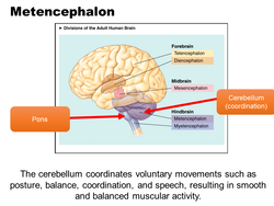
Metencephalon
|
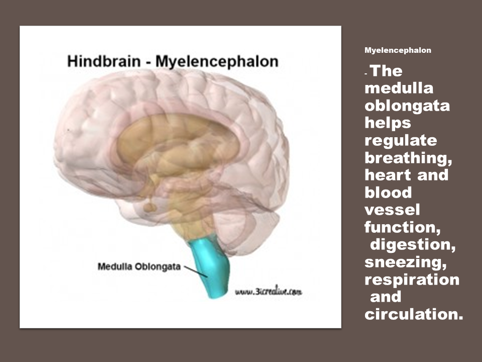
Myelencephalon
- The medulla oblongata helps regulate breathing, heart and blood vessel function, digestion, sneezing, respiration and circulation. |
Cell Differentiation
DNA has all of the instruction needed for the cell INCLUDING... What type of cell it will become All of the functions of the cell All of the cells of your body (almost all) have the same DNA. So HOW do cells develop from stem cells without an identity, to different types of cells (skin cells, nerve cells, blood cells, muscle cells, etc.)
DNA has all of the instruction needed for the cell INCLUDING... What type of cell it will become All of the functions of the cell All of the cells of your body (almost all) have the same DNA. So HOW do cells develop from stem cells without an identity, to different types of cells (skin cells, nerve cells, blood cells, muscle cells, etc.)
|
•Cellular differentiation
Cellular differentiation is the process where a cell changes from one cell type to another. The cell changes to a more specialized type. Differentiation occurs numerous times during the development of a multicellular organism as it changes from a simple zygote to a complex system of tissues and cell types. |
|
Embryonic stem cells
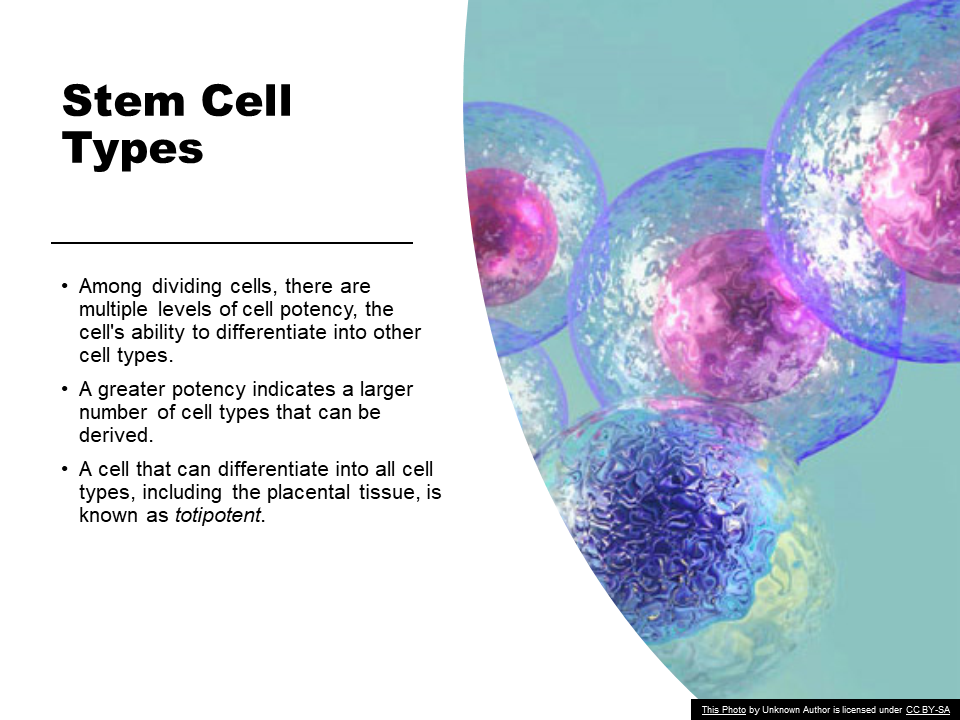
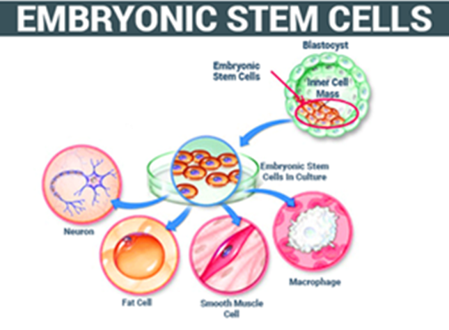
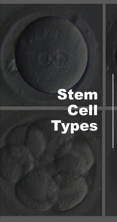
Embryonic stem cells (ES cells or ESCs) are pluripotent stem cells derived from the inner cell mass of a blastocyst, an early-stage pre-implantation embryo. Human embryos reach the blastocyst stage 4–5 days post fertilization, at which time they consist of 50–150 cells. Stem Cell Types Among dividing cells, there are multiple levels of cell potency, the cell's ability to differentiate into other cell types. A greater potency indicates a larger number of cell types that can be derived. A cell that can differentiate into all cell types, including the placental tissue, is known as totipotent.
|
HUMAN FETAL STEM CELLS IN A NUTSHELL
Human development begins when a sperm fertilizes an egg and the resulting fertilized egg creates a single totipotent cell, a zygote. In the first hours after fertilization, this zygote divides into identical totipotent cells, which can later develop into any of the three germ layers of a human (endoderm, mesoderm, or ectoderm), or into cells of the placenta Stem Cell Types After reaching a 16-cell stage, the totipotent cells of the morula differentiate into cells that will eventually become either the blastocyst's Inner cell mass or the outer trophoblasts. Approximately four days after fertilization and after several cycles of cell division, these totipotent cells begin to specialize. The inner cell mass, the source of embryonic stem cells, becomes pluripotent. |
|
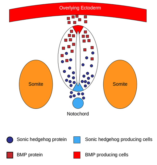
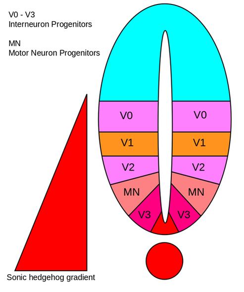
Sonic Hedgehog SHH is the best studied ligand of the hedgehog signaling pathway. It plays a key role in regulating vertebrate organogenesis, such as in the growth of digits on limbs and organization of the brain. Sonic hedgehog is a molecule that diffuses to form a concentration gradient and has different effects on the cells of the developing embryo depending on its concentration.
TAXIS: Cells can be attracted to certain chemicals or stimuli |
|
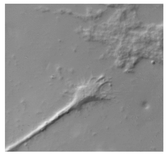
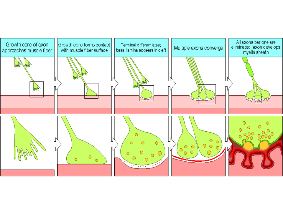
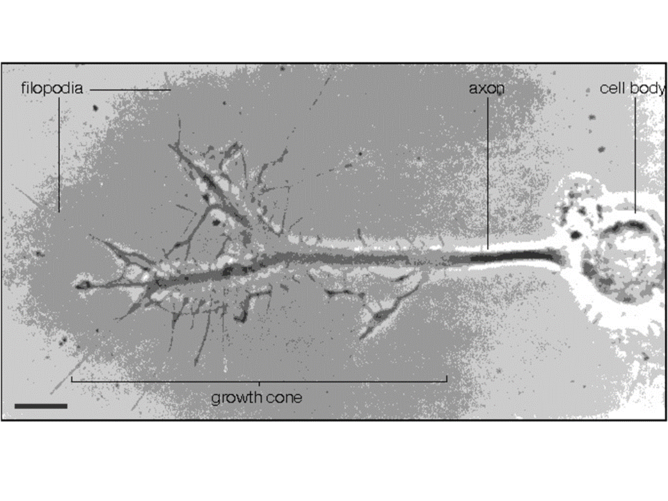
Neurons in early development, behave in such a way to either approach or avoid certain chemical signals. But rather than the whole cell moving, neural growth involves the outreach of the cell’s connecting pathway (axon) towards its downstream partner neurons.
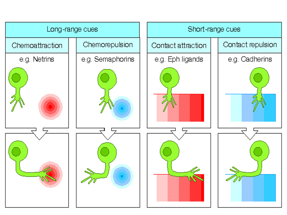
Axonal growth is led by growth cones Filopodia (growing from axons) are able to sense the environment ahead for chemical markers and cues. Mechanisms are fairly old in evolutionary terms. Intermediate chemical markers Guideposts studied in invertebrates Short and long range cues Short range chemo-attraction and chemo-repulsion Long range chemo-attraction and chemo-repulsion.
|
|
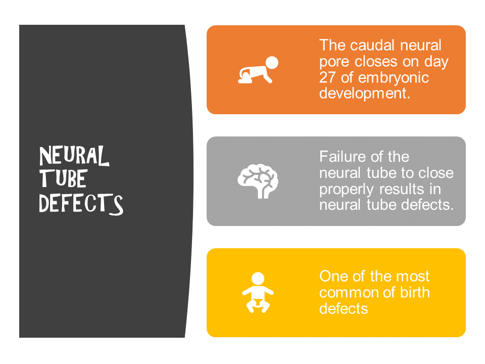
Neural tube defects can occur due to…
- Chromosomal disorders
- Environmental exposure
- Folic acid deficiency
- Certain drugs that block folic acid
- Carbamazepine
- Phenytoin
- Trimethoprim
- Carbamazepine
Anencephaly
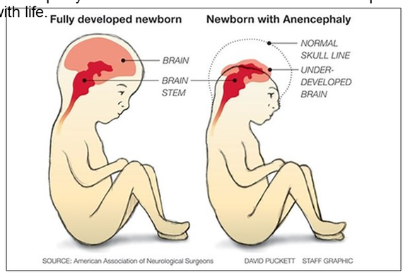
Anencephaly is the failure of the neural tube to spontaneously close at the cranial end hence the brain does not develop. Anencephaly means “no brain”. This condition is incompatible with life.
Microcephaly
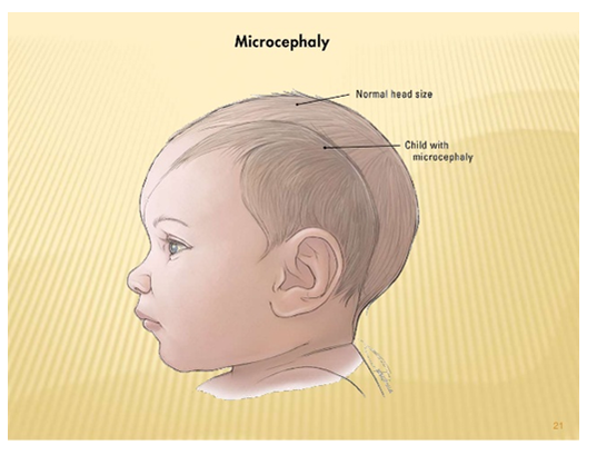
-Microcephaly-
Some infants with congenital Zika virus infection who do not have microcephaly at birth may later experience slowed head growth and develop postnatal microcephaly. Recognizing that Zika is a cause of certain birth defects does not mean that every pregnant woman infected with Zika will have a baby with a birth defect.
Some infants with congenital Zika virus infection who do not have microcephaly at birth may later experience slowed head growth and develop postnatal microcephaly. Recognizing that Zika is a cause of certain birth defects does not mean that every pregnant woman infected with Zika will have a baby with a birth defect.
Spina bifida
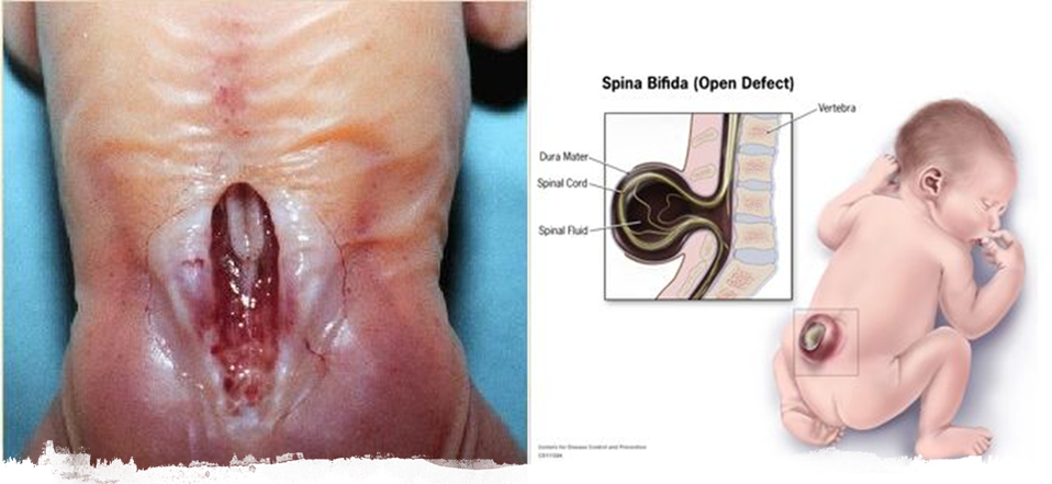
Spina bifida is the failure of the neural tube to spontaneously close at the caudal end. Vertebra overlying the area of the defect do not fully develop causing the vertebral arch to remain open.

|
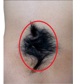
Spina Bifida Occulta
Spina bifida occulta is an asymptomatic defect caused by failure of the two halves of the vertebral arch to fuse at the midline. The only evidence of its presence may be a small tuft of hair over the defect. Spina bifida occulta only affects the vertebral arch leaving the spinal cord intact. A hair tuft is often visible on the skin just above the vertebra with a bony defect. This is the most common sign of spina bifida occulta. |
Folic acid

Folic Acid (folate) can prevent many neural tube birth defects.
Neural tube defects occur in the earliest weeks of pregnancy – before many women even know they're pregnant. It's important for women to begin taking folic acid before you start trying to conceive. Folic acid It's critically important to get enough folic acid because it helps prevent neural tube defects (NTDs), such as…
Neural tube defects occur in the earliest weeks of pregnancy – before many women even know they're pregnant. It's important for women to begin taking folic acid before you start trying to conceive. Folic acid It's critically important to get enough folic acid because it helps prevent neural tube defects (NTDs), such as…
- Spina Bifida
- Spina Bifida Occulta
- Anencephaly
- Microcephaly
- The neural tube is the part of the embryo from which your baby's spine and brain develop.

Neural tube defects According to the Centers for Disease Control (CDC), Neural tube defects affect about 3,000 pregnancies a year in the United States. Women who take the recommended daily dose of folic acid starting at least one month before conception and during the first trimester of pregnancy can reduce their baby's risk of neural tube defects by up to 70 percent.
Folic acid Folic acid is a form of vitamin B9, also known as folate. This vitamin helps prevent certain birth defects. When the nutrient comes directly from food sources, it's called folate. When it's manufactured for use as a supplement or to fortify foods, it's called folic acid.
Folic acid Folic acid is needed to make normal red blood cells and prevent a type of anemia called folate-deficiency anemia. It's essential for the production, repair, and functioning of DNA – our genetic map and a basic building block of cells.