GENETICS LAB
LAB EXERCISE:
The Cell Cycle and Human Genetics
The Cell Cycle and Human Genetics
Vocabulary
|

|
Goals
- Describe the cell cycle.
- Describe mitosis.
- Describe meiosis.
- Describe the process of crossing over and resulting chromosomal abnormalities when crossing over does not occur properly.
- Describe the process of nondisjunction I and nondisjunction II and resulting gametes.
- Describe various genetic tests used to test a fetus (NIPT, amniocentesis, CVS).
- Learn to read a Karyotype.
Lab Exercises:
- Let’s pretend that you just found out that you (or your partner) are pregnant, you have Advanced Maternal Age (35 years and older) and that this is your first OB/GYN visit. Pretend that you have never taken a college level biology course.
Equipment
- Genetic Testing pamphlets
- Your instructor will lecture about this topic
- Your instructor may provide videos demonstrating the techniques
Protocol:
- The first thing they gave you are these pamphlets to read. You are allotted 15 minutes to review the pamphlets before the OB/GYN comes in to check your embryo and order tests. You have to make a decision about running these genetic tests or not at week 10 of pregnancy.
- Read the pamphlets and circle any concepts or words that you may not understand. Add any questions that you think you would have. Put yourself in your patient’s shoes, what would they struggle understanding if they had limited biological knowledge?
- Identify what a Noninvasive Prenatal Test (NIPT) (a screening test) is used for and how is it performed.
- Identify what an Amniocentesis Diagnostic Test is used for and how is it performed.
- Identify what a Chorionic Villus Sampling (CVS) Diagnostic Test is used for and how is it performed.
- In order for you to understand this pamphlet, you must understand the cell cycle, mitosis, meiosis, nondisjunction and chromosomal abnormalities. Let’s learn about these below.
- As your postlab, put yourself in someone’s shoes.
- Why would someone want to know these results?
- Why would someone not want to know these results?
- As your postlab, let’s pretend that you or your partner are pregnant today. Your instructor will assign you a fetal karyotype. Cut out the pieces and tape them to a separate piece of paper in order. Determine if your fetus is aneuploid and if so what is the genetic abnormality called. Determine the assigned biological sex of the fetus. You will share these results next week.
- Think about what you would do if you found out that your fetus had an aneuploidy? What would you do if you were a carrier for a genetic disease? There is no right or wrong answer.
The Cell Cycle: Describe the Life Cycle of a Cell
Equipment
- Textbook/resources
- Your instructor will lecture about this topic
- Your instructor may provide videos demonstrating this process
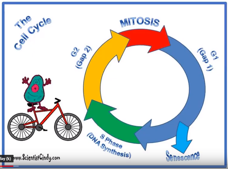
- Label the cell cycle: Interphase (G1, S, G2), Mitotic Phase (PMAT), and Cytokinesis
- Identify the function and major events of each phase.
Tutorial - The Cell Cycle
|
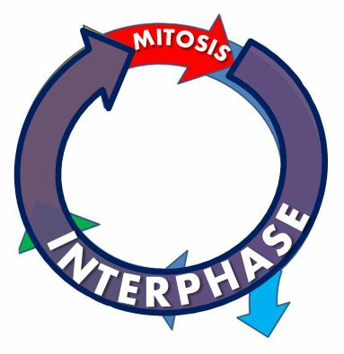
The Cell Cycle Consists of 2 MAIN PHASES
1) INTERPHASE 2) MITOSIS During interphase, the cell is preparing for cell division which occurs during the mitosis phase of the cell cycle. |
|
INTERPHASE
Interphase is subdivided into 3 phases:
Gap 1 (G1), S-Phase (or synthesis phase) and Gap 2 (G2).
Gap 1 (G1), S-Phase (or synthesis phase) and Gap 2 (G2).
|
The first phase of the cell cycle is Gap 1 or G1.
G1, is where the cell cycle BEGINS! It is a period of rapid growth of the cell. Here, the cell duplicates organelles and proteins to get ready for the next steps of the cell cycle! |
|
|
S-Phase is also known as the Synthesis Phase.
S-Phase, is where the genetic material gets duplicated or copied. The DNA is not visible under a microscope at this point of the cell cycle. The DNA is still in the form of a double helix, but it is unwound and is too thin to be seen. The DNA must be unwound at this point in order for the molecules that duplicate the DNA to get in close and do their job! 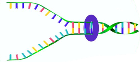
|
G2 is also known as Gap 2.
G2, is the final stage before mitosis begins. The cell grows even larger and makes the final proteins and organelles it needs to begin mitosis! After G2, the process of mitosis will begin.
|
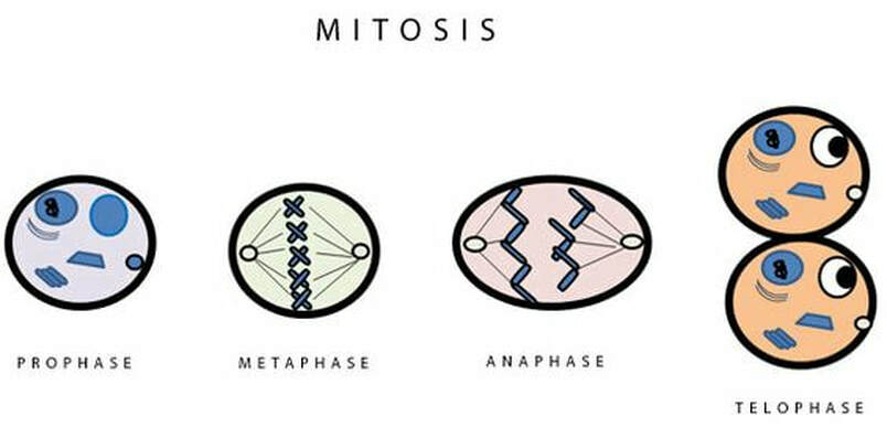
Mitosis
Mitosis is separated into 4 phases.
|
|
PROPHASE
|
Prophase is the first stage of mitosis. In prophase,
|
|
Late in prophase, a lot of important events occur. Occasionally, late prophase is referred to as its own unique phase called prometaphase. In late prophase, or prometaphase, chromosomes continue to condense. A complex of proteins called kinetochores, will form at the centromere region of the chromosomes. The kinetochores serve as attachment points that connect to the microtubules of the mitotic spindle. The mitotic spindle grows out of the centrosomes that migrate to opposite ends of the cell.
|
Chromosome - by ScientistCindy
|
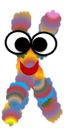
METAPHASE
|
|
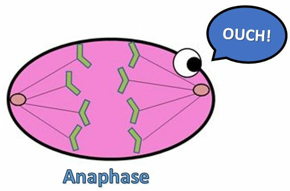
ANAPHASE
|
Anaphase is the next step in mitosis. In anaphase, sister chromatids are pulled toward opposite sides of the cell.
|
|
Telophase
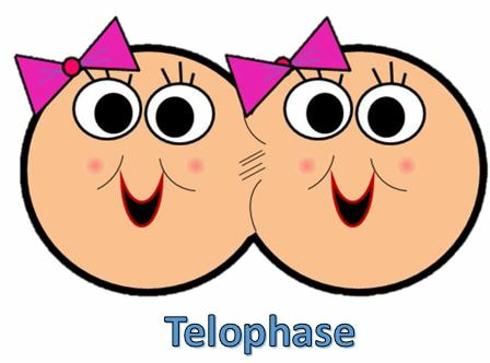
|
Telophase is the next phase in mitosis. In telophase, chromosomes arrive at opposite poles and begin to decondense into chromatin. The nuclear envelope reforms and the mitotic spindle breaks down.
|
|
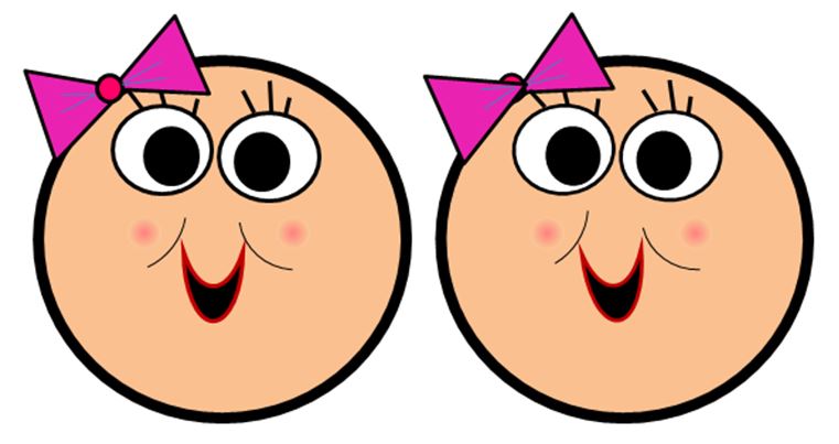
Telophase ends in the final step of separation called cytokinesis. In cytokinesis,
|
|
There is one more thing we need to know about the cell cycle. The cell cycle is not always going, because sometimes cells are not actively dividing. Cells that are not dividing, are stuck in the Resting Phase of the cell cycle, called G0 .
MITOSIS - The Nursery Rhyme
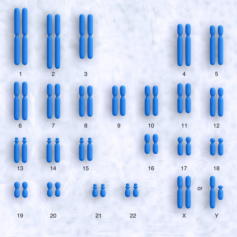
Karyotyping
|
Karyotyping is the process of pairing and ordering all the chromosomes of an organism, thus providing a genome-wide snapshot of an individual's chromosomes. Karyotypes are prepared using standardized staining procedures that reveal characteristic structural features for each chromosome. Clinical cytogeneticists analyze human karyotypes to detect gross genetic changes—anomalies involving several megabases or more of DNA. Karyotypes can reveal changes in chromosome number associated with aneuploid conditions, such as trisomy 21 (Down syndrome). Careful analysis of karyotypes can also reveal more subtle structural changes, such as chromosomal deletions, duplications, translocations, or inversions. In fact, as medical genetics becomes increasingly integrated with clinical medicine, karyotypes are becoming a source of diagnostic information for specific birth defects, genetic disorders, and even cancers.
|
NONDISJUNCTION
Nondisjunction occurs in cell division when chromosomes do not divide properly. Chromosomes contain all of a cell’s DNA, which it needs in order to function and reproduce. Normally, when a cell divides, the chromosomes line up in an orderly way and then separate from each other before cell division. When these chromosomes fail to separate properly, nondisjunction has occurred. The resulting daughter cells have an incorrect number of chromosomes; one may have too many, while another may have too few. This causes problems in cell function because a cell cannot function normally without the right amount of chromosomes.
|
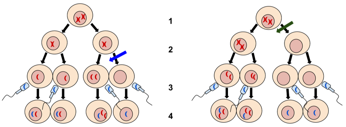
1. Meiosis I
2. Meiosis II 3. Fertilization 4. Zygote |
|
- Nondisjunction is when chromosomes fail to separate normally resulting in a gain or loss of chromosomes. This can happen during mitosis or meiosis.
- Primary nondisjunction occurs during meiosis I and the result is both members of a homologous pair go into the same daughter cell. This has the eggs have one more or one less number of chromosomes. Once the sperm fertilizes the egg then there is an abnormal number of chromosomes.
- Secondary nondisjunction happens during meiosis II and this results when the sister chromatids fail to separate, ending up with both daughter chromosomes going into the same gamete. During this, one egg will have one more or one less chromosome. Once these eggs are fertilized there will be two zygotes with an abnormal number of chromosomes. It is less harmful to have secondary nondisjunction since you can still have two normal gametes while in primary nondisjunction there are no normal gametes.
Chromosomal Disorders
Aneuploidy
The most common type of chromosomal abnormality is known as aneuploidy. Aneuploidy is a condition in which an individual has an abnormal chromosome number due to either having an extra chromosome or amissing chromosome. Most people with aneuploidy have trisomy (three copies of a chromosome) instead of monosomy (single copy of a chromosome).
Monosomy
The only known survivable monosomy in humans is Turner syndrome, where the affected individual is monosomic for the X chromosome. Other monosomies are usually lethal during early fetal development, and survival is only possible if not all the cells of the body are affected in case of a mosaicism (see below), or if the normal number of chromosomes is restored via duplication of the single monosomic chromosome ("chromosome rescue").
Turner syndrome (X monosomy) (45, X0).
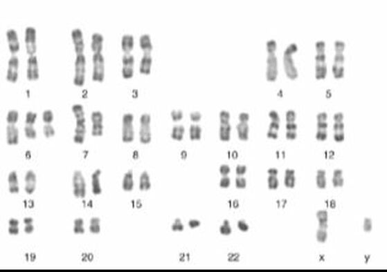
Karyotype of X monosomy (Turner syndrome)
Karyotype of X monosomy (Turner syndrome)
This condition is characterized by the presence of only one X chromosome and no Y chromosome (see bottom right corner). Source: https://en.wikipedia.org/wiki/Nondisjunction#Monosomy
|
This condition is characterized by the presence of only one X chromosome and no Y chromosome (see bottom right corner). The complete loss of an entire X chromosome accounts for about half the cases of Turner syndrome. The majority (>99%) of fetuses with only one X chromosome (karyotype 45, X0) are spontaneously aborted. |
Autosomal trisomy
The term autosomal trisomy means that a chromosome other than the sex chromosomes X and Y is present in 3 copies instead of the normal number of 2 in diploid cells.
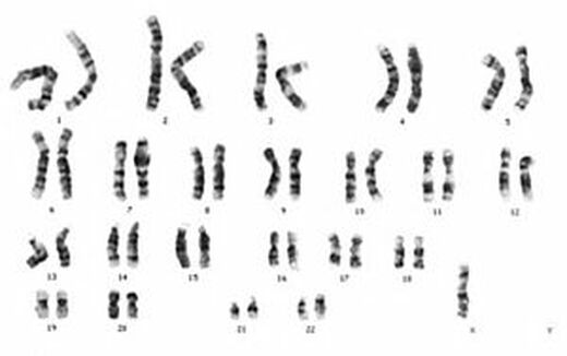
Down syndrome (trisomy 21).
The term autosomal trisomy means that a chromosome other than the sex chromosomes X and Y is present in 3 copies instead of the normal number of 2 in diploid cells.
Down syndrome (trisomy 21).
Karyotype of trisomy 21 (Down syndrome)
|
Trisomy 21 is the most common chromosomal anomaly in humans, affecting about 5,000 babies born each year and more than 350,000 people in the United States. Also known as Down syndrome, trisomy 21 is a genetic condition caused by an extra chromosome. Note that chromosome 21 is present in 3 copies, while all other chromosomes show the normal diploid state with 2 copies. Most cases of trisomy of chromosome 21 are caused by a nondisjunction event during meiosis I (see text). Down syndrome, a trisomy of chromosome 21, is the most common anomaly of chromosome number in humans. The majority of cases results from nondisjunction during maternal meiosis I. Trisomy occurs in at least 0.3% of newborns and in nearly 25% of spontaneous abortions. It is the leading cause of pregnancy wastage and is the most common known cause of mental retardation. It is well documented that advanced maternal age is associated with greater risk of meiotic nondisjunction leading to Down syndrome. This may be associated with the prolonged meiotic arrest of human oocytes potentially lasting for more than four decades.
|
Edwards syndrome (trisomy 18) and Patau syndrome (trisomy 13)
Human trisomies compatible with live birth, other than Down syndrome (trisomy 21), are Edwards syndrome (trisomy 18) and Patau syndrome (trisomy 13). Complete trisomies of other chromosomes are usually not viable and represent a relatively frequent cause of miscarriage.
Human trisomies compatible with live birth, other than Down syndrome (trisomy 21), are Edwards syndrome (trisomy 18) and Patau syndrome (trisomy 13). Complete trisomies of other chromosomes are usually not viable and represent a relatively frequent cause of miscarriage.
|
Sex chromosome aneuploidy
The term sex chromosome aneuploidy summarizes conditions with an abnormal number of sex chromosomes, i.e. other than XX (female) or XY (male). Formally, X chromosome monosomy (Turner syndrome, see above) can also be classified as a form of sex chromosome aneuploidy. |
|
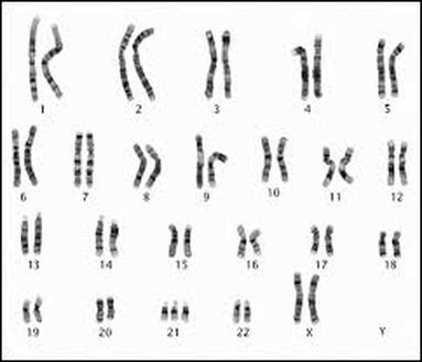
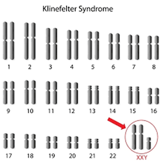
Klinefelter syndrome (47, XXY)
|
Klinefelter syndrome is the most common sex chromosome aneuploidy in humans. It represents the most frequent cause of hypogonadism and infertility in men. Most cases are caused by nondisjunction errors in paternal meiosis I. About eighty percent of individuals with this syndrome have one extra X chromosome resulting in the karyotype XXY. The remaining cases have either multiple additional sex chromosomes (48,XXXY; 48,XXYY; 49,XXXXY), mosaicism (46,XY/47,XXY), or structural chromosome abnormalities.
|
|
XYY Male (47, XYY)
The incidence of XYY syndrome is approximately 1 in 800-1000 male births. Many cases remain undiagnosed because of their normal appearance and fertility, and the absence of severe symptoms. The extra Y chromosome is usually a result of nondisjunction during paternal meiosis II.
Trisomy X (47,XXX)
Trisomy X is a form of sex chromosome aneuploidy where females have three instead of two X chromosomes. Most patients are only mildly affected by neuropsychological and physical symptoms. Studies examining the origin of the extra X chromosome observed that about 58-63% of cases were caused by nondisjunction in maternal meiosis I, 16-18% by nondisjunction in maternal meiosis II, and the remaining cases by post-zygotic, i.e. mitotic, nondisjunction.
Uniparental disomy
Uniparental disomy denotes the situation where both chromosomes of a chromosome pair are inherited from the same parent and are therefore identical. This phenomenon most likely is the result of a pregnancy that started as a trisomy due to nondisjunction. Since most trisomies are lethal, the fetus only survives because it loses one of the three chromosomes and becomes disomic. Uniparental disomy of chromosome 15 is, for example, seen in some cases of Prader-Willi syndrome and Angelman syndrome.