THE JOINTS
Joints can be classified into structural categories or they can be classified into functional categories.
The structural classification of joints takes in account what types of tissues are present in the joint and/or if a joint cavity exists.
The 3 Structural Categories of Joints Are...
The structural classification of joints takes in account what types of tissues are present in the joint and/or if a joint cavity exists.
The 3 Structural Categories of Joints Are...
- fibrous joints
- cartilaginous joints
- synovial joints
The 5 Functional Categories of Joints Are...
- synarthroses (syn = together, arthro = joint), which are immovable joints
- amphiarthroses (amphi = on both sides), slightly movable joints
- diarthroses (dia = through, apart), or freely movable joints
- cartilaginous joints
- synovial joints (Only synovial joints have a joint cavity)
- Freely movable joints predominate in the limbs.
- Synovial joints are freely movable.
- Immovable and slightly movable joints are largely restricted to the axial skeleton.
- Immovable and slightly movable joints are much more stable than moveable joints. This means they are less prone to injury.
- In general, fibrous joints (such as cranial sutures) are immovable.
- However, cartilaginous joints have both rigid and slightly movable examples.
Fibrous Joints
There are three types of fibrous joints.
- sutures
- syndesmoses
- gomphoses
Properties of fibrous joints.
- Most fibrous joints are "fixed" or "immovable".
- They have no joint cavity.
- They are connected via fibrous connective tissue.
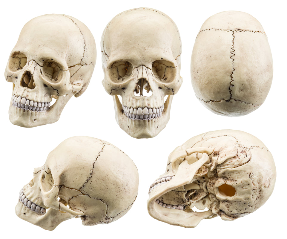
Sutures are immovable joints that are completely filled with very short, interconnecting fibers, that appear like stitches or sutures. This type of joint is found only in the skull and nowhere else in the body. The immovable nature of the sutures "fixes" the cranial bones in position as a protective adaptation that protects the brain.
|
|
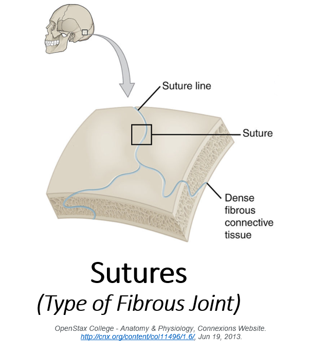
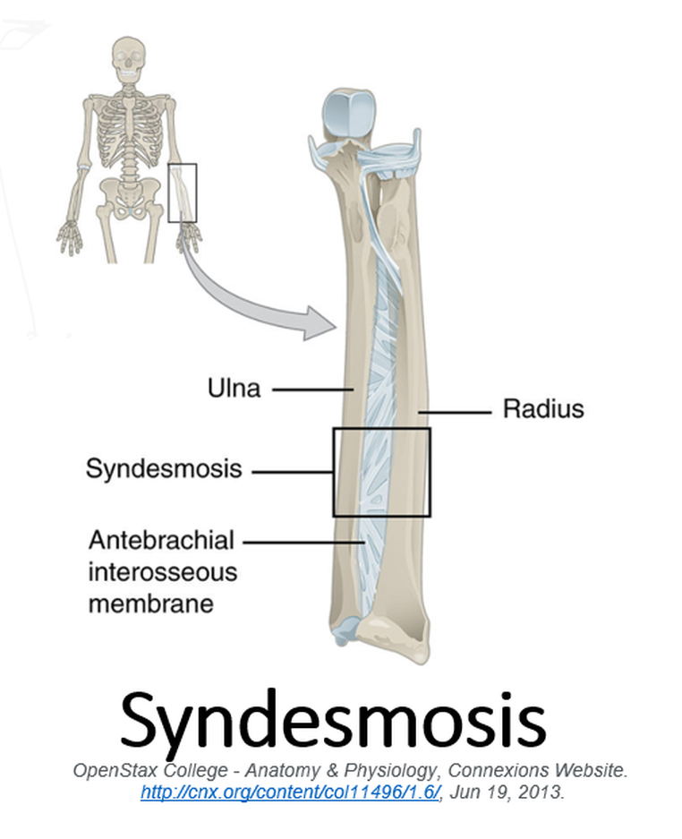
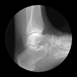
Syndesmoses are immovable joints that are held together with ligaments. Between the fibula and tibia at the ankle.
Some of the long bones in the body such as he radius and ulna in the forearm and the tibia and fibula of the lower leg are joined by a syndemosis (singular) or syndemoses (plural). Syndesmoses are considered slightly movable (or amphiarthrodial). In syndesmoses, the bones are connected by ligaments and bands of fibrous tissue. The syndesmoses may have some very slight movability , depending on the length of the cords or bands of fibrous tissue that it is composed of. The longer the fibrous tissue, the more movability is possible. The cords and bands of fibrous tissue found in syndesmoses are always shorter than those found in sutures.
|
|
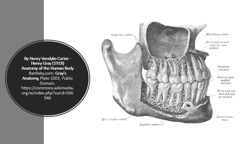
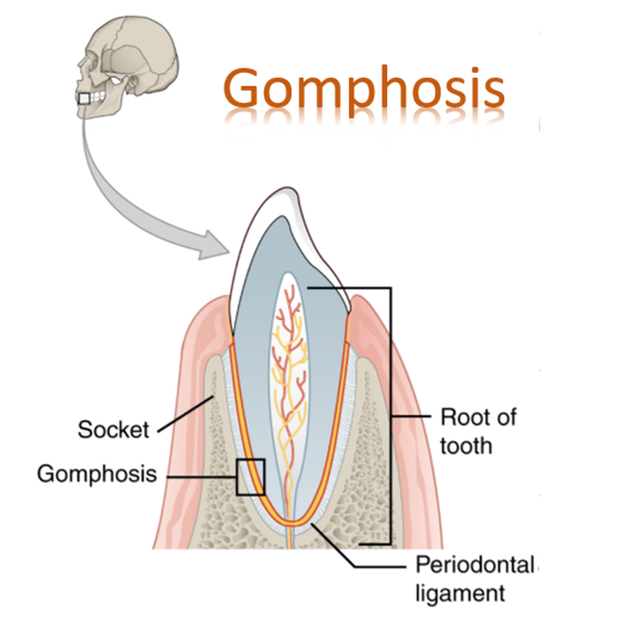
The only example is the articulation of a tooth with its bony alveolar socket. The term gomphosis comes from the Greek gompho, meaning “nail” or “bolt,” and refers to the way teeth are embedded in their sockets (as if hammered in). The fibrous connection in this case is the short periodontal ligament
|
A gomphosis is a "peg-in-socket" fibrous joint. The term gomphosis comes from a word meaning “nail”. This type of articulation found between our teeth and the bony sockets they sit in. The tooth is held into place in its socket through the periodontal ligament. |
Cartilaginous Joints
As you might expect, cartilaginous joints contain cartilage. They do not have a cavity and are slightly mobile.
There are two types of cartilaginous joints.
1) Synchondroses
2) Symphyses
Synchondrosis
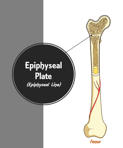
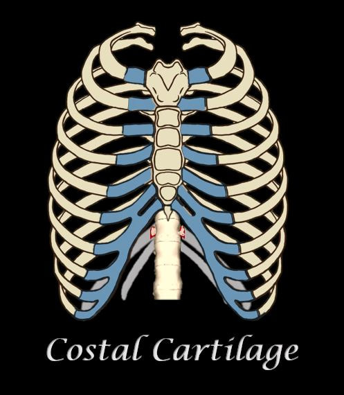
The term "synchondrosis" means “junction of cartilage”. These joints are joined via plates of hyaline cartilage. Virtually all synchondrosis joints are synarthrotic (immovable).
Examples of synchondroses include...
Examples of synchondroses include...
- The epiphyseal plates (epiphyseal lines) in long bones of children. Epiphyseal plates are temporary joints and eventually become synostoses.
- Another example of a synchondrosis is the immovable joint between the costal cartilage of the first rib and the manubrium of the sternum.
|
|
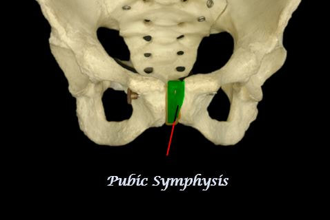
Symphyses
|
The term symphysis means “growing together". The bone of these joints are connected using fibrocartilage. Fibrocartilage is strong. resistant and is a good shock absorber. These joint permit only a limited amount of movement. The surface of the fibrocartilage is covered with a layer of hyaline cartilage, to act as articular cartilage for the bony articulation points.
Examples of symphases include...
|
|
Synovial Joints
|
Synovial joints are joints in which the articulating bones are separated by a fluid-filled joint cavity.
|
|
6 Types of Synovial Joints
|
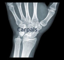
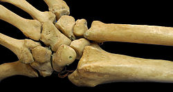
Carpals Have Plane Joints
The distal radioulnar articulation pivot-joint formed between the head of ulna and the ulnar notch on the lower extremity of radius.
|
The superior radioulnar joint is a synovial pivot joint between the circumference of the head of the radius
|
Plane or Gliding Joint
A plane joint or gliding joint, is formed from flattened articulation surfaces. This structures allows the bones to glide past one another in any direction.
Gliding joints are found between the carpal bones of the wrist.
|
Gliding joints are found between the tarsal bones of the ankle; and between the tarsals and the metatarsals of the foot.
|
|
Gliding Movements : Gliding motion is permitted by joints that have with relatively flat articulating surfaces. These are the "plane joints" or "gliding" joints of the body. The movements arises from the nearly flat articulating surfaces of bone that "glide" across each other. The direction of the "gliding" motion can be either back-and-forth or side-to-side.
|
|
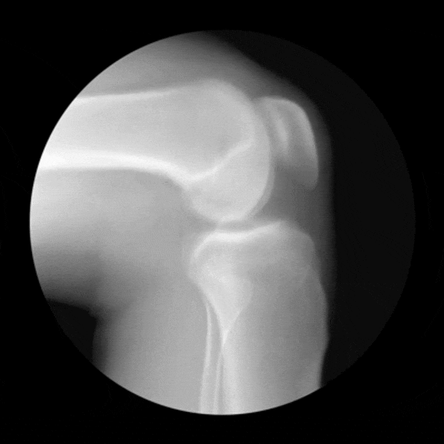
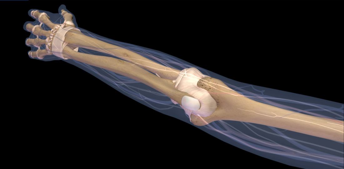
Hinge Joints
|
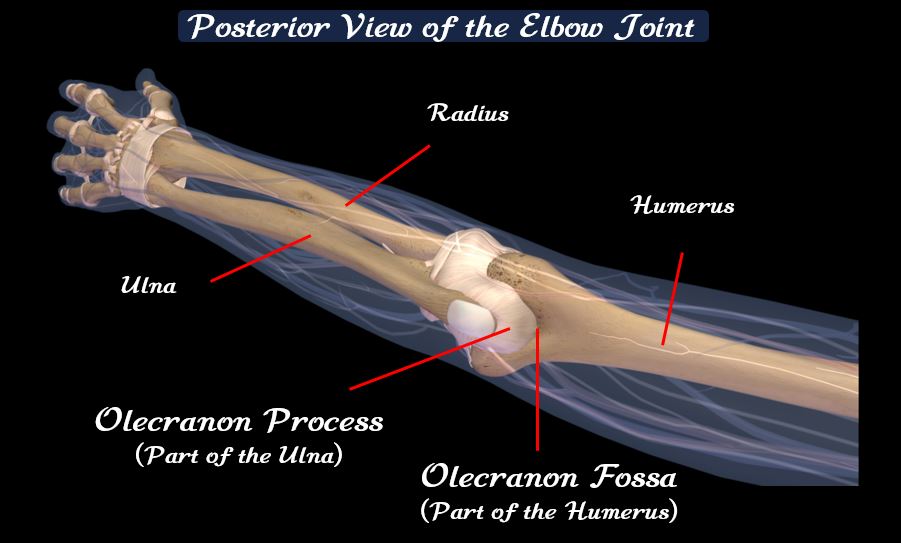
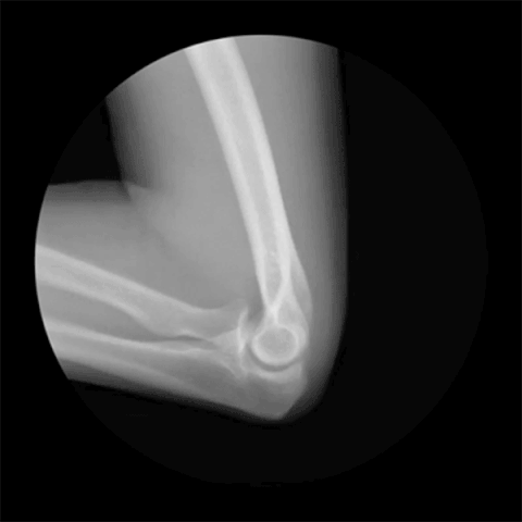
Hinge joints are located in our phalanges, ankles, elbows, and knees. Hinge joints are formed between two or more bones where the bones can only move along one axis to flex or extend.
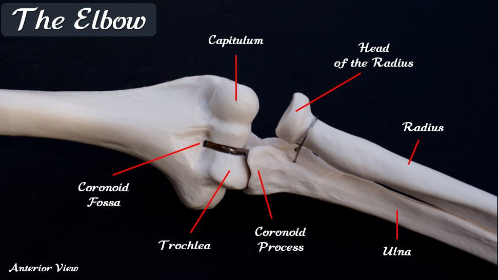
The elbow joint has a hinge joint formed between the distal end of the humerus the proximal end of the ulna. |
The elbow joint has a hinge joint formed between the distal end of the humerus the proximal end of the ulna.
|
Knees are "Hinge Joints"
|
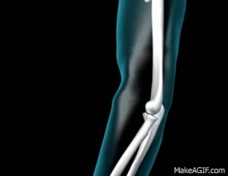
The elbow joint has a hinge joint formed between the distal end of the humerus the proximal end of the ulna.
|
Hinge Joints Allow for Flexion and Extension
Hinge joints provides a stable and smooth structure that allows for the motions of flexion and extension.
- Flexion = Flexion is the bending movement that decreases the angle of the joint and brings the articulating bones closer together.
- Extension = Extension is the reverse of flexion and occurs at the same joints. It increases the angle of the joint . It typically straightens a flexed limb or body part.
Pivot Joints
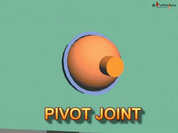

In addition to the hinge joint formed between the ulna and the humerus, the elbow joint also has a pivot joint that is formed between the humerus and the radius.
Pivot joints allow for rotational motion. The pivot joint between the radius and the humerus, allows for 2 unique movements that are only possible in the forearms and hands, and are not possible anywhere else in the body. These movements are called pronation and supination..
Pivot joints allow for rotational motion. The pivot joint between the radius and the humerus, allows for 2 unique movements that are only possible in the forearms and hands, and are not possible anywhere else in the body. These movements are called pronation and supination..
|
Pronation and supination are movements only found in the forearms and hands, that allow for the rotation of the hands.
|

The elbow joint has a pivot joint formed between the distal end of the humerus the proximal end of the radius.
|
Ball-and-Socket Joints
|
Ball-and-socket have the most freedom of motion. There are only two ball-and-socket joints in the entire body.
Ball-and-Socket Joints Allow for Circumduction of the Limbs.
The circular (or, more precisely, conical) movement of a body part, such as a ball-and-socket joint or the eye. It consists of a combination of flexion, extension, adduction, and abduction. "Windmilling" the arms and rotating the hand from the wrist are examples of circumductive movement.
CIRCUMDUCTION OF THE ARM
|
CIRCUMDUCTION OF TH LEG
|
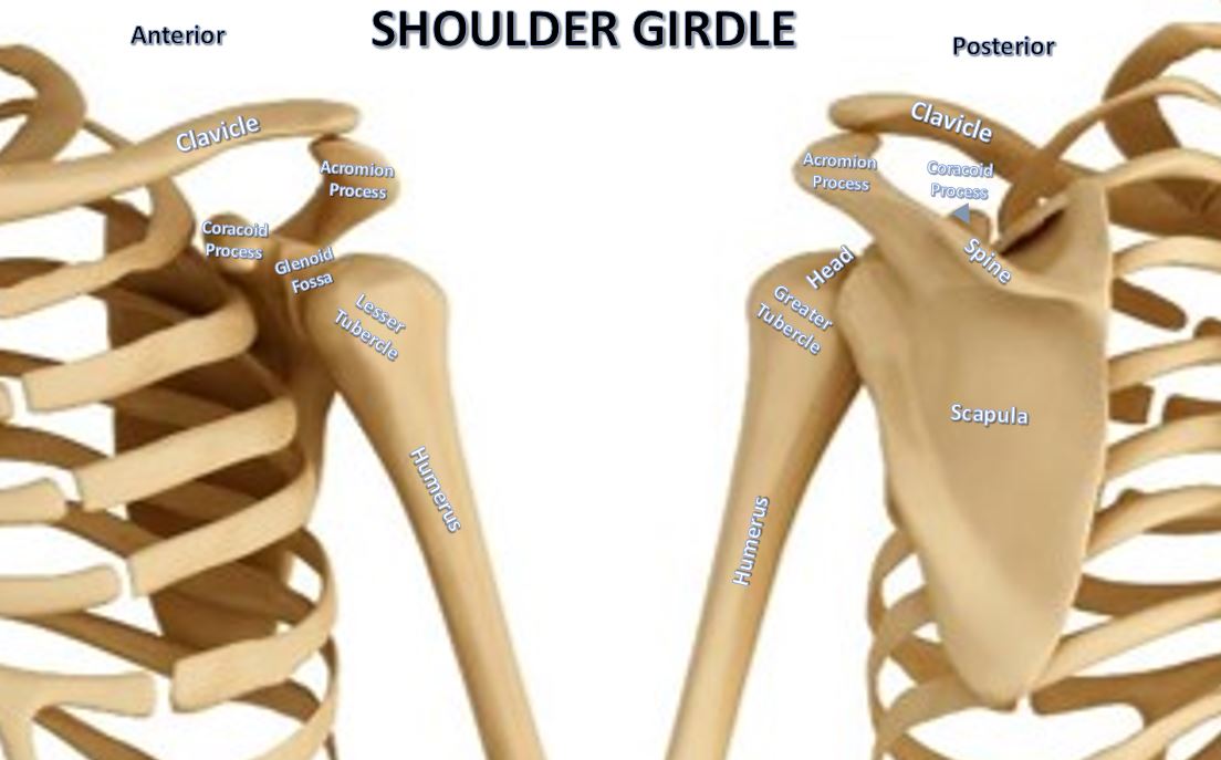
1. Shoulder (Glenohumeral) Joint
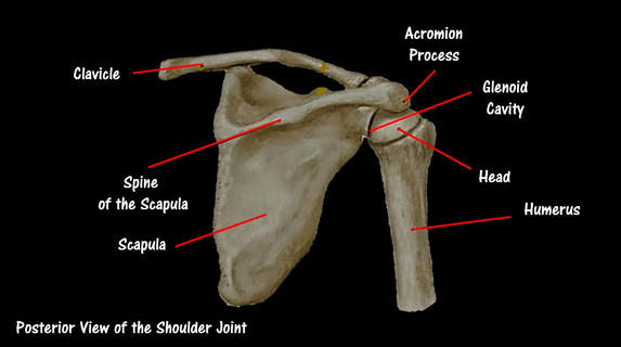
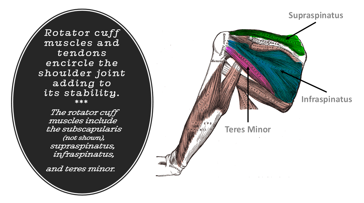
The shoulder or glenohumeral joint is the most freely moving joint of the body. However, in order for this freedom of movement to exist, the shoulder joint itself has sacrificed stabilization. Even though ball-and-socket joints have the potential to be very stable, in the case of the glenohumeral joint, the glenoid cavity of the scapulae is much too shallow to stabilize the large head of the humerus it articulates with. In fact, the glenoid cavity is only about one-third the size of the humeral head and adds only minimal stability to the joint.
- The coracohumeral ligament provides helps support the weight of the upper limb anteriorly.
- Three glenohumeral ligaments provide minor support.
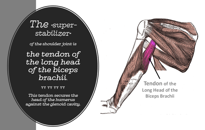
- The “superstabilizer” is the tendon of the long head of the biceps brachii muscle of the arm. This tendon secures the head of the humerus against the glenoid cavity.
Rotator cuff muscles and tendons encircle the shoulder joint adding to its stability. The rotator cuff muscles include the subscapularis, supraspinatus, infraspinatus, and teres minor. The rotator cuff is a common area of injury due to vigorous circumduction.
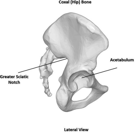
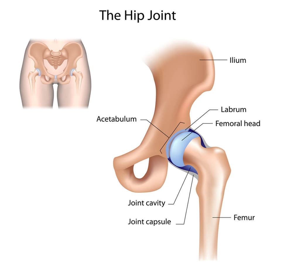
The Hip (Coxal) Joint
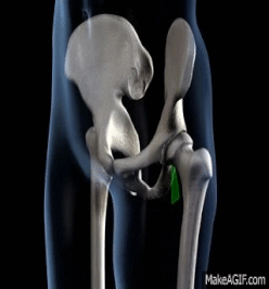
The hip (coxal) joint is a much more stable ball-and-socket joint, compared to the shoulder joint. In the hip joint, the acetabulum creates a deeply cupped surface of articulation for the head of the femur, which gives the joint added stability. However, the tradeoff is that the range of motion is more limited than that of the shoulder joint.
|
|
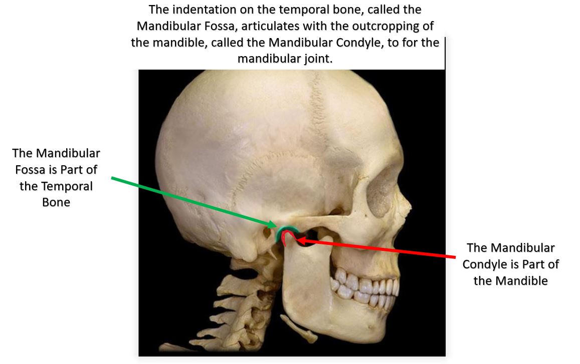
Temporomandibular Joint
The temporomandibular joint (TMJ), or jaw joint, is the articulation of the mandibular condyles (located on the mandible) and the mandibular fossa (located on the temporal bones). This joint is a modified hinge joint that lies just anterior to the ear.
Temporomandibular joint dysfunction (TMD or, TMJD) is any number of dysfunctions causing pain emanating from the TMJ area. 20 to 30% of people will experience some sort of TMJ discomfort in their lifetime. One potential cause of TMJ dysfunction is the grinding of the teeth while sleeping. Sufferers experience pain, restricted movement of the jaw and "popping" sounds upon movement of the jaw. noises from the temporomandibular joints (TMJ) during jaw movement. Although TMD is not life-threatening, it can be detrimental to quality of life,[3] because the symptoms can become chronic and difficult to manage.
condylar (or ellipsoid) joints
- condylar (or ellipsoid) joints
There are 6 features common to all synovial joints.
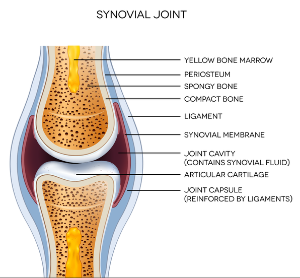
- The articulate cartilage, which is composed of hyaline cartilage. It provides a smooth gliding surface.
- The joint cavity (articular cavity) contains a small amount of synovial fluid.
- They have an articular capsule. which encloses the joint. The articulate capsule is composed of dense irregular connective tissue that strengthens the joint so that the bones are not pulled apart.
- Synovial fluid is a slippery substance lying within joint capsule. Synovial fluid provides a slippery, weight-bearing film that reduces friction between the cartilages. Without this lubricant, rubbing would wear away joint surfaces and excessive friction could overheat and destroy the joint tissues.
- Reinforcing ligaments. Synovial joints are reinforced and strengthened by a number of bandlike ligaments, usually capsular ligaments, which are thickened parts of the fibrous layer.
- Nerves and blood vessels. Synovial joints are richly supplied with sensory nerve fibers that innervate the capsule. Synovial joints are also richly supplied with blood vessels, most of which supply the synovial membrane.
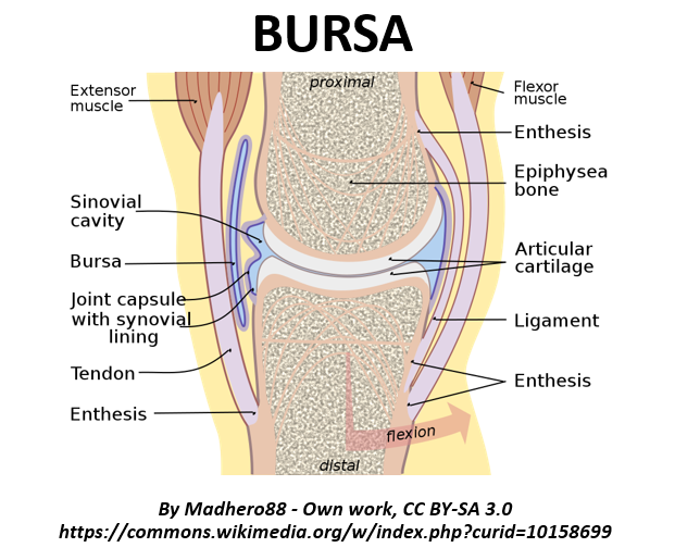
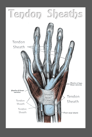
Bursae and Tendon Sheaths
Bursae and tendon sheaths are not actually part of the joints, but they are associated structures that lie just external to the joint. They are sacs filled with synovial fluid. that function to reduce friction during joint activity.
The difference between a bursa and a tendon sheath is that tendon sheath more elongated. Tendon sheaths wrap around areas with multiple crowded tendons such as the wrists.
The difference between a bursa and a tendon sheath is that tendon sheath more elongated. Tendon sheaths wrap around areas with multiple crowded tendons such as the wrists.
|
|
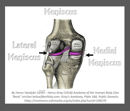
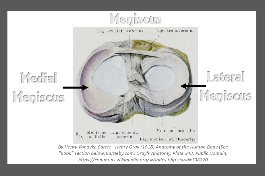
Meniscus
Some joints have articulate discs or wedges of fibrocartilage separating the articular surfaces. The meniscus improves the complementary fit between the articulation points of the bone. This makes the joint more stable and reduces wear and tear on the joint surfaces.
|
|
Cartilage Tears
|
Most cartilage injuries involve tearing of the meniscus. This type of injury can come as the result of overuse or excessive physical activity. Tears and overuse damage to the articular cartilages of other joints is becoming increasingly common in young athletes. Cartilage tears heal extremely slowly due to their avascular nature. Surgery is sometime needed for repair.
|
|
Factors Influencing the Stability of Synovial Joints
|
Because joints are constantly stretched and compressed, they must be stabilized so that they do not dislocate (come out of alignment). The stability of a synovial joint depends on three factors:
|
|
1. Articular Surfaces
|

|
2. Ligaments
|
The capsules and ligaments of synovial joints unite the bones and prevent excessive or undesirable motion.
3. Muscle Tone
|

TendonsFor most joints, the muscle tendons that cross the joint are the most important stabilizing factor. These tendons are kept under tension by the tone of their muscles. |
|
Movements Allowed by Synovial Joints
|
|
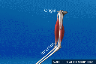
Every skeletal muscle of the body is attached to bone or other connective tissue structures at no fewer than two points. The muscle’s origin is attached to the immovable (or less movable) bone. Its other end, the insertion, is attached to the movable bone. Body movement occurs when muscles contract across joints and their insertion moves toward their origin.
There are three general types of movements:
- gliding
- angular movements
- rotation
|
Gliding Movements : Gliding motion is permitted by joints that have with relatively flat articulating surfaces. These are the "plane joints" or "gliding" joints of the body. The movements arises from the nearly flat articulating surfaces of bone that "glide" across each other. The direction of the "gliding" motion can be either back-and-forth or side-to-side.
Angular Movements : Angular movements increase or decrease the angle between two bones. These movements may occur in any plane of the body and include the following
Flexion = bending movement that decreases the angle of the joint and brings the articulating bones closer together.
|

|
Extension Extension is the reverse of flexion and occurs at the same joints. It increases the angle between the articulating bones and typically straightens a flexed limb or body part.
- Examples include
- straightening a flexed neck, body trunk, elbow, or knee.
ABDUCTION AND ADDUCTION
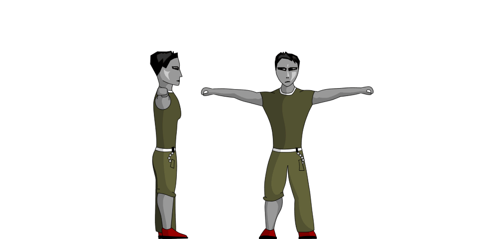
Abduction Abduction (“moving away”) is movement of a limb away from the midline or median plane of the body, along the frontal plane.
|
Adduction Adduction (“moving toward”) is the opposite of abduction, so it is the movement of a limb toward the body midline or, in the case of the digits, toward the midline of the hand or foot.
|
|
Circumduction Circumduction is moving a limb so that it describes a cone in space (circum = around; duco = to draw). The distal end of the limb moves in a circle, while the point of the cone (the shoulder or hip joint) is more or less stationary.
Rotation Rotation is the turning of a bone around its own long axis. It is the only movement allowed between the first two cervical
Supination and Pronation The terms supination (soo″pĭ- na′shun; “turning backward”) and pronation (pro-na′shun; “turning forward”) refer to the movements of the radius around the ulna.
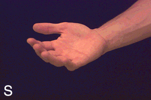
Supination and Pronation
|
Circumduction of the Arms
Rotation
|
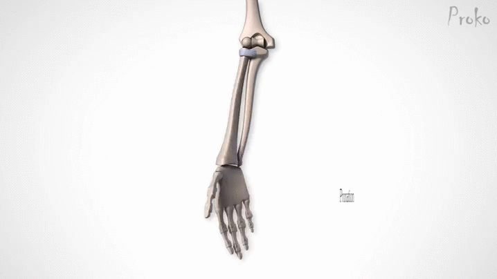
Supination and Pronation
|
Dorsiflexion and Plantar Flexion of the Foot The up-and- down movements of the foot at the ankle are given more specific names.
|
|
|
|
Inversion and Eversion Inversion and eversion are special movements of the foot.
|
|
Protraction and Retraction Nonangular anterior and posterior movements in a transverse plane are called protraction and retraction, respectively (Figure 8.6d).
|
|
|
|
Elevation and Depression
|
Sprains
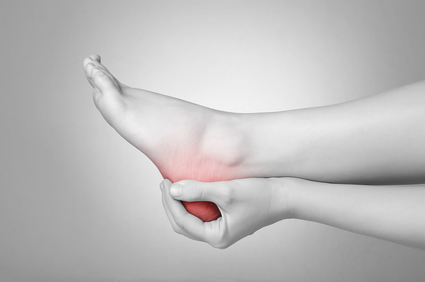
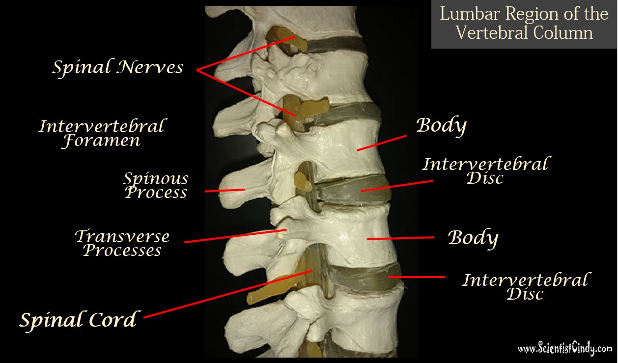
Sprains occur when the ligaments that surround a joint are stretched or torn. Common sites of sprains are the ankle, the knee, and the lumbar region of the spine. Sprains are slow to heal because ligaments are poorly vascularized. Sprains are painful and movement to the injured area should be restricted during recovery.
Arthritis
The term arthritis describes over 100 different types of inflammatory or degenerative diseases that damage the joints. In all its forms, arthritis is the most widespread crippling disease in the U.S. One out of every five persons in North America will suffer from some sort of arthritic ailment in their lifetime.
Osteoarthritis Osteoarthritis (OA)
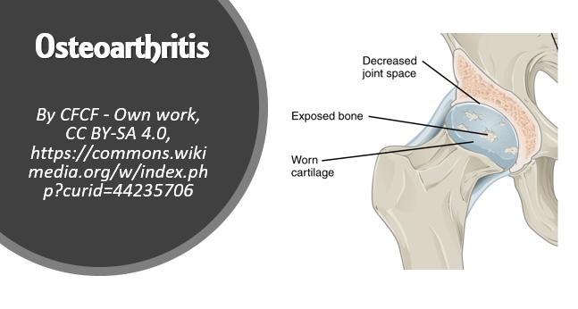
Osteoarthritis Osteoarthritis (OA) is the most common form of chronic arthritis. It is a chronic, age-related, degenerative condition. More women develop OA than men, and nearly all people develop some level of OA by the age of 80.
It is believed that OA may be caused by excessive amounts of the cartilage-destroying enzymes being released. The result is damaged, eroded articular cartilages that loose their smooth surface. As the disease progresses, the exposed bone tissue thickens and forms bony spurs (osteophytes) that lead to restricting the movement of the joint. Aching and stiffness of the cervical and lumbar spine, fingers, knuckles, knees, and hips are most affected.
Glucosamine and chondroitin sulfate, are natural supplements that some believe may function to replace the these substances that are normally present in healthy cartilage.
Rheumatoid Arthritis
Rheumatoid Arthritis (RA) is an autoimmune disease in which the body’s immune system attacks its own tissues.
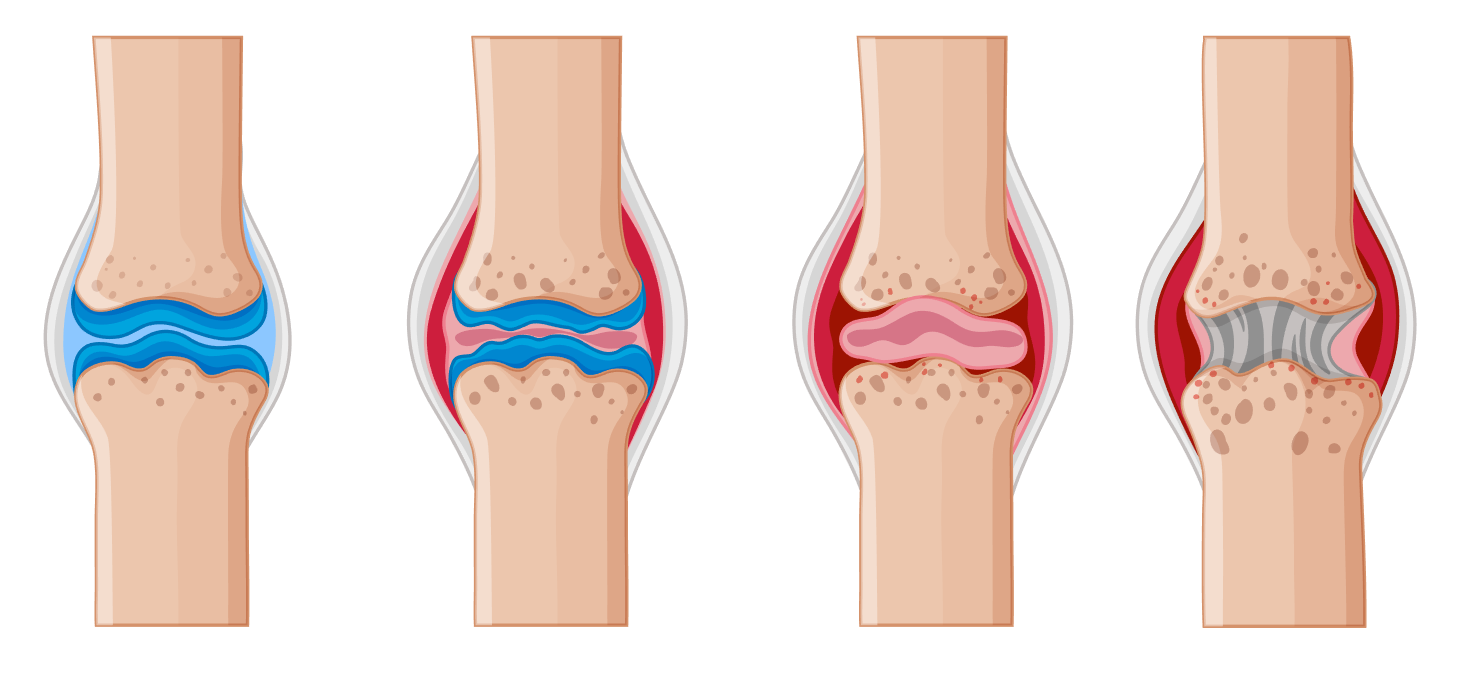
Rheumatoid arthritis (RA) is a chronic inflammatory disorder that usually has its onset between the ages of 30 and 50. It affects three times as many women as men. People with RA initially experience bilateral joint tenderness and stiffness in the small joints of the fingers, wrists, ankles, and feet. Later advanced stages of RA can result in muscle weakness, pain and swelling.
RA treatments include anti-inflammatory drugs, immunosuppressants, and pain relievers.
- Inflammation - RA begins with inflammation of the synovial membrane (synovitis) of the affected joints due to an abnormal accumulation of synovial fluid.
- Thickening of the synovial membrane - Synovial fluid that becomes inflamed for a prolonged period of time can lead to a thickening of the synovial membrane.
- Development of a pannus - The thickened synovial membrane develops into a pannus, which is an abnormal tissue that clings to the articular cartilages.
- Scar Tissue - The pannus continues to eat away at the underlying cartilage and bone, leaving behind scar tissue.
- Ossification - This scar tissue can ossify with time, fusing the bones together and immobilizing the joint.
- Ankylosis - This severe late-stage of RA is called ankylosis which translates to “stiff condition”.
RA treatments include anti-inflammatory drugs, immunosuppressants, and pain relievers.